
Fundamentals
You have noticed changes on your thighs, an alteration in the texture of your skin that brings you here seeking clarity. The appearance of cellulite is a deeply personal experience, one that is tied to the very fabric of female biology. It is a valid concern, reflecting an outward sign of intricate, internal biological processes. The question of whether hormonal protocols can influence this condition is astute.
It points toward a sophisticated understanding that our body’s systems are interconnected, and what appears on the surface is often a map of our inner world. Your question moves the conversation from a superficial level to a much more meaningful exploration of physiology.
To begin this exploration, we must first establish a shared language for what cellulite is from a biological standpoint. The term describes a topographical change in the skin’s surface, creating a dimpled or undulating appearance. This happens primarily in areas where fat is distributed in women, such as the thighs, buttocks, and hips. The visual effect arises from the unique architecture of the subcutaneous tissue in females.
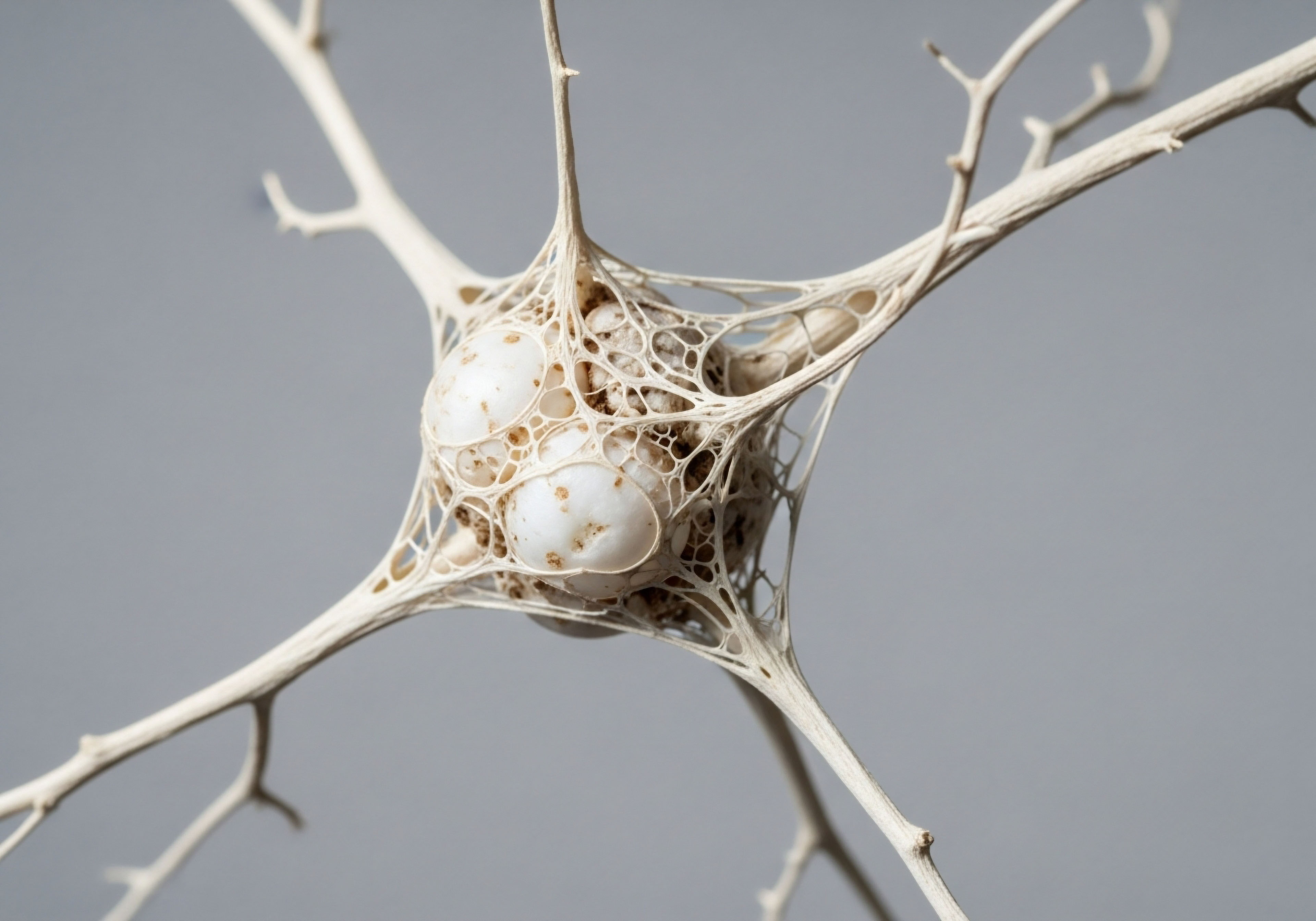
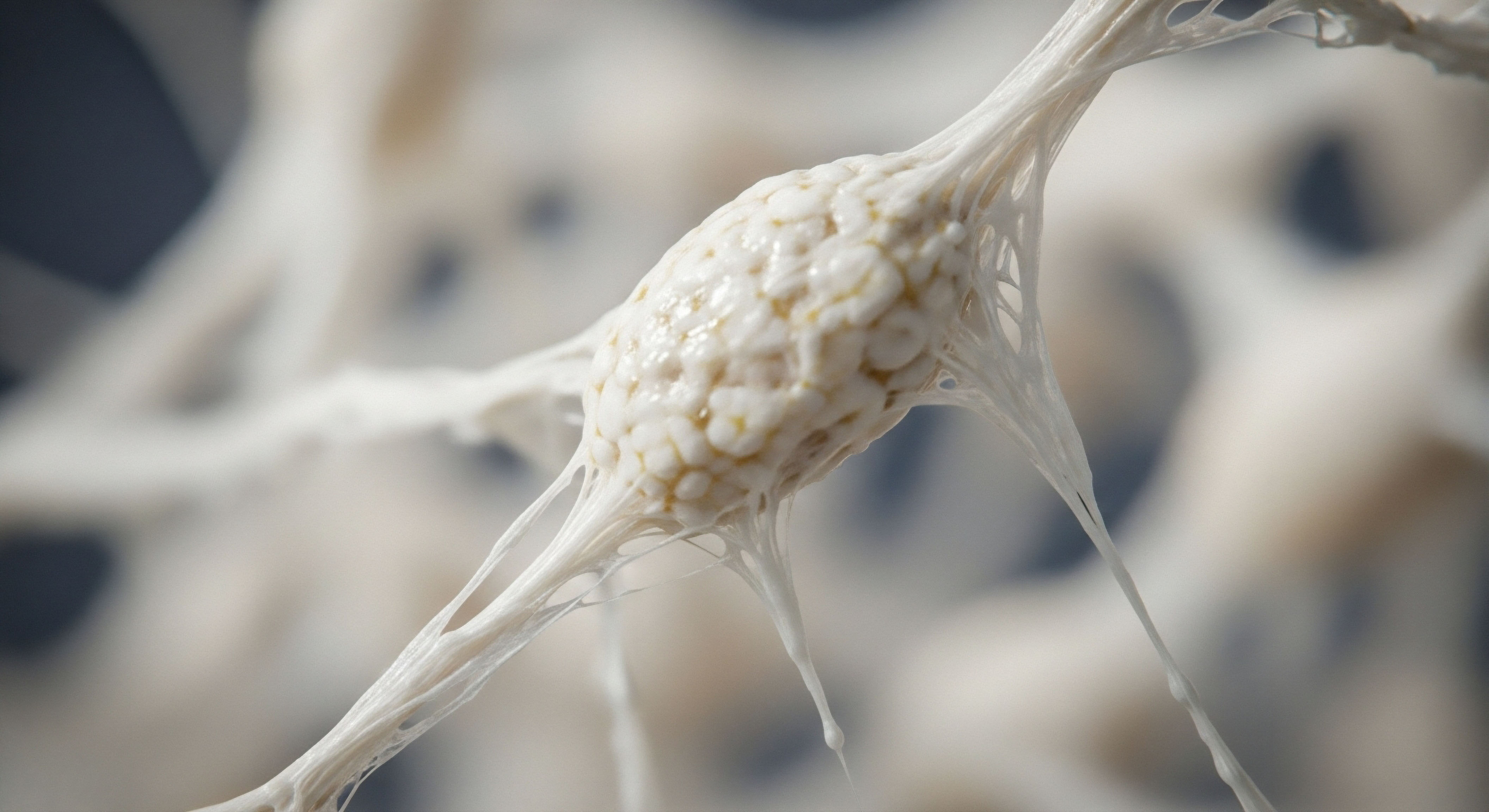
Imagine the skin as the roof of a house and the deeper muscle layer as the foundation. In between lies a layer of fat, compartmentalized by walls of connective tissue Meaning ∞ Connective tissue is a fundamental tissue type providing structural support, connection, and separation for various body components. called fibrous septae. In women, these septae are predominantly oriented perpendicular to the skin’s surface. These fibrous tethers anchor the skin to the deeper tissues.
When fat cells (adipocytes) within these compartments increase in volume, or when the septae themselves become less flexible, they pull the skin down, causing the fat to bulge between them. This dynamic creates the characteristic dimpling.
Cellulite’s appearance is a result of the structural relationship between fat cells and the connective tissue bands that anchor the skin.

The Hormonal Conductor
The entire system of skin, fat, and connective tissue operates under the influence of the endocrine system. Hormones act as chemical messengers, regulating cellular function throughout the body. Estrogen, the primary female sex hormone, is a powerful conductor of this orchestra.
It has profound effects on skin physiology, influencing everything from hydration and thickness to the production of key structural proteins. Estrogen receptors are found on the very cells responsible for building the skin’s framework ∞ the fibroblasts, which produce collagen, and the keratinocytes in the epidermis.
Estrogen’s role is fundamentally constructive in this context. It stimulates fibroblasts to synthesize collagen and elastin, the proteins that give skin its strength and elasticity. It also promotes the production of hyaluronic acid, which helps the skin retain water, contributing to its plumpness and turgor.
During the reproductive years, when estrogen levels are optimal, this system functions efficiently, maintaining the structural integrity of the dermis and the flexibility of the fibrous septae. The observation that cellulite often appears or becomes more pronounced during times of hormonal fluctuation, such as puberty, pregnancy, and particularly during the approach to menopause, is a direct testament to this connection.

Why Is Female Anatomy a Factor?
The predisposition of women to cellulite is rooted in anatomical and physiological differences that are hormonally driven. The vertical arrangement of the fibrous septae Meaning ∞ Fibrous septae are partitions or walls formed by dense connective tissue, primarily composed of collagen fibers, which divide and support various tissues and organs within the body. in women is a key distinction from the crisscrossing, more supportive network found in men. This structural difference means that any expansion of fat cells or loss of skin integrity is more likely to result in the surface dimpling characteristic of cellulite. These sex-specific traits are established during development and are maintained by the hormonal environment.
Therefore, understanding cellulite requires an appreciation of it as a feature of female biology, influenced heavily by the hormonal signals that define that biology. The journey to addressing its appearance begins with understanding the systems that create it.


Intermediate
Having established the foundational relationship between female anatomy, connective tissue, and hormonal influence, we can now examine the specific mechanisms through which hormonal shifts impact the tissues involved in cellulite. The transition into perimenopause and menopause marks a significant alteration in the body’s endocrine environment. The decline in ovarian production of estrogen sets off a cascade of changes that directly affect the skin’s structural integrity and the dynamics of the subcutaneous layer.
The menopausal transition is associated with a quantifiable decrease in skin quality. Studies have shown that skin collagen can decrease by as much as 30% in the first five years following menopause. This loss of collagen, which provides the primary scaffolding for the dermis, leads to thinner, less elastic skin.
A thinner, weaker dermal layer provides less resistance to the pressure from the underlying fat compartments, making the topography of the fibrous septae more visible. This process is a direct consequence of reduced estrogen signaling to the fibroblast cells responsible for collagen synthesis.
Declining estrogen levels during menopause directly reduce the skin’s ability to produce collagen, compromising its structural support system.

Hormonal Optimization and Connective Tissue
This brings us to the core of your question ∞ how can hormonal optimization protocols Meaning ∞ Hormonal Optimization Protocols are systematic clinical strategies designed to restore or maintain optimal endocrine balance. influence this process? The goal of such therapies in this context is to restore the physiological signaling that supports tissue integrity. By replenishing the body’s levels of key hormones, we can potentially mitigate the accelerated decline in connective tissue health associated with menopause.
A well-designed hormonal protocol for a woman experiencing these changes often includes bioidentical estradiol and progesterone. These are the primary hormones we aim to balance to support systemic health, which includes the integumentary system (the skin).
- Estradiol ∞ This is the most potent form of estrogen. Administered via transdermal creams, patches, or pellets, estradiol directly interacts with estrogen receptors in the skin. This interaction stimulates fibroblasts to increase the production of Type I and Type III collagen, the two most abundant types in the dermis. It also upregulates hyaluronic acid production, improving skin hydration and thickness. By reinforcing the dermal structure, estradiol can improve the skin’s firmness and elasticity, creating a stronger barrier against the herniation of subcutaneous fat.
- Progesterone ∞ Often prescribed in conjunction with estrogen, particularly for women with an intact uterus, progesterone also plays a role in skin health. It can help modulate the effects of androgens (male hormones) on the skin and may have a calming effect on skin inflammation. Its primary role in a balanced protocol is to ensure the healthy regulation of the endocrine system as a whole.
- Testosterone ∞ While primarily considered a male hormone, testosterone is also vital for women’s health, albeit in much smaller quantities. Low-dose testosterone therapy for women, often administered via subcutaneous injections or pellets, can have beneficial effects on connective tissue. Androgen receptors are also present on fibroblasts. Testosterone can contribute to increased skin thickness and collagen production. Some clinical observations suggest that testosterone may also play a role in improving the tone and integrity of the fibrous septae themselves.

What Are the Limits of Hormonal Intervention for Cellulite?
It is important to calibrate expectations. Hormonal optimization Meaning ∞ Hormonal Optimization is a clinical strategy for achieving physiological balance and optimal function within an individual’s endocrine system, extending beyond mere reference range normalcy. is a systemic approach aimed at restoring the physiological environment that supports healthy tissue. It is not a targeted cosmetic treatment that surgically cuts or dissolves the fibrous septae. The primary benefit of hormonal therapy in this context is preventative and restorative at a cellular level.
It works to improve the overall quality, thickness, and hydration of the skin and the underlying dermal matrix. This can lead to a visible improvement in the skin’s smoothness and a reduction in the appearance of dimpling. The therapy addresses the declining integrity of the container (the skin), making the underlying architecture less apparent.
The table below outlines the targeted effects of key hormones used in female optimization protocols on the components of the skin and subcutaneous tissue.
| Hormone | Target Cell/Component | Primary Biological Action |
|---|---|---|
| Estradiol | Fibroblasts | Increases synthesis of Type I & III Collagen, Elastin, Hyaluronic Acid |
| Estradiol | Keratinocytes | Improves epidermal thickness and barrier function |
| Progesterone | Sebaceous Glands | Modulates sebum production |
| Testosterone | Fibroblasts | Contributes to collagen synthesis and skin thickness |
By understanding these mechanisms, we can see that addressing the hormonal component of skin aging is a logical strategy for anyone concerned with changes like cellulite. It is an approach that supports the body’s own systems of maintenance and repair, leading to improvements in tissue quality from the inside out.
Academic
A sophisticated analysis of the potential for hormonal replacement therapy (HRT) to mitigate the appearance of gynoid lipodystrophy, or cellulite, requires a deep examination of the molecular biology of the skin’s extracellular matrix Meaning ∞ The Extracellular Matrix, often abbreviated as ECM, represents the non-cellular component present within all tissues and organs, providing essential physical scaffolding for cellular constituents and initiating crucial biochemical and biomechanical signals. (ECM) and the profound regulatory influence exerted by steroid hormones. The visual manifestation of cellulite is the macroscopic outcome of microscopic changes within the dermis and the adipose-septal architecture. These changes are intricately linked to the decline of ovarian hormone production, particularly 17β-estradiol, during the menopausal transition. The efficacy of HRT, therefore, hinges on its ability to reverse or slow these cellular and molecular alterations.

Estrogen Receptor Signaling in Dermal Fibroblasts
The primary mechanism through which estrogen mediates its effects on the skin is via binding to its cognate receptors, Estrogen Receptor Meaning ∞ Estrogen receptors are intracellular proteins activated by the hormone estrogen, serving as crucial mediators of its biological actions. α (ERα) and Estrogen Receptor β (ERβ), which are located in the nuclei of dermal fibroblasts, keratinocytes, and endothelial cells. Fibroblasts are the key effector cells in this system, responsible for synthesizing and remodeling the ECM. The ECM is a complex network of proteins and glycosaminoglycans, with Type I and Type III collagen forming its principal structural backbone.
Upon binding estradiol, the estrogen receptor acts as a ligand-activated transcription factor. It translocates to the nucleus, binds to specific DNA sequences known as Estrogen Response Elements (EREs) in the promoter regions of target genes, and modulates their transcription. Crucially, the genes encoding procollagen Type I and Type III are positively regulated by this pathway.
Clinical studies have demonstrated that the administration of estrogen, both systemically and topically, leads to a measurable increase in the messenger RNA (mRNA) for these collagens, with a corresponding increase in their protein expression in the dermis. One study documented a 6.49% increase in skin collagen content after six months of oral estrogen therapy.
Estrogen directly stimulates the genetic machinery within fibroblast cells to produce more collagen, reinforcing the skin’s structural matrix.

The Role of Transforming Growth Factor Beta
The signaling cascade is more complex than direct gene transcription alone. Estrogen also cross-talks with other critical signaling pathways, notably the Transforming Growth Factor-beta (TGF-β) pathway. TGF-β is a potent stimulator of fibroblast proliferation and ECM deposition. Research has shown that estrogen can upregulate the expression of both TGF-β and its receptors on fibroblasts.
This amplification of the TGF-β signal creates a powerful pro-collagen synthesis environment. This is a critical point ∞ estrogen does not simply turn on a switch; it sensitizes the cellular machinery to other growth signals, creating a robust and sustained effect on tissue maintenance.
Conversely, estrogen has been shown to downregulate the expression of certain Matrix Metalloproteinases Meaning ∞ Matrix Metalloproteinases, commonly abbreviated as MMPs, are a family of zinc-dependent enzymes responsible for the controlled breakdown of components within the extracellular matrix, including various collagens, elastin, and fibronectin, facilitating tissue turnover and structural adaptation. (MMPs), particularly MMP-1 (collagenase). MMPs are enzymes responsible for the degradation of ECM components. By simultaneously increasing collagen synthesis (via direct ERE binding and TGF-β upregulation) and decreasing collagen degradation (by suppressing MMPs), estrogen shifts the homeostatic balance of the ECM toward net accumulation. This results in a thicker, denser, and more structurally sound dermis, which is better able to resist the mechanical stresses exerted by the underlying adipose tissue.

How Does HRT Affect the Fibrous Septae?
The fibrous septae themselves are composed primarily of Type I collagen. While direct studies on the effect of HRT on septal tissue specifically are limited, the established mechanisms of estrogen action on dermal collagen are logically extensible to these structures. As the septae are also composed of collagen synthesized by fibroblasts, they are subject to the same hormonal regulation. A systemic environment rich in the signals that promote collagen synthesis Meaning ∞ Collagen synthesis is the precise biological process by which the body constructs collagen proteins, its most abundant structural components. and inhibit its degradation would theoretically lead to stronger, more robust septae.
This biochemical recalibration could improve their ability to function as supportive structures, potentially reducing the severity of dimpling over time. This is an area where further research is warranted to fully elucidate the direct histological changes within the septae following long-term hormonal therapy.
The table below details the molecular-level effects of estrogen on the key cellular processes involved in maintaining the dermal extracellular matrix.
| Molecular Target | Effect of Estrogen Signaling | Net Physiological Outcome |
|---|---|---|
| Procollagen I & III Genes (COL1A1, COL3A1) | Upregulation of transcription via ERE binding | Increased synthesis of collagen fibers |
| TGF-β Pathway | Upregulation of TGF-β and its receptors | Amplified signal for fibroblast proliferation and ECM production |
| Matrix Metalloproteinase-1 (MMP-1) Gene | Downregulation of transcription | Decreased degradation of existing collagen fibers |
| Hyaluronic Acid Synthase Gene | Upregulation of transcription | Increased production of hyaluronic acid, improving hydration |
In conclusion, from an academic and mechanistic perspective, the rationale for using HRT to improve skin quality and potentially the appearance of cellulite is robust. It is grounded in the fundamental principles of molecular endocrinology and cell biology. By restoring estrogenic signaling, HRT directly counteracts the primary drivers of age-related dermal atrophy ∞ decreased collagen synthesis and increased degradation. This leads to a quantitative and qualitative improvement in the skin’s extracellular matrix, which forms the basis for any visible aesthetic improvement.
- Receptor Activation ∞ The process begins with estradiol binding to estrogen receptors within fibroblast cells, initiating a cascade of intracellular events.
- Transcriptional Regulation ∞ The activated receptor directly influences the genetic expression of proteins essential for the skin’s structure, promoting the creation of new collagen.
- Enzymatic Modulation ∞ Simultaneously, the hormonal signals reduce the activity of enzymes that break down the existing collagen network, preserving the skin’s integrity.
References
- Calleja-Agius, Jean, and Mark P. Brincat. “Effects of hormone replacement therapy on connective tissue ∞ why is this important?” Best Practice & Research Clinical Obstetrics & Gynaecology, vol. 23, no. 1, 2009, pp. 121-7.
- Piérard-Franchimont, C. et al. “Hormone replacement therapy for skin ageing in post-menopausal women ∞ a review.” Gynecological and Reproductive Endocrinology and Metabolism, vol. 5, no. 1, 2024, pp. 34-37.
- Stevenson, S. and J. Thornton. “Effect of estrogens on skin aging and the potential role of SERMs.” Clinical Interventions in Aging, vol. 2, no. 3, 2007, pp. 283-97.
- Bass, Lawrence S. and Michael S. Kaminer. “Insights Into the Pathophysiology of Cellulite ∞ A Review.” Dermatologic Surgery, vol. 46, 2020, pp. S77-S85.
- Verdier-Sévrain, S. and F. Bonte. “Skin hydration ∞ a review on its molecular mechanisms.” Journal of Cosmetic Dermatology, vol. 6, no. 2, 2007, pp. 75-82.
- Brincat, M. P. et al. “Skin collagen changes in postmenopausal women receiving different regimens of estrogen therapy.” Obstetrics and Gynecology, vol. 70, no. 6, 1987, pp. 123-7.
- Nürnberger, F. and G. Müller. “So-called cellulite ∞ an invented disease.” Journal of Dermatologic Surgery and Oncology, vol. 4, no. 3, 1978, pp. 221-9.
- Rossi, A.B.R. and A.L. Vergnanini. “Cellulite ∞ a review.” Journal of the European Academy of Dermatology and Venereology, vol. 14, no. 4, 2000, pp. 251-62.
Reflection
The information presented here provides a biological framework for understanding the connection between your internal hormonal environment and the visible texture of your skin. You began with a question about appearance, and through this exploration, we have arrived at a deeper appreciation for the body’s intricate systems of maintenance and repair. The knowledge that you can influence these foundational systems is a powerful starting point. This is your biology, and understanding its language is the first step on any path toward personal wellness.
Consider how this information reframes your perspective, shifting the focus from a single aesthetic concern to a holistic view of your body’s long-term health and vitality. The path forward is one of personalized strategy, guided by your unique physiology and goals.


