


Fundamentals
The feeling is unmistakable. After discontinuing a hormonal protocol, the body can feel as though its internal engine has been switched off. Vitality wanes, mental clarity clouds over, and a profound sense of inertia sets in. This experience is a direct reflection of a system in transition, a conversation that has been temporarily silenced and is now struggling to resume.
Understanding this process begins with appreciating the elegant biological architecture that governs masculine hormonal health ∞ the Hypothalamic-Pituitary-Gonadal (HPG) axis. This system operates as a continuous, dynamic feedback loop, a conversation between the brain and the testes designed to maintain hormonal equilibrium.
At the highest level of command sits the hypothalamus, a specialized region of the brain that acts as the primary sensor for the body’s hormonal state. When it detects a need for testosterone, it releases Gonadotropin-Releasing Hormone (GnRH). This is the initial instruction, a precise chemical message sent directly to the pituitary gland.
The pituitary, acting as the system’s amplifier and dispatcher, receives the GnRH signal and responds by secreting two critical messenger hormones into the bloodstream ∞ Luteinizing Hormone (LH) and Follicle-Stimulating Hormone (FSH). These hormones travel throughout the body, yet they are specifically designed to be received by the testes.
The health of the HPG axis is defined by the clarity of its signals and the capacity of the testes to respond.
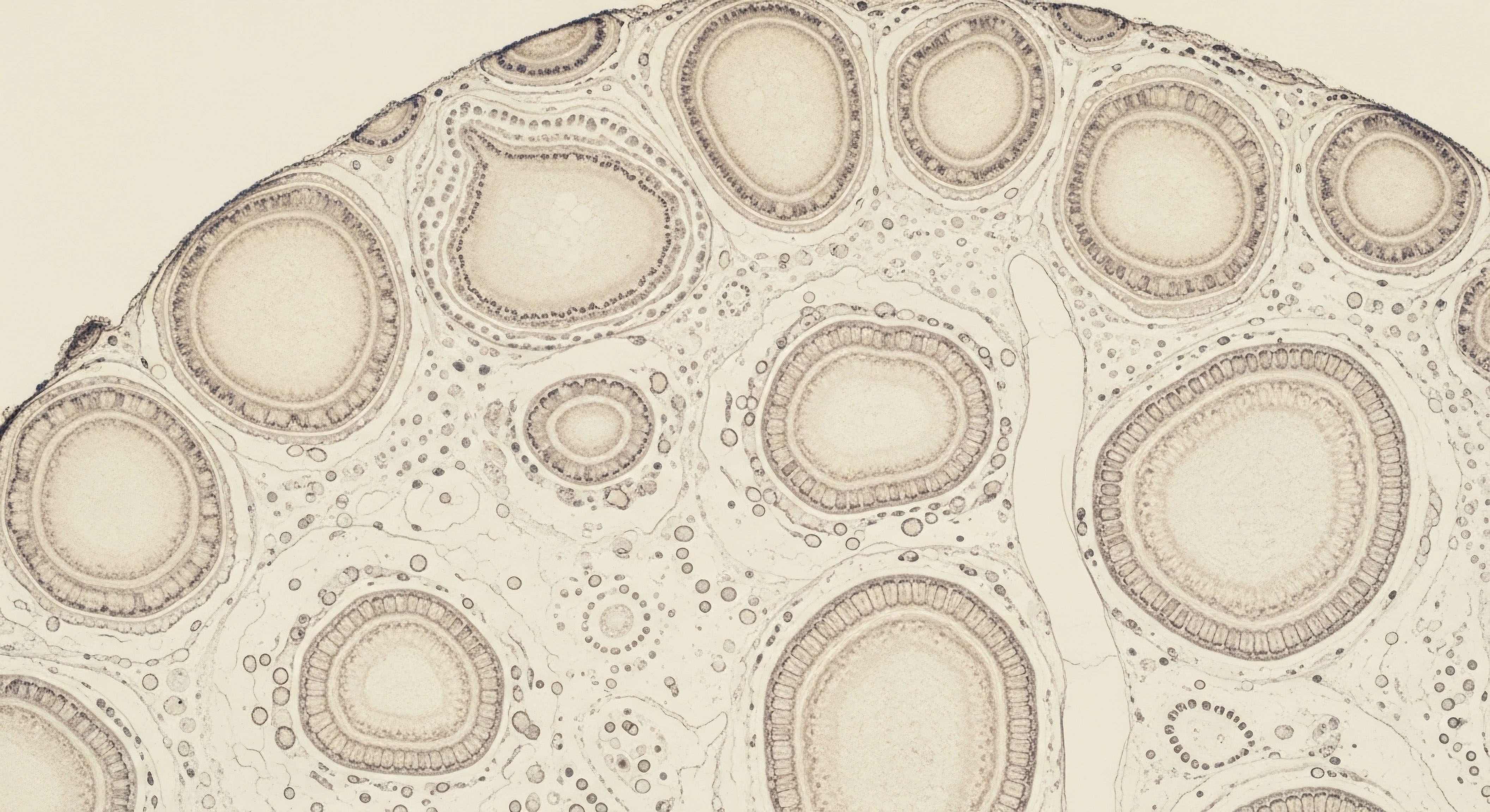
The testes represent the manufacturing center of this entire operation. They are a highly specialized factory with two primary production lines, each managed by a distinct cell type. LH signals the Leydig cells, which are responsible for the production and secretion of testosterone.
This is the hormone that governs libido, muscle mass, energy levels, and cognitive function. Concurrently, FSH communicates with the Sertoli cells. These are the master regulators of spermatogenesis, the intricate process of sperm production. Sertoli cells create a nurturing environment for developing sperm and also produce a hormone called inhibin B, which signals back to the brain that sperm production is active.
The system’s elegance lies in its self-regulation. Rising levels of testosterone and inhibin B travel back to the brain, signaling to the hypothalamus and pituitary to decrease their output of GnRH, LH, and FSH. This negative feedback ensures hormonal levels remain within a healthy, functional range.

The Testicular Endowment
The capacity for the HPG axis to recover its function following the cessation of external hormone administration is fundamentally linked to the condition of this testicular factory before the protocol began. Pre-existing testicular function is the baseline, the inherent operational capacity and structural integrity of the Leydig and Sertoli cells.
This ‘testicular endowment’ is a product of genetics, age, and lifelong health. A system that was robust, efficient, and healthy possesses a high degree of functional reserve. Its cellular machinery is intact, its signaling pathways are clear, and its cell populations are dense and vigorous.
Conversely, a testicular environment that was already compromised will have a much more challenging path to recovery. Factors like advanced age, chronic inflammation, metabolic dysfunction, or environmental exposures can diminish the health and number of Leydig and Sertoli cells. This creates a state of reduced functional reserve.
When the brain’s signals (LH and FSH) are reintroduced after a period of suppression, this compromised factory struggles to respond. The machinery is sluggish, the workforce is depleted, and the production lines cannot easily come back online. Therefore, the journey to restoring endogenous hormonal balance is a direct reflection of the health of the foundational tissues responsible for hormone production.


Intermediate
When an individual undertakes a protocol involving exogenous testosterone, the HPG axis interprets the elevated serum hormone levels as a signal that the testicular factory is over-producing. In response to this perceived surplus, the hypothalamus and pituitary initiate a system-wide shutdown of their own signals to maintain homeostasis.
GnRH secretion diminishes, which in turn halts the release of LH and FSH. This cessation of the brain’s stimulating messengers is known as central suppression. The testes, deprived of their operational commands, enter a state of dormancy. Leydig cells, lacking the LH signal, cease testosterone production. Sertoli cells, without FSH, slow the machinery of spermatogenesis. This physiological response is the reason for the testicular atrophy often observed during such therapies; the factory is temporarily decommissioned.
The process of recovery, initiated upon the discontinuation of external androgens, is the reversal of this sequence. As the exogenous testosterone clears from the bloodstream, the brain’s sensors detect declining levels. The hypothalamus begins to cautiously send out GnRH pulses once more. The pituitary responds, releasing LH and FSH.
These initial signals are the wake-up call to the dormant testes. The central question at this stage is whether the testes are capable of mounting a response. The speed and completeness of this reactivation are directly governed by the pre-existing health and resilience of the testicular tissue. A system with a high functional reserve, characterized by healthy and plentiful Leydig and Sertoli cells, will respond more promptly and robustly to the returning hormonal signals.

What Determines Recovery Potential?
The trajectory of HPG axis recovery is influenced by a collection of interconnected factors. Each one contributes to the timeline and ceiling of the system’s restoration. A comprehensive understanding of these elements is vital for setting realistic expectations and designing effective recovery protocols.
- Baseline Testicular Health The absolute number and functional quality of Leydig and Sertoli cells present before suppression began is the primary determinant of recovery potential. A higher baseline predicts a more efficient restart.
- Duration of Suppression Longer periods of HPG axis shutdown can lead to a more profound state of testicular dormancy. The cellular machinery may require more time and stronger signals to reactivate fully.
- Age of the Individual Biological aging is associated with a natural decline in the number and efficiency of testicular cells. An older individual inherently possesses a lower functional reserve, which can extend the recovery timeline.
- Underlying Health Conditions Metabolic syndrome, chronic inflammation, and poor vascular health can all impair testicular function. These conditions create a suboptimal environment for cellular recovery and hormone production.
- Genetic Predisposition Individual genetic factors can influence the sensitivity of the hypothalamus and pituitary as well as the inherent robustness of the testicular cells themselves.

Strategic Interventions for Axis Reactivation
For individuals seeking to facilitate HPG axis recovery, specific clinical protocols are designed to stimulate different components of the system. These interventions act as strategic catalysts, intended to shorten the period of hormone deficiency and encourage the restoration of the body’s natural production rhythm. The selection and application of these agents depend on the specific goals, whether for fertility or for the normalization of endogenous testosterone.
Clinical protocols for HPG axis recovery aim to amplify the body’s own signaling to restart testicular function.
These protocols are not a universal solution; their efficacy is deeply intertwined with the individual’s baseline testicular health. A protocol can amplify a signal, but there must be a healthy and responsive production facility to receive that signal and act upon it. The table below outlines the mechanisms of common agents used in post-suppression recovery protocols.
| Agent | Primary Mechanism of Action | Target Component |
|---|---|---|
| Gonadorelin (or hCG) | Acts as a Luteinizing Hormone (LH) analog, directly stimulating the Leydig cells in the testes to produce testosterone and support spermatogenesis. | Testes (Leydig Cells) |
| Clomiphene Citrate | A Selective Estrogen Receptor Modulator (SERM) that blocks estrogen receptors in the hypothalamus, preventing estrogen’s negative feedback and prompting an increase in GnRH, LH, and FSH production. | Hypothalamus / Pituitary |
| Tamoxifen Citrate | Another SERM that functions similarly to Clomiphene, blocking estrogen feedback at the pituitary to increase the output of LH and FSH. It is often used for its distinct binding profile. | Pituitary |
| Anastrozole | An Aromatase Inhibitor (AI) that reduces the conversion of testosterone to estrogen throughout the body. Lowering systemic estrogen reduces negative feedback on the pituitary and hypothalamus, thereby increasing LH and FSH. | Systemic (Aromatase Enzyme) |
The choice of agent or combination of agents is a clinical decision based on laboratory markers, individual health status, and the degree of suppression. For instance, using a SERM like Clomiphene aims to restart the entire axis from the top down, while using Gonadorelin provides direct stimulation to the testes, a bottom-up approach. Both strategies ultimately depend on the foundational capacity of the testicular cells to perform their designated functions once the signal arrives.


Academic
A granular analysis of HPG axis recovery moves beyond systemic feedback loops and into the realm of cellular biology and paracrine signaling within the testicular microenvironment. The ultimate success of a recovery protocol is contingent upon the cytological integrity and responsiveness of the gonadal cell populations.
Pre-existing testicular function is a clinical term for the accumulated health of these cells. The administration of exogenous androgens induces a state of functional quiescence, and the recovery is a measure of the cells’ ability to emerge from this state and resume complex steroidogenic and gametogenic activities.
The Leydig cell is the primary locus of testosterone synthesis. Its function is inextricably linked to its morphology, particularly the abundance of its smooth endoplasmic reticulum (SER), the organelle housing the enzymes essential for steroidogenesis. Research demonstrates a direct correlation between LH suppression and a quantifiable reduction in SER volume within Leydig cells.
The recovery of steroidogenic capacity post-suppression follows a parallel track with the restoration of this intricate cellular architecture. Therefore, a Leydig cell population that was robust and structurally sound prior to suppression retains a greater capacity for rapid morphological and functional reconstitution upon the reintroduction of endogenous LH.
How Does Sertoli Cell Health Govern Recovery?
The Leydig cell does not operate in isolation. Its persistence and function are critically dependent on a healthy relationship with the Sertoli cell population. Sertoli cells, which form the structural framework of the seminiferous tubules, provide essential support for spermatogenesis and also engage in complex paracrine signaling that sustains the entire testicular ecosystem.
Studies involving the selective ablation of Sertoli cells in animal models reveal a profound impact on the Leydig cell population. In the absence of Sertoli cell-derived support factors, Leydig cells undergo apoptosis, leading to a significant decline in their numbers. One study demonstrated that the loss of Sertoli cells precipitated a 75% reduction in the Leydig cell population.
This interdependence establishes a critical principle ∞ the potential for testosterone recovery is directly linked to the health of the cells responsible for sperm production. A man’s pre-existing Sertoli cell function, therefore, is a powerful predictor of his ability to restore Leydig cell function.
A compromised Sertoli population, diminished by age or metabolic disease, cannot provide the necessary local signaling to maintain Leydig cell health during a period of suppression or to support their reactivation afterward. This systems-biology perspective clarifies that testicular reserve is a property of the entire organ, not just the testosterone-producing cells.

What Are the Key Biomarkers of Testicular Reserve?
Assessing pre-existing testicular function involves analyzing a panel of biomarkers that, together, paint a picture of the health of the HPG axis and the testicular factory. These laboratory values provide objective data on the integrity of both the signaling and manufacturing components of the system.
| Biomarker | Origin | Clinical Significance |
|---|---|---|
| Luteinizing Hormone (LH) | Pituitary Gland | Indicates the strength of the pituitary’s signal to the Leydig cells. Low levels suggest central suppression, while high levels with low testosterone suggest primary testicular failure. |
| Follicle-Stimulating Hormone (FSH) | Pituitary Gland | Reflects the pituitary’s signal to the Sertoli cells. Elevated levels often point to impaired spermatogenesis and Sertoli cell dysfunction. |
| Inhibin B | Sertoli Cells | A direct product of Sertoli cells that provides negative feedback to the pituitary regarding FSH. It is considered a primary marker of spermatogenic activity and Sertoli cell health. |
| Anti-Müllerian Hormone (AMH) | Sertoli Cells (pre-pubertal) | While primarily a marker of ovarian reserve in females, in males it is produced by Sertoli cells and can be an indicator of their functional status, particularly in certain clinical contexts. |
| Total and Free Testosterone | Leydig Cells | The primary output of the Leydig cells. The relationship between testosterone levels and LH levels is fundamental to diagnosing the location of axis dysfunction. |
| Testicular Volume | Testes | A physical measurement that correlates with the mass of seminiferous tubules and, by extension, the Sertoli and germ cell populations. Atrophy indicates suppression or dysfunction. |
The measurement of inhibin B is particularly insightful. As a direct product of the Sertoli cells, its level offers a window into the health of the spermatogenic engine. Low inhibin B levels in the context of high FSH are a strong indicator of Sertoli cell stress or damage.
This finding has profound implications for HPG axis recovery, suggesting that the very cells needed to support the Leydig cell population are themselves compromised. This deepens the understanding that a successful recovery requires a dual competency ∞ the brain must re-learn how to signal, and the testicular environment must be healthy enough to respond and rebuild.
The resilience of the testicular microenvironment, particularly the symbiotic relationship between Sertoli and Leydig cells, is the bedrock of hormonal recovery.
Furthermore, the process of cellular senescence contributes to the age-related decline in testicular reserve. As men age, a subset of both Leydig and Sertoli cells enters a senescent state, characterized by a cessation of division and the secretion of pro-inflammatory cytokines.
This creates an inflammatory local environment that degrades the function of neighboring cells and compromises the integrity of the blood-testis barrier. An individual beginning a hormone protocol with a higher burden of senescent cells has a significantly lower functional reserve, predisposing them to a slower, more difficult, and potentially incomplete recovery of their HPG axis.

References
- Zirkin, B. R. & Chen, H. (2000). Regulation of Leydig cell steroidogenic function during aging. Biology of Reproduction, 63(4), 977 ∞ 981.
- Raman, J. D. & Schlegel, P. N. (2002). Aromatase inhibitors for male infertility. The Journal of Urology, 167(2 Pt 1), 624 ∞ 629.
- Rebourcet, D. O’Shaughnessy, P. J. Monteiro, A. Milne, L. Cruickshanks, L. Jeffrey, N. & Smith, L. B. (2014). Sertoli cells maintain Leydig cell number and peritubular myoid cell activity in the adult mouse testis. PLoS One, 9(8), e105687.
- Coward, R. M. Rajanahally, S. Kovac, J. R. Smith, R. P. Pastuszak, A. W. & Lipshultz, L. I. (2013). Anabolic-androgenic steroid-induced hypogonadism ∞ diagnosis and treatment. Postgraduate Medicine, 125(6), 127-134.
- Drobnis, E. Z. & Stoddart, A. (2019). Sa-022 ∞ Exogenous testosterone and male fertility. Journal of Andrology, 30(s1), 1-22.
- Welsh, M. Moffat, L. Belling, K. de Franca, L. R. Segatelli, T. M. Saunders, P. T. & Smith, L. B. (2009). Androgen receptor signalling in Sertoli cells is required for the maintenance of adult Leydig cells. International Journal of Andrology, 32(4), 385-391.
- Rastrelli, G. Corona, G. & Maggi, M. (2018). The role of testosterone in managing the metabolic syndrome. Current Opinion in Endocrinology, Diabetes and Obesity, 25(3), 199 ∞ 220.
- Lykhonosov, M. P. Fedotov, Y. N. & Kalinchenko, S. Y. (2020). Peculiarity of recovery of the hypothalamic-pituitary-gonadal (hpg) axis, in men after using androgenic anabolic steroids. Problems of Endocrinology, 66(4), 59-66.

Reflection
The information presented here maps the biological terrain of hormonal recovery, tracing the intricate connections from the brain’s highest command centers to the cellular machinery within the testes. This knowledge transforms the abstract feeling of ‘being offline’ into a comprehensible physiological process.
It reveals that the body’s capacity to restore its natural rhythm is not a matter of chance, but a direct consequence of the foundational health established over a lifetime. This understanding is the first step. The path forward involves looking inward, assessing the personal landscape of your own health, and recognizing that true optimization is a dialogue between targeted clinical strategies and the body’s inherent, cultivated vitality.