


Fundamentals
You may have found yourself on a frustrating plateau. You follow the protocols, you are diligent with your nutrition and training, yet the results you anticipate, the vitality you seek, remain just out of reach. This experience, this feeling of being stalled despite your best efforts, is a valid and common starting point for a deeper investigation into your own unique biology.
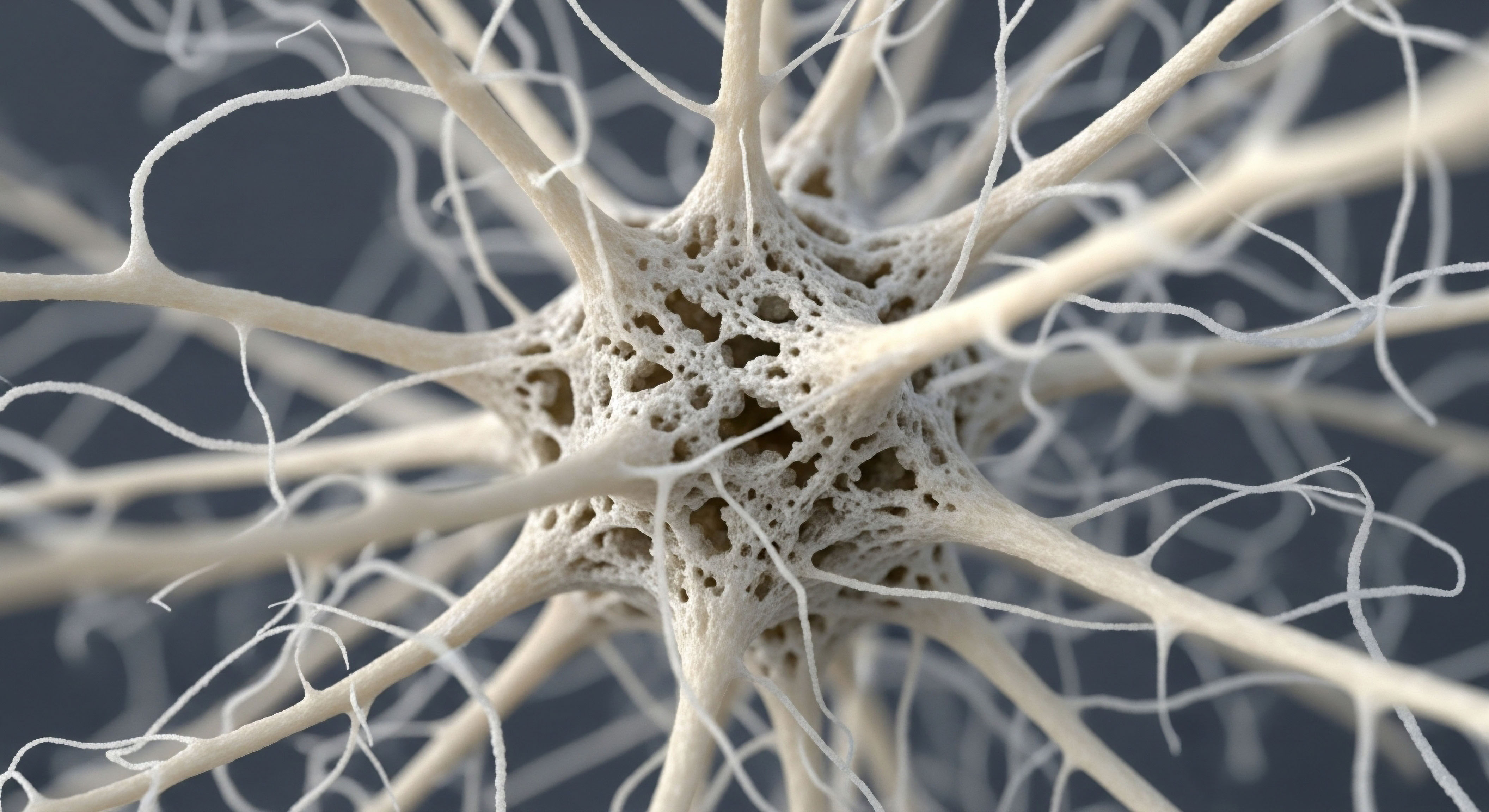
The answer often resides within the complex and dynamic environment of your body’s internal architecture, specifically its composition of muscle and adipose tissue. Your body is a finely tuned system of communication, and the tissues that comprise it are the broadcasting stations and receivers that determine how well those communications are sent and received. Understanding this internal landscape is the first step toward understanding why a standardized protocol might require personalized adjustments.
We can begin by looking at the very nature of body composition. It is the precise makeup of your physical self, categorized primarily into lean body mass and fat mass. Lean body mass includes muscle, bone, organs, and water. Fat mass, or adipose tissue, is the body’s primary energy reserve.
For many years, scientific thought viewed adipose tissue as a passive storage depot, a simple container for excess calories. We now understand this view is profoundly incomplete. Adipose tissue is a sophisticated and highly active endocrine organ, a critical signaling hub that produces and secretes a multitude of powerful biochemical messengers.
These messengers, known as adipokines, travel throughout your body and influence everything from appetite and metabolism to immune function and inflammation. The quantity and, most importantly, the type of adipose tissue you carry directly dictates the hormonal and inflammatory signals your body produces, creating the specific biological environment in which any therapeutic peptide or hormone must operate.

The Two Faces of Adipose Tissue
To grasp the influence of body composition, we must differentiate between the main types of fat. Your body contains several types, but for this discussion, we will focus on the two most significant players in metabolic health ∞ subcutaneous adipose tissue (SAT) and visceral adipose tissue (VAT).
- Subcutaneous Adipose Tissue (SAT) ∞ This is the fat stored directly beneath your skin, the tissue you can pinch. While excess SAT can be a cosmetic concern, it is generally considered less metabolically harmful than its deeper counterpart. It participates in hormone production and energy storage, but its inflammatory output is comparatively lower. In some contexts, it can even have protective metabolic characteristics.
- Visceral Adipose Tissue (VAT) ∞ This is the fat stored deep within the abdominal cavity, surrounding vital organs like the liver, pancreas, and intestines. You cannot see or pinch VAT. Its accumulation is a central factor in metabolic dysfunction. VAT is highly metabolically active and is a potent source of pro-inflammatory signals that can disrupt systemic communication.
- Brown Adipose Tissue (BAT) ∞ A specialized form of fat that burns energy to produce heat. While less abundant in adults, its activation is a subject of intense research for its potential metabolic benefits. Its role in peptide responsiveness is less direct but is part of the overall metabolic picture.
The ratio of VAT to SAT is a far more meaningful predictor of metabolic health than body mass index (BMI) alone. An individual can have a “normal” BMI yet carry a dangerous amount of visceral fat, a condition sometimes called “thin-on-the-outside, fat-on-the-inside” or TOFI. This internal environment, rich in inflammatory signals from VAT, is often the unseen obstacle that can blunt the effectiveness of even the most well-designed wellness protocols.

Peptides as Precision Messengers
Now, let’s consider the other side of the equation ∞ the therapeutic peptides themselves. Peptides are short chains of amino acids that act as highly specific signaling molecules. Think of them as keys designed to fit into specific locks (receptors) on the surface of cells.
When a peptide like Sermorelin or Ipamorelin binds to its receptor on the pituitary gland, it instructs that gland to produce and release growth hormone. When a hormone like testosterone is administered, it binds to androgen receptors throughout the body, initiating a cascade of effects related to muscle growth, libido, and red blood cell production.
The effectiveness of these therapies depends on three core factors:
- The integrity of the signal itself (the quality and dosage of the peptide or hormone).
- The number and sensitivity of the receptors (the locks on the cells).
- The clarity of the signaling environment (the absence of interfering noise).
Body composition, particularly the amount of visceral fat, profoundly impacts the second and third factors. A body with high visceral adiposity is a body filled with systemic, low-grade inflammation. This inflammation is like static on a radio channel, making it difficult for the precise signals from peptides and hormones to be heard and acted upon.
The pro-inflammatory molecules released by VAT can directly interfere with receptor function, effectively changing the locks so the keys no longer fit as well as they should. This creates a state of “resistance,” where the body becomes less responsive to both its own endogenous hormones and to exogenous therapeutic agents.
Therefore, your body composition is not a passive backdrop to your health journey; it is an active, influential force that shapes your biological reality and your response to any intervention designed to improve it.


Intermediate
Understanding that visceral adipose tissue (VAT) functions as an active endocrine organ is the foundation. Now, we must examine the specific mechanisms through which it exerts its powerful influence, creating a biochemical environment that can either support or hinder the goals of hormonal optimization and peptide therapy.
The challenges in achieving desired outcomes are frequently rooted in the molecular crosstalk between hypertrophied, dysfunctional adipocytes and the rest of the body’s signaling systems. This crosstalk generates a state of chronic, low-grade inflammation and hormonal dysregulation that directly impacts the efficacy of protocols like Testosterone Replacement Therapy (TRT) and Growth Hormone Secretagogue (GHS) use.

The Inflammatory Cascade from Visceral Fat
When visceral adipocytes become overfilled with lipids (hypertrophy), they experience cellular stress. This stress triggers a protective response that, when sustained, becomes detrimental. These stressed fat cells begin to secrete a cocktail of pro-inflammatory cytokines, chief among them being Tumor Necrosis Factor-alpha (TNF-α) and Interleukin-6 (IL-6). These molecules are not contained within the adipose tissue; they spill into the bloodstream and circulate throughout the body, systemically altering cellular function.
This inflammatory state has direct consequences for peptide and hormone responsiveness:
- Insulin Resistance ∞ TNF-α is a primary driver of insulin resistance. It directly interferes with the insulin receptor signaling pathway in muscle and liver cells. When cells become less sensitive to insulin, the pancreas must produce more to manage blood glucose. The resulting hyperinsulinemia is itself a pro-inflammatory state and is linked to reduced growth hormone secretion, creating a negative feedback loop that promotes further fat accumulation.
- Receptor Desensitization ∞ Chronic exposure to inflammation can downregulate or desensitize critical hormone receptors. For example, inflammation can impair the function of the Growth Hormone Releasing Hormone (GHRH) receptor on the pituitary gland. This means that even if a GHS like Sermorelin or CJC-1295 is administered, the pituitary’s ability to respond and release growth hormone is blunted. The signal is sent, but the receiver is compromised.
- Systemic Stress ∞ The inflammatory messengers from VAT contribute to the overall allostatic load on the body, stimulating the hypothalamic-pituitary-adrenal (HPA) axis and increasing cortisol levels. Elevated cortisol can suppress the hypothalamic-pituitary-gonadal (HPG) axis, leading to lower natural testosterone production and further complicating TRT protocols.
The chronic inflammatory state generated by excess visceral fat acts as a systemic communication disruptor, directly impairing the ability of cells to receive and respond to both natural and therapeutic hormonal signals.

How Does Body Fat Alter Testosterone Therapy?
One of the most direct ways body composition affects hormonal therapy is through the activity of an enzyme called aromatase. Adipose tissue is a primary site of aromatase expression in the body, particularly in men. This enzyme is responsible for converting androgens (like testosterone) into estrogens (like estradiol). While a certain level of this conversion is necessary for health, excess adipose tissue creates an environment of over-conversion.
Consider a male patient on a standard TRT protocol. An individual with a higher body fat percentage, especially high VAT, will have significantly more aromatase activity. This has two major clinical implications:
- Reduced Testosterone Efficacy ∞ A larger portion of the administered testosterone is converted into estradiol before it can exert its full androgenic effects on muscle, bone, and brain tissue. This can lead to suboptimal results in terms of lean mass gain, fat loss, and improvements in well-being, even with what appears to be an adequate dose of testosterone.
- Increased Estrogenic Side Effects ∞ The elevated estradiol levels can lead to unwanted side effects such as gynecomastia (breast tissue development), water retention, and mood swings. This is why an aromatase inhibitor like Anastrozole is often a necessary component of TRT protocols, especially for individuals with higher body fat. The need for Anastrozole is a direct consequence of the endocrine activity of the adipose tissue.

Table of TRT Protocol Considerations Based on Body Composition
The following table illustrates how clinical thinking must adapt to an individual’s body composition for a successful outcome.
| Biometric Profile | Primary Hormonal Challenge | Protocol Consideration | Therapeutic Goal |
|---|---|---|---|
| Lean Individual (Low Visceral Fat) | Primary hypogonadism; low testosterone production with minimal peripheral conversion. | Standard TRT dosage (e.g. Testosterone Cypionate) may be sufficient. Lower dose or less frequent Anastrozole may be needed. | Restore testosterone to optimal physiological levels with minimal side effect management. |
| High Body Fat Individual (High Visceral Fat) | Hypogonadism compounded by high aromatase activity and inflammation-induced receptor resistance. | TRT dosage may need careful titration. Proactive use of an aromatase inhibitor (Anastrozole) is often required. Lifestyle interventions to reduce VAT are critical for long-term success. | Restore testosterone while actively managing estrogen conversion and improving the underlying metabolic environment to enhance receptor sensitivity. |
The Impact on Growth Hormone Secretagogues
The effectiveness of GHS peptides like Sermorelin, Ipamorelin, and Tesamorelin is profoundly influenced by an individual’s metabolic health, which is dictated by their body composition. Research has shown that obesity and high levels of visceral fat are associated with a blunted growth hormone response to GHS administration. The mechanisms are multifaceted.
Firstly, high circulating levels of insulin and free fatty acids, both consequences of VAT accumulation, are known to increase the secretion of somatostatin. Somatostatin is the body’s natural “off switch” for growth hormone release. It acts on the pituitary to inhibit GH secretion.
Therefore, a person with high visceral fat has a stronger inhibitory signal (somatostatin) that the stimulatory signal from a peptide like Ipamorelin must overcome. Secondly, as mentioned, the inflammation generated by VAT can reduce the sensitivity of the GHRH receptors themselves. The result is a diminished pulse of growth hormone for a given dose of the peptide.
This is why protocols targeting fat loss, such as those using Tesamorelin, are designed to specifically reduce VAT, which in turn improves the body’s own GH pulsatility and its responsiveness to therapy over time. The peptide helps reduce the very thing that was inhibiting its effectiveness, creating a positive feedback loop.


Academic
A sophisticated analysis of peptide responsiveness requires moving beyond systemic inflammation and into the specific molecular signaling pathways governed by adipokines. Adipose tissue, particularly VAT, is the source of numerous signaling molecules, but two stand out for their opposing and powerful roles in metabolic regulation ∞ leptin and adiponectin.
The balance, or more accurately, the imbalance, of these two hormones in states of high visceral adiposity creates a complex web of cellular and neuroendocrine dysregulation. This dysregulation provides a precise molecular explanation for the variable and often blunted responses observed in individuals undergoing peptide and hormone therapies. Understanding this dynamic is essential for translating clinical protocols into predictable, personalized outcomes.

Leptin Resistance a Central Mediator of Therapeutic Failure
Leptin is a hormone secreted by adipocytes, and its circulating levels are generally proportional to total fat mass. In a healthy system, leptin acts on the hypothalamus to signal satiety and permit increased energy expenditure, forming a classic negative feedback loop to maintain energy homeostasis.
In individuals with significant visceral obesity, a paradoxical state known as leptin resistance occurs. Despite having very high circulating levels of leptin, the brain, particularly the hypothalamus, fails to recognize and respond to the signal. This is analogous to the insulin resistance seen in type 2 diabetes.
The molecular underpinnings of leptin resistance are complex, involving impaired transport of leptin across the blood-brain barrier and defects in the intracellular signaling cascade downstream of the leptin receptor (LEPR), specifically the Janus kinase 2/Signal Transducer and Activator of Transcription 3 (JAK2/STAT3) pathway. Chronic inflammation, driven by cytokines like TNF-α from VAT, can induce the expression of proteins like Suppressor of Cytokine Signaling 3 (SOCS3), which directly inhibits JAK2 phosphorylation, effectively blocking the leptin signal within the neuron.
This state of central leptin resistance has profound implications for peptide therapies:
- Dysregulation of the HPG Axis ∞ Leptin has a permissive effect on the reproductive axis. It provides a signal of energy sufficiency to the hypothalamus, which is necessary for the proper pulsatile release of Gonadotropin-Releasing Hormone (GnRH). In a state of leptin resistance, the hypothalamus interprets the situation as one of energy deficit, despite ample stored energy. This can lead to a suppression of GnRH pulses, which in turn reduces the pituitary’s release of Luteinizing Hormone (LH) and Follicle-Stimulating Hormone (FSH). The result is diminished endogenous testosterone production in men and menstrual irregularities in women. This underlying suppression means that a TRT protocol is initiated in a system that is already fighting against itself, complicating efforts to find a stable and effective dosage.
- Blunting of Growth Hormone Secretion ∞ The neuroendocrine circuits that control appetite and growth are deeply intertwined. The same hypothalamic nuclei that sense leptin, such as the arcuate nucleus (ARC), also play a role in regulating the balance between Growth Hormone-Releasing Hormone (GHRH) and somatostatin. Leptin resistance disrupts this delicate balance, often favoring an increase in somatostatin tone. This elevated inhibitory signal directly counteracts the stimulatory input of GHS peptides like Sermorelin or CJC-1295/Ipamorelin, leading to a diminished GH pulse amplitude and reduced IGF-1 production for a given therapeutic dose.
Central leptin resistance, driven by visceral adiposity, creates a neuroendocrine environment of perceived starvation that actively suppresses the very hormonal axes that peptide therapies aim to support.

Adiponectin Deficiency the Loss of a Protective Signal
In stark contrast to leptin, adiponectin is an adipokine whose levels are inversely correlated with visceral fat mass. Lean individuals have high levels of adiponectin, while those with high VAT have low levels. Adiponectin is a potent insulin-sensitizing, anti-inflammatory, and anti-atherogenic molecule. Its deficiency in visceral obesity removes a critical layer of metabolic protection.

Table of Adipokine Influence on Metabolic Pathways
| Adipokine | Effect of High VAT | Primary Action | Impact on Peptide/Hormone Therapy |
|---|---|---|---|
| Leptin | Levels Increase (Resistance develops) | Pro-inflammatory; signals satiety (when functional). | Resistance suppresses HPG and HPT axes, increasing somatostatin tone and blunting GHS response. |
| Adiponectin | Levels Decrease | Anti-inflammatory; enhances insulin sensitivity. | Deficiency worsens insulin resistance and systemic inflammation, reducing receptor sensitivity for both androgens and GH. |
| TNF-α | Levels Increase | Pro-inflammatory; induces insulin and leptin resistance. | Directly interferes with insulin and leptin receptor signaling, exacerbating the negative feedback loops. |
| Resistin | Levels Increase | Pro-inflammatory; linked to insulin resistance. | Contributes to the overall inflammatory load, further impairing cellular responsiveness to therapeutic signals. |
Adiponectin exerts its beneficial effects by activating the AMP-activated protein kinase (AMPK) pathway in tissues like the liver and skeletal muscle. AMPK activation promotes fatty acid oxidation and glucose uptake, thereby improving insulin sensitivity and reducing lipotoxicity. The low adiponectin state seen in visceral obesity means this protective pathway is downregulated. This has several consequences relevant to peptide therapy:
- Worsening Insulin Resistance ∞ The lack of adiponectin’s insulin-sensitizing effects compounds the insulin resistance caused by TNF-α. This makes blood glucose management more challenging and can be particularly problematic with certain peptides like MK-677, which can independently raise blood glucose. A body with low adiponectin has a reduced capacity to buffer the metabolic side effects of some therapies.
- Increased Endothelial Dysfunction ∞ Adiponectin helps maintain the health of the vascular endothelium. Its absence, coupled with the presence of pro-inflammatory cytokines, contributes to endothelial dysfunction. This impairs blood flow and the delivery of therapeutic peptides and hormones to their target tissues, representing a physical, flow-based limitation on therapeutic efficacy.
- Enhanced Hepatic Fat Accumulation ∞ By promoting fatty acid oxidation in the liver, adiponectin protects against non-alcoholic fatty liver disease (NAFLD). In its absence, lipotoxicity worsens, impairing liver function. The liver is the primary site of Insulin-like Growth Factor 1 (IGF-1) production in response to growth hormone. A dysfunctional, fatty liver may have a compromised ability to produce IGF-1, even in the presence of a strong GH signal generated by a GHS.

What Is the Integrated Pathophysiological Consequence?
The combined effect of high leptin (with resistance) and low adiponectin creates a self-perpetuating cycle of metabolic decay. This state, originating from excess visceral fat, establishes an internal milieu that is fundamentally inhospitable to hormonal optimization. The body’s signaling pathways are biased towards energy storage, inflammation, and a stress response.
Introducing a therapeutic peptide into this environment is like trying to plant a seed in barren, toxic soil. While the peptide provides a potent stimulatory signal, the systemic environment lacks the necessary downstream machinery and metabolic flexibility to respond appropriately.
The clinical result is a requirement for higher doses, the addition of adjunctive medications to control side effects (like Anastrozole), and ultimately, a ceiling on the achievable benefits. This underscores a critical principle ∞ successful peptide and hormone therapy requires a concurrent, aggressive strategy to improve body composition, specifically to reduce visceral adipose tissue and thereby reverse the underlying state of adipokine dysregulation and chronic inflammation.

References
- Kershaw, E. E. & Flier, J. S. (2004). Adipose tissue as an endocrine organ. The Journal of Clinical Endocrinology & Metabolism, 89(6), 2548 ∞ 2556.
- Makino, H. et al. (2006). Effects of Growth Hormone Releasing Hormone on Visceral Fat, Metabolic and Cardiovascular Indices in Human Studies. Vascular health and risk management, 2(2), 173 ∞ 181.
- Fasshauer, M. & Blüher, M. (2015). Adipose tissue communication ∞ neuronal and endocrine pathways. Nature Reviews Endocrinology, 11(7), 397 ∞ 408.
- McInnes, K. J. et al. (2012). Increased adipose tissue aromatase activity improves insulin sensitivity and reduces adipose tissue inflammation in male mice. Diabetes, 61(2), 421 ∞ 433.
- Perry, R. J. et al. (2016). Adiponectin, Leptin, and Fatty Acids in the Maintenance of Metabolic Homeostasis Through Adipose Tissue Crosstalk. Current opinion in clinical nutrition and metabolic care, 19(4), 290 ∞ 298.
- Sigalos, J. T. & Pastuszak, A. W. (2018). Beyond the androgen receptor ∞ the role of growth hormone secretagogues in the modern management of body composition in hypogonadal males. Translational Andrology and Urology, 7(Suppl 1), S45 ∞ S53.
- Ahmed, B. et al. (2021). Altered Expression of Aromatase and Estrogen Receptors in Adipose Tissue From Men With Obesity or Type 2 Diabetes. Frontiers in Endocrinology, 12, 739707.
- Friedman, J. M. (2017). Leptin and the neural circuit regulation food intake and glucose metabolism.. National Institutes of Health.
- Kelesidis, T. et al. (2010). The Role of Adipokines in Glucose Homeostasis ∞ Focus on Leptin and Adiponectin. Current Diabetes Reports, 10(2), 122-128.
- Gleeson, M. Bishop, N. C. Stensel, D. J. Lindley, M. R. Mastana, S. S. & Nimmo, M. A. (2011). The anti-inflammatory effects of exercise ∞ mechanisms and implications for the prevention and treatment of disease. Nature Reviews Immunology, 11(9), 607-615.

Reflection

Calibrating Your Internal Environment
The information presented here provides a map of the intricate biological landscape within you. It details the messengers, the signals, and the interference that can arise from the very composition of your body. This knowledge serves a distinct purpose ∞ to shift the perspective from one of frustration with a protocol to one of curiosity about your own internal environment.
The question evolves from “Why isn’t this working for me?” to “What is my body’s current condition, and how can I optimize it to receive these signals effectively?”
This journey into personal health optimization is one of continuous calibration. The data points on a lab report, the number on a scale, and the subjective feeling of vitality are all feedback from this complex system. Viewing your body composition, particularly the unseen visceral fat, as an active participant in your hormonal health is a profound step.
It transforms the process from a passive reception of therapy into an active partnership with your own physiology. The path forward involves addressing the root cause of the signaling disruption. The true potential of any advanced therapeutic protocol is unlocked when it is introduced into an environment that is prepared to listen.

Glossary

adipose tissue

body composition

fat mass

endocrine organ

visceral adipose tissue

peptide responsiveness

visceral fat

growth hormone

sermorelin

testosterone replacement therapy

growth hormone secretagogue

insulin resistance

growth hormone releasing hormone

aromatase activity

side effects

anastrozole

adiponectin

leptin resistance

chronic inflammation
