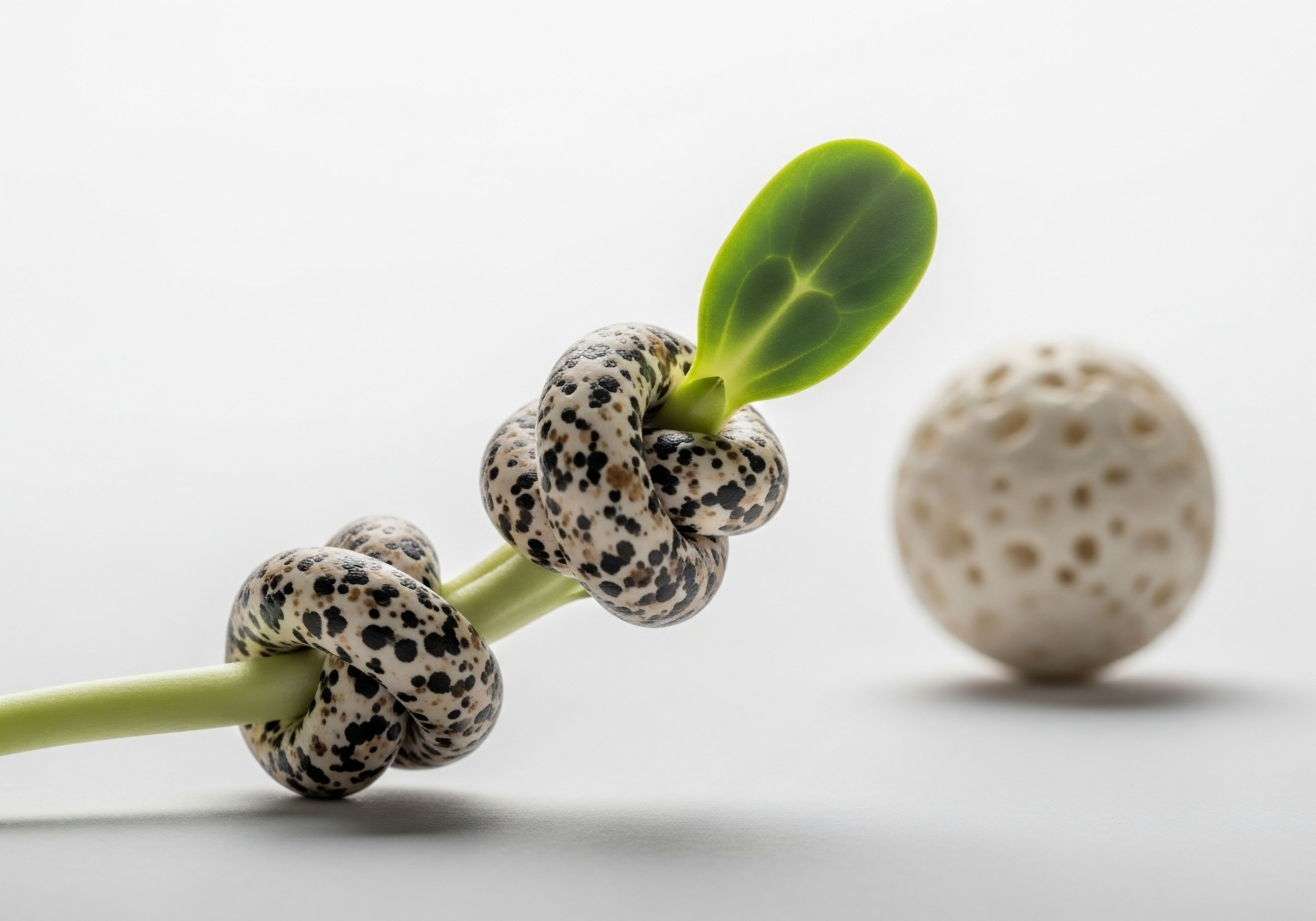


Fundamentals
You feel it as a subtle shift over time. The energy that once felt boundless now seems to have a more definite limit. The resilience you took for granted requires more conscious effort to maintain. This lived experience is a universal aspect of the human condition, a biological reality rooted deep within our cells.
For decades, we have understood intuitively that disciplined exercise and mindful eating are potent tools to counter this slow decline. We now have the scientific language to describe the machinery that turns these actions into tangible, biological renewal. At the heart of this process is a family of proteins called sirtuins, which function as the intelligent command center for your cellular health, translating your lifestyle choices into a direct investment in your longevity.
Think of your body as a highly complex and resource-intensive enterprise. Within every cell, sirtuins act as the master regulators, the executive team that monitors the company’s energy budget and allocates resources with precision. Their primary function is to sense the overall energy status of the cell and, in response, to orchestrate vast programs of maintenance, repair, and efficiency.
These proteins are a class of enzymes known as NAD-dependent deacetylases. This means they require a specific molecule, nicotinamide adenine dinucleotide (NAD+), to function. NAD+ is a critical coenzyme present in every cell, central to metabolism and energy production. The available pool of NAD+ is the direct fuel source for sirtuin activity. When NAD+ levels are high, sirtuins are active and executing their protective protocols. When NAD+ levels decline, their activity wanes, and cellular maintenance programs slow down.
Sirtuins are cellular sensors that translate metabolic states, such as those induced by diet and exercise, into protective, longevity-promoting actions.
This is where the well-documented benefits of caloric restriction and exercise come into play. These practices are, from a cellular perspective, forms of beneficial stress. Caloric restriction, or reducing energy intake without malnutrition, lowers the amount of glucose and fatty acids entering the cells.
This adjustment slows down metabolic processes that consume NAD+, thereby increasing the ratio of NAD+ to its reduced form, NADH. Exercise has a related yet distinct effect. Intense physical activity consumes large amounts of ATP, the cell’s immediate energy currency.
This consumption leads to a rise in AMP, a molecule signaling low energy, which in turn activates a pathway that stimulates NAD+ synthesis. Both stressors converge on the same outcome ∞ an increase in the cellular concentration of NAD+, which provides the necessary fuel to activate sirtuins.
Once activated, sirtuins initiate a cascade of events designed to protect the organism during a period of perceived scarcity or high demand. They travel along the strands of our DNA, modifying proteins called histones to tighten the DNA structure, which silences certain genes and stabilizes the genome.
They activate DNA repair mechanisms, helping to correct the damage that accumulates with age. Furthermore, they enhance mitochondrial health, improve the cell’s resistance to oxidative stress, and regulate inflammatory responses. Through these deep-seated mechanisms, sirtuins convert the stress of a workout or a period of fasting into a signal for cellular fortification and renewal, providing a clear biological basis for the profound health benefits we have observed for centuries.
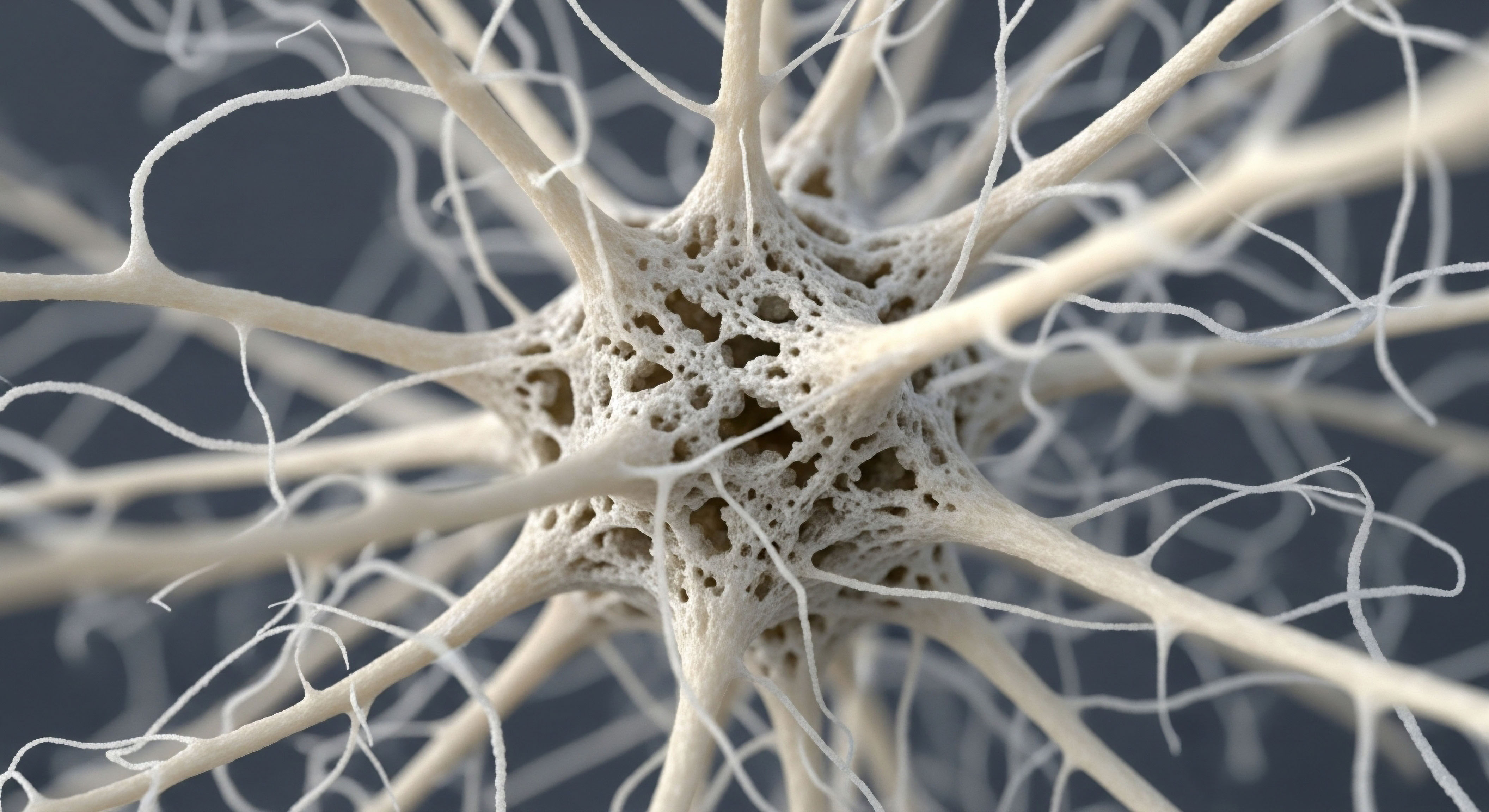


Intermediate
To truly appreciate the role of sirtuins, we must move beyond the general concept of cellular sensing and examine the specific molecular pathways they command. The connection between exercise and enhanced cellular function is beautifully illustrated by the interaction between SIRT1, the most studied sirtuin, and a transcriptional coactivator known as PGC-1α (peroxisome proliferator-activated receptor-gamma coactivator 1-alpha).
PGC-1α is often called the master regulator of mitochondrial biogenesis, the process of creating new mitochondria. The functional capacity of our mitochondria, the powerhouses of our cells, is a direct determinant of our metabolic health and energy levels.
The SIRT1 PGC-1α Axis
During exercise, the metabolic demands of muscle cells lead to a rise in the AMP/ATP ratio, activating an energy sensor called AMP-activated protein kinase (AMPK). This activation, combined with the increased NAD+ levels from metabolic shifts, creates the perfect environment for SIRT1 to become highly active.
SIRT1’s primary target in this context is PGC-1α. In its inactive state, PGC-1α is marked with acetyl groups that repress its function. SIRT1 functions as a deacetylase, meaning it chemically removes these acetyl groups from PGC-1α. This deacetylation acts like a molecular switch, activating PGC-1α and unleashing its powerful transcriptional program.
An activated PGC-1α then coordinates the expression of genes required to build new, more efficient mitochondria. This process is analogous to upgrading a city’s electrical grid, resulting in more power, less waste, and greater overall system resilience.
The activation of PGC-1α by SIRT1 is a central mechanism through which exercise directly enhances mitochondrial health and cellular energy production.

Connecting Cellular Energy to Hormonal Health
The influence of sirtuins extends well into the endocrine system, creating a bridge between our metabolic state and our hormonal balance. This is particularly relevant in the context of personalized wellness protocols, including hormonal optimization. For instance, SIRT1 plays a direct role in male hormonal health by regulating testosterone biosynthesis within the Leydig cells of the testes.
It achieves this by modulating autophagy, the cellular process of cleaning out damaged components. By controlling autophagy, SIRT1 influences the availability of cholesterol, the essential precursor for producing testosterone and other steroid hormones. A decline in SIRT1 activity can therefore contribute to reduced testosterone production, a condition that is clinically addressed through Testosterone Replacement Therapy (TRT).
Similarly, sirtuins modulate the Growth Hormone (GH) and Insulin-like Growth Factor 1 (IGF-1) axis. SIRT1 can act at the level of the hypothalamus and the liver to fine-tune GH signaling. During periods of caloric restriction, for example, SIRT1 helps to induce a state of transient GH resistance in the liver.
This adaptive response reduces IGF-1 production, conserving energy and resources when nutrients are scarce. This mechanism highlights SIRT1’s role as a master coordinator, ensuring that growth signals are appropriately balanced with the body’s overall energy status. This has implications for therapies involving growth hormone peptides like Sermorelin or Ipamorelin, as the underlying cellular energy environment can influence their efficacy.
Below is a summary of key cellular outcomes mediated by sirtuin activation.
- Mitochondrial Biogenesis ∞ SIRT1 activates PGC-1α, leading to the creation of new mitochondria, which is particularly prominent in muscle tissue after exercise.
- DNA Repair and Stability ∞ Sirtuins help maintain genomic integrity by deacetylating histones and recruiting DNA repair factors to sites of damage.
- Inflammation Control ∞ SIRT1 can deacetylate components of the NF-κB signaling pathway, a central regulator of inflammation, thereby dampening chronic inflammatory responses.
- Autophagy Regulation ∞ Sirtuins promote cellular cleanup by initiating autophagy, which removes dysfunctional proteins and organelles, a process linked to testosterone production.
- Metabolic Efficiency ∞ Sirtuins improve insulin sensitivity and regulate glucose and fat metabolism in tissues like the liver, muscle, and adipose tissue.
The table below compares the primary mechanisms through which exercise and caloric restriction activate sirtuin pathways.
| Stimulus | Primary Cellular Trigger | Key Sirtuins Implicated | Primary Physiological Outcome |
|---|---|---|---|
| Exercise | Increased AMP/ATP ratio; Increased NAD+ consumption and subsequent synthesis | SIRT1, SIRT3 | Enhanced mitochondrial biogenesis in muscle; Improved cardiovascular function |
| Caloric Restriction | Reduced glucose/fatty acid influx; Increased NAD+/NADH ratio | SIRT1, SIRT6 | Systemic reduction in inflammation; Enhanced DNA repair and cellular stress resistance |


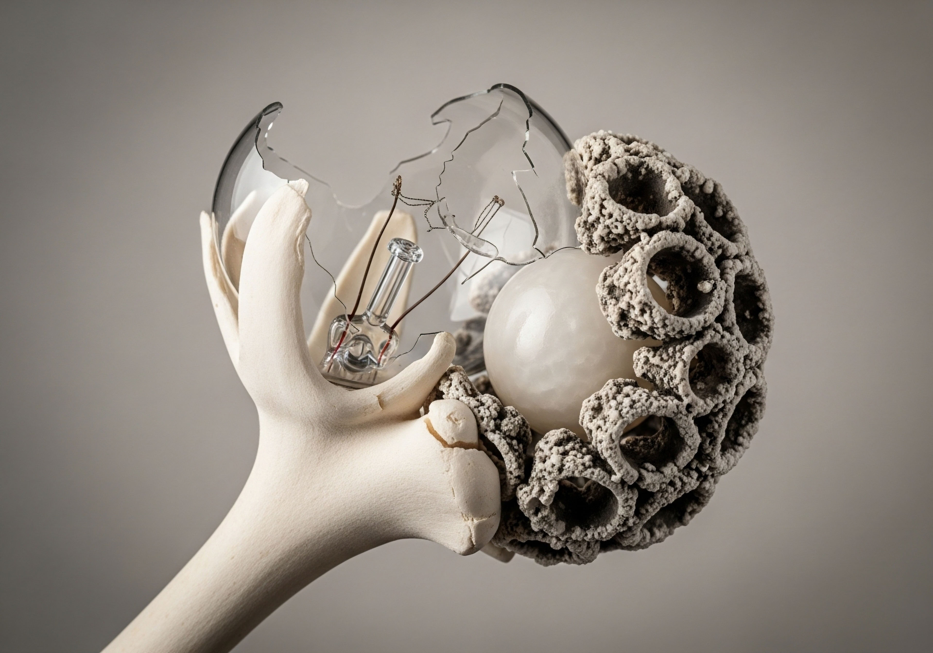
Academic
A systems-biology perspective reveals the sirtuin network as a critical integration point between environmental inputs and organismal homeostasis. The activity of this network is governed by the bioavailability of NAD+, making the regulation of the intracellular NAD+ pool a paramount control mechanism for longevity pathways.
The NAD+ salvage pathway, where NAD+ is recycled from its breakdown product, nicotinamide, is the primary source of cellular NAD+. The enzyme nicotinamide phosphoribosyltransferase (NAMPT) is the rate-limiting step in this pathway, positioning it as a central regulator of sirtuin function. The expression and activity of NAMPT are, in turn, influenced by exercise, diet, and circadian rhythms, creating a tightly regulated feedback system.

How Does Cellular NAD+ Competition Affect Hormonal Regulation?
The cellular pool of NAD+ is not exclusively reserved for sirtuins. It is also consumed by other enzymes, most notably PARPs (Poly polymerases) and CD38. PARPs are activated by DNA damage, and their function in DNA repair requires vast amounts of NAD+.
Chronic inflammation and accumulating DNA damage, both hallmarks of aging, can lead to PARP over-activation, which severely depletes the NAD+ pool. Similarly, the cell-surface enzyme CD38, a major NADase in mammalian tissues, increases its expression with age and contributes to the age-related decline in NAD+ levels.
This creates a state of competitive NAD+ consumption. When PARPs and CD38 are chronically active, they effectively sequester NAD+ away from sirtuins, diminishing their capacity to regulate metabolism, inflammation, and endocrine function. This depletion can directly impact hormonal axes. For example, a reduction in SIRT1 activity due to NAD+ depletion can impair the autophagic flux in Leydig cells necessary for efficient testosterone synthesis, contributing to age-related hypogonadism.
The age-related decline in NAD+ levels, exacerbated by chronic inflammation and DNA damage, creates a competitive disadvantage for sirtuins, impacting metabolic and endocrine resilience.
This systems-level view provides a mechanistic rationale for protocols aimed at supporting hormonal health. Therapies such as TRT for men or progesterone support for women address the downstream effects of hormonal decline. Interventions that support NAD+ levels, either through lifestyle modifications like exercise and fasting or through supplementation with NAD+ precursors, address the upstream cause of that decline, potentially restoring the capacity of sirtuins like SIRT1 to properly regulate the Hypothalamic-Pituitary-Gonadal (HPG) and Growth Hormone axes.
The seven mammalian sirtuins have distinct subcellular locations and functions, allowing them to coordinate cellular responses across different compartments.
| Sirtuin | Cellular Location | Primary Function and Key Targets | Relevance to Exercise/CR |
|---|---|---|---|
| SIRT1 | Nucleus, Cytoplasm | Deacetylates PGC-1α, FOXO proteins, NF-κB, histones. Regulates metabolism, inflammation, DNA repair, and autophagy. | Central mediator of both exercise and CR benefits. Links NAD+ sensing to mitochondrial and endocrine function. |
| SIRT2 | Cytoplasm | Regulates cell cycle, mitosis, and gluconeogenesis. | Involved in metabolic regulation and genomic stability. |
| SIRT3 | Mitochondria | Major mitochondrial deacetylase. Regulates metabolic enzymes, antioxidant defenses (via FOXO3), and mitochondrial protein synthesis. | Crucial for the mitochondrial adaptations to exercise and fasting. Reduces oxidative stress. |
| SIRT4 | Mitochondria | Regulates amino acid metabolism and insulin secretion from pancreatic beta-cells. | Fine-tunes mitochondrial metabolism in response to nutrient availability. |
| SIRT5 | Mitochondria | Removes other acyl groups (succinyl, malonyl). Regulates urea cycle and fatty acid oxidation. | Participates in metabolic reprogramming during fasting states. |
| SIRT6 | Nucleus | Critical for DNA repair (base excision repair), telomere maintenance, and suppression of genomic instability. Regulates glucose metabolism. | A key longevity-assurance sirtuin activated by CR, protecting the genome. |
| SIRT7 | Nucleolus | Regulates ribosomal DNA transcription and protein synthesis. Protects against cardiac hypertrophy. | Modulates protein synthesis in response to cellular stress, contributing to cardiac health. |
The molecular interplay is precise. In the context of the GH/IGF-1 axis, SIRT1’s ability to deacetylate STAT5 (Signal Transducer and Activator of Transcription 5) is a prime example of its regulatory sophistication. GH signaling in the liver proceeds through the JAK2-STAT5 pathway to stimulate IGF-1 synthesis.
During fasting, elevated SIRT1 activity leads to STAT5 deacetylation, which inhibits its ability to promote IGF-1 gene transcription. This action is a highly specific, adaptive mechanism to uncouple growth from an energy-deprived state, preventing hypoglycemia and conserving resources. Understanding this level of detail clarifies why simply administering growth-promoting peptides may yield variable results without first optimizing the underlying metabolic environment and NAD+ status that govern sirtuin activity.

References
- Guarente, Leonard. “Calorie restriction and sirtuins revisited.” Genes & development vol. 27,19 (2013) ∞ 2072-85.
- Grabowska, Wioleta, et al. “Sirtuins at the Service of Healthy Longevity.” Frontiers in Physiology, vol. 12, 2021, p. 794935.
- Kane, Ann E. and David A. Sinclair. “Sirtuins and NAD+ in the Development and Treatment of Metabolic and Cardiovascular Diseases.” Circulation Research, vol. 123, no. 7, 2018, pp. 848-863.
- Canto, Carles, and Johan Auwerx. “PGC-1alpha, SIRT1 and AMPK, an energy sensing network that controls energy expenditure.” Current opinion in lipidology vol. 20,2 (2009) ∞ 98-105.
- Gurd, Brendon J. “Deacetylation of PGC-1α by SIRT1 ∞ importance for skeletal muscle function and exercise-induced mitochondrial biogenesis.” Applied Physiology, Nutrition, and Metabolism, vol. 36, no. 5, 2011, pp. 589-597.
- Lu, Lu, et al. “Sirt1 regulates testosterone biosynthesis in Leydig cells via modulating autophagy.” Protein & Cell, vol. 12, no. 1, 2021, pp. 73-78.
- Stawerska, Renata, and Andrzej Lewiński. “Involvement of Sirtuin 1 in the Growth Hormone/Insulin-like Growth Factor 1 Signal Transduction and Its Impact on Growth Processes in Children.” International Journal of Molecular Sciences, vol. 24, no. 20, 2023, p. 15421.
- Schrauwen-Hinderling, Vera B. et al. “The role of the sirtuin-PGC-1α axis in muscle mitochondrial biogenesis and function.” The Journal of physiology vol. 589,Pt 18 (2011) ∞ 4413-20.
- Haigis, Marcia C. and David A. Sinclair. “Mammalian sirtuins ∞ biological insights and disease relevance.” Annual review of pathology vol. 5, 2010, pp. 253-95.
- Imai, Shin-ichiro, and Leonard Guarente. “NAD+ and sirtuins in aging and disease.” Trends in cell biology vol. 24,8 (2014) ∞ 464-71.

Reflection
The biological mechanisms we have explored are not abstract concepts occurring in a laboratory. They are active within you at this very moment. The sirtuin network is your own internal system for translating intention into physiology, a direct link between the choices you make and the vitality you experience.
Understanding these pathways offers a new lens through which to view your own health. The fatigue, the subtle changes in recovery, the shifts in metabolic function ∞ these experiences can be understood as signals from a complex system requesting a change in inputs.
The knowledge that specific actions can directly fuel the core machinery of cellular maintenance and repair is a powerful realization. It shifts the perspective from passively managing symptoms to proactively cultivating the biological environment for optimal function. This understanding is the first step. The next is to consider how these universal principles apply to your unique biology, your personal history, and your future goals.

Glossary

sirtuins

caloric restriction

dna repair

pgc-1α

sirt1

mitochondrial biogenesis

ampk

testosterone biosynthesis

autophagy

growth hormone
