


Fundamentals
The sense of moving through your days with a pervasive feeling of resistance, as if you are wading through water, is a tangible biological reality. This experience of diminished vitality, persistent fatigue, mental fog, or a body that no longer responds the way it once did is your physiology communicating a profound disruption.
These feelings are the perceptible outcomes of a breakdown in your body’s intricate internal messaging system, a network governed by hormones. When this sophisticated communication falters, the metabolic machinery that dictates your energy, mood, and physical form begins to operate inefficiently. Understanding the metabolic consequences of these unaddressed hormonal shifts is the first step toward reclaiming your biological sovereignty.
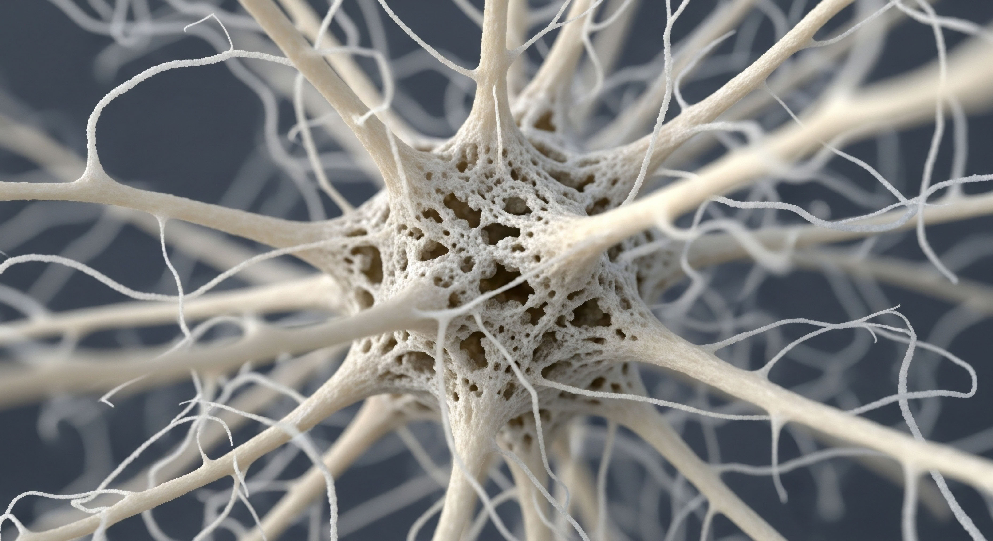
Your body operates as a meticulously coordinated system where hormones act as chemical messengers, delivering precise instructions to cells, tissues, and organs. These signals regulate everything from your heart rate to your appetite, and critically, how your body utilizes and stores energy. Key messengers like testosterone, estrogen, and growth hormone are central to this metabolic conversation.
Testosterone, for instance, sends powerful signals that support the maintenance of lean muscle mass, a metabolically active tissue that functions like an engine for burning calories. Estrogen plays a complex and crucial part in directing how cells respond to insulin, the primary hormone responsible for managing blood sugar.
When these hormonal signals become weak, intermittent, or absent, the receiving cells can no longer execute their metabolic duties correctly. The result is a cascade of systemic dysfunction that manifests as the very symptoms that disrupt your quality of life.
Untreated hormonal imbalances directly impair the body’s ability to manage energy, leading to systemic metabolic dysfunction.

The Architecture of Hormonal Communication
To appreciate the consequences of a breakdown, one must first understand the architecture of the system itself. The endocrine system is not a collection of isolated glands; it is a highly integrated network. The Hypothalamic-Pituitary-Gonadal (HPG) axis, for example, is a primary command-and-control pathway.
The hypothalamus in the brain releases Gonadotropin-Releasing Hormone (GnRH) in carefully timed pulses. This signal instructs the pituitary gland to release Luteinizing Hormone (LH) and Follicle-Stimulating Hormone (FSH). These hormones, in turn, travel to the gonads ∞ the testes in men and the ovaries in women ∞ and direct them to produce testosterone and estrogen.
This is a delicate feedback loop. The brain listens for the return signal of sex hormones in the bloodstream to determine if it needs to send more or fewer instructions. When production at the gonadal level falters due to age or other factors, the entire communication circuit is disrupted. The brain may send more signals, but the receiving end cannot respond, leading to a state of hormonal deficiency that echoes throughout the body’s metabolic processes.

Testosterone and Its Metabolic Mandate
In men, a decline in testosterone, or hypogonadism, sets off a predictable series of metabolic disturbances. One of the most immediate consequences is a shift in body composition. Testosterone actively promotes the development of muscle precursor cells and simultaneously inhibits the formation of fat cells. When testosterone levels fall, this directive weakens.
The body loses its stimulus to maintain metabolically costly muscle tissue, leading to sarcopenia, or age-related muscle loss. Concurrently, the body becomes more permissive to the storage of adipose tissue, particularly visceral adipose tissue (VAT), the deep abdominal fat that surrounds vital organs.
This type of fat is not merely a passive storage depot; it is a metabolically active organ in its own right, secreting inflammatory molecules that further disrupt metabolic health and create a vicious cycle of worsening hormonal and metabolic function. The loss of muscle and gain of visceral fat directly contributes to a lower metabolic rate, making weight management increasingly difficult.

Estrogen and Progesterone the Female Metabolic Regulators
In women, the hormonal transition of perimenopause and menopause introduces a different yet equally impactful set of metabolic challenges. Estrogen is a key regulator of insulin sensitivity, glucose uptake, and fat distribution. As estrogen levels fluctuate and ultimately decline, cells in the muscle and liver become less responsive to insulin’s signal to absorb glucose from the blood.
This condition, known as insulin resistance, forces the pancreas to produce more insulin to achieve the same effect, leading to high circulating levels of both glucose and insulin. This state promotes fat storage, particularly in the abdominal region, shifting body composition from a “pear” shape to a more metabolically unfavorable “apple” shape.
Progesterone also plays a role in this metabolic equation, influencing fluid balance, mood, and in some contexts, interacting with insulin signaling pathways. The withdrawal of these hormones creates a metabolic environment ripe for the development of metabolic syndrome, a cluster of conditions including high blood pressure, high blood sugar, and abnormal cholesterol levels that dramatically increases cardiovascular risk.


Intermediate
The progression from feeling “off” to developing a clinically recognized metabolic disorder is a journey from subtle signaling errors to a full-blown systemic crisis. At the intermediate level of understanding, we move beyond identifying the key hormonal players and begin to dissect the specific mechanisms through which their absence or dysregulation drives pathology.
This involves a closer examination of the cellular and systemic feedback loops and an introduction to the clinical protocols designed to intelligently restore this broken communication. The goal of such interventions is to re-establish the body’s metabolic equilibrium, addressing the root cause of the dysfunction.

The Vicious Cycle of Hypogonadism and Metabolic Derangement
In men, the relationship between low testosterone and metabolic disease is bidirectional. Low testosterone initiates the process by promoting the accumulation of visceral adipose tissue (VAT). This metabolically active fat then becomes a source of chronic, low-grade inflammation. VAT releases inflammatory cytokines and reduces the secretion of adiponectin, a beneficial hormone that enhances insulin sensitivity.
This inflammatory state directly impairs the ability of insulin to do its job in muscle and liver cells, leading to insulin resistance. Concurrently, an enzyme within this excess fat tissue, called aromatase, converts a portion of the body’s remaining testosterone into estradiol.
While some estrogen is vital for men, excessive aromatization further lowers testosterone levels and can disrupt the delicate hormonal balance, creating a self-perpetuating cycle of low testosterone, increased fat, and worsening insulin resistance. This cascade predictably increases the risk for developing type 2 diabetes and metabolic syndrome.
Therapeutic interventions for hormonal decline are designed to restore physiological signaling and interrupt the cycles that lead to metabolic disease.

Restoring the Signal Male Hormonal Optimization
For men with diagnosed hypogonadism, Testosterone Replacement Therapy (TRT) is a direct intervention designed to restore physiological levels of this critical hormone. The standard protocol often involves weekly intramuscular or subcutaneous injections of Testosterone Cypionate. This approach provides a stable level of testosterone in the bloodstream, directly addressing the deficiency. However, a sophisticated protocol does more than just replace testosterone. It aims to manage the entire HPG axis communication system.
- Gonadorelin ∞ This peptide is a synthetic version of GnRH. By administering it, typically twice per week via subcutaneous injection, the protocol continues to send a signal to the pituitary gland, prompting it to release LH and FSH. This maintains the natural signaling pathway, which helps preserve testicular function and size, preventing the testicular atrophy that can occur when the testes are no longer receiving a stimulus from the brain.
- Anastrozole ∞ This is an aromatase inhibitor. It is used judiciously to manage the conversion of testosterone to estradiol. In men with significant adipose tissue, this conversion can be excessive, leading to side effects. Anastrozole blocks the aromatase enzyme, helping to maintain a healthy testosterone-to-estrogen ratio and mitigating risks associated with elevated estrogen.
- Enclomiphene ∞ This compound may be included to selectively block estrogen receptors at the pituitary gland. This action reduces the negative feedback from estrogen, which can further support the pituitary’s output of LH and FSH, thereby supporting the body’s own testosterone production machinery.

Perimenopause as a Catalyst for Metabolic Disruption
For women, the hormonal fluctuations of perimenopause are the primary drivers of metabolic change. The decline in estrogen production directly impacts glucose metabolism in several ways. Estrogen helps maintain the health and number of insulin receptors on cells. As estrogen wanes, cells become less sensitive to insulin.
Furthermore, estrogen influences the function of pancreatic beta cells, the cells that produce insulin. The loss of its supportive role can contribute to the dysregulation of insulin secretion. This developing insulin resistance is a central feature of the menopausal transition and is a primary driver of the increased risk for metabolic syndrome and cardiovascular disease seen in postmenopausal women. The body’s set point for fat storage also changes, favoring accumulation around the midsection, which further fuels insulin resistance.

A Tailored Approach Female Hormonal Recalibration
Hormonal optimization for women is a nuanced process tailored to their specific symptoms and menopausal status. The goal is to buffer the metabolic consequences of hormonal decline.
The following table outlines typical therapeutic approaches for both men and women, highlighting the targeted nature of modern hormonal protocols.
| Protocol Aspect | Male TRT Protocol | Female Hormone Protocol |
|---|---|---|
| Primary Hormone | Testosterone Cypionate (e.g. 100-200mg weekly) | Testosterone Cypionate (low dose, e.g. 10-20 units weekly) and/or Estradiol |
| Key Objective | Restore testosterone to optimal physiological range to improve muscle mass, metabolic function, and well-being. | Alleviate menopausal symptoms, protect bone density, and mitigate metabolic shifts like insulin resistance. |
| HPG Axis Support | Gonadorelin to maintain testicular signaling and function. | Not typically required in the same manner, as ovarian function naturally declines. |
| Estrogen Management | Anastrozole to control excessive aromatization of testosterone into estradiol. | Progesterone is prescribed (if the woman has a uterus) to balance estrogen’s effects on the endometrium. Anastrozole may be used with testosterone pellets if needed. |
| Delivery Methods | Intramuscular/subcutaneous injections, gels, pellets. | Subcutaneous injections, transdermal creams, oral progesterone, pellets. |


Academic
A sophisticated analysis of the metabolic consequences of hormonal imbalances requires a shift in perspective from systemic observation to molecular mechanics. At this level, we investigate the precise biochemical pathways and gene-level instructions that are altered when hormonal signals are lost.
The body is revealed as a landscape of cellular receptors, signaling cascades, and transcription factors that are exquisitely sensitive to hormonal input. The pathologies of untreated hypogonadism and menopause are, at their core, the cumulative result of countless molecular conversations gone awry. Here, we will dissect the specific molecular actions of key hormones on target tissues and explore advanced therapeutic peptides that modulate these pathways with high specificity.

Molecular Endocrinology of Testosterone Action on Body Composition
Testosterone’s profound effects on muscle and fat are mediated through its interaction with the androgen receptor (AR), a protein located inside cells. When testosterone binds to the AR, the resulting complex travels to the cell’s nucleus and acts as a transcription factor, directly influencing the expression of specific genes. This genomic action orchestrates a dual program of myogenesis (muscle building) and lipolysis (fat breakdown) while inhibiting adipogenesis (fat creation).

How Does Testosterone Influence Muscle Growth at the Cellular Level?
The myotrophic effects of testosterone are multifaceted. First, it stimulates the proliferation of satellite cells, which are muscle stem cells. These cells can fuse with existing muscle fibers, donating their nuclei and increasing the myonuclear domain. This enhances the muscle fiber’s capacity for protein synthesis, leading to hypertrophy (growth).
Second, testosterone upregulates the expression of key anabolic signaling molecules like Insulin-like Growth Factor 1 (IGF-1) within the muscle tissue itself. This creates a powerful local growth-promoting environment. Third, it directly increases the rate of muscle protein synthesis by influencing the machinery responsible for building contractile proteins. The result is a direct command to build and maintain lean, metabolically active tissue.

The Adipose Tissue Regulation Pathway
Simultaneously, testosterone sends inhibitory signals to adipose tissue. It influences the fate of mesenchymal pluripotent stem cells, directing them toward a myogenic (muscle) lineage and away from an adipogenic (fat) lineage. In mature fat cells, testosterone enhances their sensitivity to catecholamines (like adrenaline), which are potent stimulators of lipolysis, the process of breaking down stored triglycerides into free fatty acids that can be used for energy.
It also appears to inhibit lipid uptake by adipocytes. Therefore, the loss of testosterone removes both the “build muscle” command and the “burn fat” command, leading to the characteristic shift in body composition toward less muscle and more fat, a state that is inherently pro-inflammatory and insulin-resistant.
Advanced peptide therapies work by precisely targeting specific hormonal receptor systems to restore physiological function.

Advanced Modalities Growth Hormone Peptide Therapy
Beyond direct hormone replacement, a more sophisticated approach involves using peptides to stimulate the body’s own endocrine pathways. Growth Hormone (GH) is a critical metabolic hormone that declines with age. It promotes lipolysis, supports tissue repair, and influences insulin sensitivity. Directly administering GH can be effective but can also lead to side effects and disrupt the natural pulsatile release. Growth Hormone Releasing Peptides (GHRPs) and Growth Hormone Releasing Hormone (GHRH) analogs offer a more nuanced way to enhance GH secretion.
This table details some of the key peptides used in clinical protocols to support metabolic health through the GH axis.
| Peptide | Mechanism of Action | Primary Metabolic Effects | Clinical Application Context |
|---|---|---|---|
| Sermorelin | A GHRH analog. It binds to GHRH receptors on the pituitary gland, stimulating the natural, pulsatile release of GH. | Increases endogenous GH levels, leading to elevated IGF-1, which supports tissue repair and anabolism. Can improve body composition over time. | Used for anti-aging, improving sleep, and supporting recovery. It works within the body’s natural feedback loops. |
| Ipamorelin / CJC-1295 | Ipamorelin is a GHRP (ghrelin mimetic) that stimulates a strong pulse of GH release. CJC-1295 is a long-acting GHRH analog. The combination provides a powerful synergistic effect on GH secretion. | Potent stimulation of GH and IGF-1, leading to enhanced lipolysis, muscle growth, and improved recovery. Ipamorelin is selective and does not significantly impact cortisol. | Popular among athletes and those seeking significant improvements in body composition, fat loss, and muscle gain. |
| Tesamorelin | A potent GHRH analog specifically studied and approved for reducing visceral adipose tissue in certain populations. | Directly targets and reduces deep abdominal fat, which is a key driver of metabolic syndrome and insulin resistance. | A highly targeted therapy for individuals with significant visceral adiposity contributing to metabolic dysfunction. |
| MK-677 (Ibutamoren) | An orally active, non-peptide ghrelin mimetic and GH secretagogue. | Sustained increases in GH and IGF-1 levels, which can increase lean body mass and improve sleep quality. | An oral alternative to injectable peptides for raising GH levels, often used for muscle gain and recovery. |

The Molecular Crosstalk of Female Hormones and Insulin Signaling
The link between menopause and insulin resistance is grounded in the molecular interplay between estrogen and the insulin signaling cascade. Estrogen, acting through the Estrogen Receptor Alpha (ERα), has a permissive and enhancing effect on the insulin signaling pathway.
Specifically, estrogen can increase the expression and functional activity of key proteins in the insulin pathway, including the insulin receptor itself and Insulin Receptor Substrate 1 (IRS-1). When insulin binds to its receptor, it triggers a cascade of phosphorylation events, activating pathways like PI3K/Akt, which ultimately leads to the translocation of GLUT4 glucose transporters to the cell surface, allowing glucose to enter the cell.
Estrogen signaling runs in parallel, supporting the efficiency of this process. The withdrawal of estrogen during menopause removes this supportive influence, making the entire insulin signaling pathway less efficient and contributing directly to cellular insulin resistance.
Progesterone’s role is more complex. At high concentrations, it can sometimes antagonize insulin’s effects, potentially by inhibiting steps in the PI3-kinase pathway or reducing the expression of IRS-1. The fluctuating and eventual loss of both hormones during the menopausal transition creates a state of profound metabolic instability at the cellular level, providing a clear molecular basis for the increased risk of type 2 diabetes and cardiovascular disease in this population.

References
- Bhasin, Shalender, et al. “Testosterone Therapy in Men with Hypogonadism ∞ An Endocrine Society Clinical Practice Guideline.” The Journal of Clinical Endocrinology & Metabolism, vol. 103, no. 5, 2018, pp. 1715 ∞ 1744.
- Saad, Farid, et al. “The role of testosterone in the metabolic syndrome ∞ a review.” The Journal of Steroid Biochemistry and Molecular Biology, vol. 114, no. 1-2, 2009, pp. 40-43.
- Traish, Abdulmaged M. et al. “The dark side of testosterone deficiency ∞ I. Metabolic syndrome and erectile dysfunction.” Journal of Andrology, vol. 30, no. 1, 2009, pp. 10-22.
- Mauvais-Jarvis, Franck, et al. “Estrogen and androgen receptors ∞ regulators of fuel homeostasis and emerging targets for diabetes and obesity.” Trends in Endocrinology & Metabolism, vol. 24, no. 5, 2013, pp. 225-234.
- Lizcano, Fernando, and Diego Guarin. “Estrogen deficiency and the origin of obesity during menopause.” BioMed research international, vol. 2014, 2014.
- Kadi, Fawzi. “Cellular and molecular mechanisms responsible for the action of testosterone on human skeletal muscle. A basis for illegal performance enhancement.” British journal of pharmacology, vol. 154, no. 3, 2008, pp. 522-528.
- Kalkhoff, R. K. “Metabolic effects of progesterone.” American journal of obstetrics and gynecology, vol. 142, no. 6 Pt 2, 1982, pp. 735-738.
- Veldhuis, Johannes D. and Ali Iranmanesh. “Physiological regulation of the human growth hormone (GH)-insulin-like growth factor type I (IGF-I) axis ∞ predominant impact of age, obesity, gonadal function, and sleep.” Sleep, vol. 19, no. 10 Suppl, 1996, pp. S221-4.
- Merriam, G. R. and K. Y. T. D. Wagner. “The somatopause ∞ clinical and physiological aspects.” Endocrine, vol. 7, no. 1, 1997, pp. 69-72.
- Corpas, E. S. M. Harman, and M. R. Blackman. “Human growth hormone and human aging.” Endocrine reviews, vol. 14, no. 1, 1993, pp. 20-39.

Reflection

Translating Knowledge into Personal Strategy
The information presented here offers a detailed map of the biological territory connecting your hormonal state to your metabolic function. It provides a language to describe the lived experience of fatigue, weight gain, and diminished capacity, grounding these feelings in concrete physiological processes. This knowledge transforms abstract symptoms into understandable, addressable mechanisms.
The purpose of this deep exploration is to equip you with a new level of understanding, allowing you to engage in a more informed and collaborative dialogue with your healthcare provider. Your personal health narrative is unique, written in the language of your own biochemistry and life experiences.

What Is Your Body Communicating?
Consider the symptoms you experience not as isolated problems, but as signals from a complex, integrated system. What is the overarching message your body is sending? Is it a message of energy scarcity, of inflammatory stress, or of a breakdown in cellular communication? Viewing your health through this systemic lens allows for a more holistic perspective.
The journey toward optimized health begins with listening intently to these signals and seeking a clinical partner who can help you interpret them. This framework is your starting point for asking deeper questions and moving toward a personalized strategy that restores your body’s innate capacity for vitality and function.

Glossary

growth hormone

pituitary gland

body composition

hypogonadism

visceral adipose tissue

adipose tissue

perimenopause

insulin resistance

metabolic syndrome
insulin signaling

testosterone replacement therapy

hpg axis

aromatase inhibitor
