


Fundamentals
The experience of noticing a change in your body, particularly in an area so central to male identity, can be deeply unsettling. When one or both testicles decrease in size, a condition known as testicular atrophy, it is a direct signal from your body that a significant physiological shift is occurring.
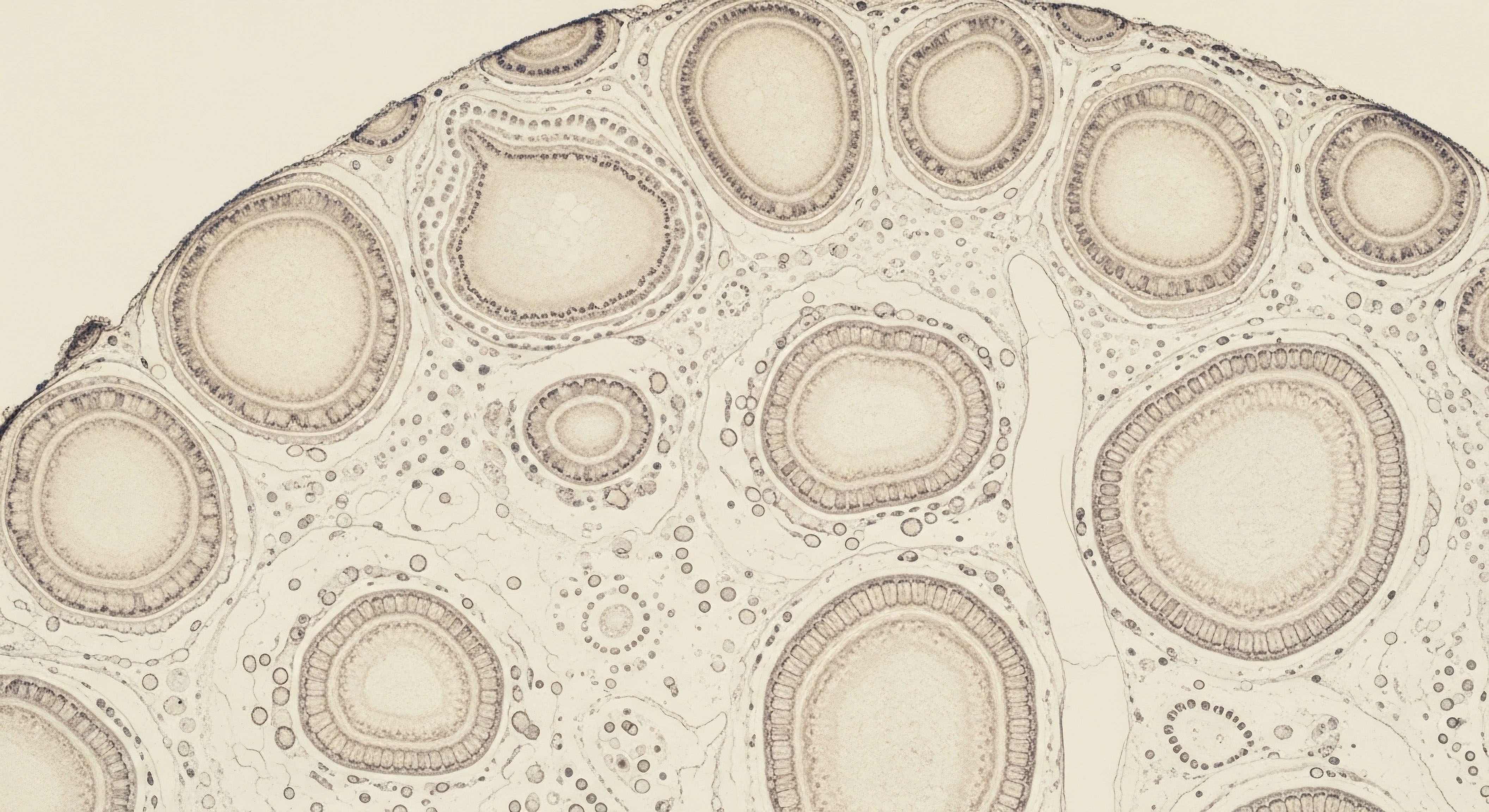
This is a physical manifestation of cellular change, specifically the loss of the specialized cells responsible for the core functions of the testicles ∞ the germ cells that produce sperm and the Leydig cells Meaning ∞ Leydig cells are specialized interstitial cells within testicular tissue, primarily responsible for producing and secreting androgens, notably testosterone. that generate testosterone. Understanding this process is the first step toward addressing the root cause and reclaiming your sense of well-being.
The primary and most immediate consequence of this cellular loss is a decline in testicular function. This presents in two critical ways. First, the capacity for sperm production is diminished, which directly impacts fertility. For many, this is a profound concern, touching upon fundamental aspects of life planning and legacy.
Second, the body’s main production line for testosterone is compromised. Testosterone is a systemic hormone, a vital messenger that regulates functions far beyond the reproductive system. Its decline initiates a cascade of effects that can be felt throughout the body, from energy levels and mood to muscle mass and mental clarity.
Testicular atrophy signifies a reduction in the specialized cells that produce both sperm and testosterone, directly impacting fertility and hormonal balance.
Recognizing the symptoms associated with testicular atrophy Meaning ∞ Testicular atrophy refers to the clinical condition characterized by a measurable decrease in the size and volume of one or both testicles from their normal adult dimensions. is essential. Beyond the visible change in size, you might experience a constellation of symptoms that point toward low testosterone, or hypogonadism. These can include a persistent sense of fatigue that sleep doesn’t resolve, a noticeable drop in libido or sexual desire, and difficulties with erectile function.
Some men also report changes in mood, a loss of muscle mass despite consistent exercise, or an increase in body fat. These are not isolated issues; they are interconnected signals that the body’s hormonal equilibrium has been disturbed. Addressing testicular atrophy involves looking at this complete picture to understand the full scope of its impact on your health.


What Are the Immediate Health Concerns?
The immediate health concerns stemming from testicular atrophy are centered on the dual roles of the testes. The reduction in volume is a direct reflection of a loss of internal machinery. This loss impairs the seminiferous tubules, where sperm are created, and the Leydig cells, the body’s primary testosterone factories. The direct implications are:
- Fertility Issues ∞ A reduction in the germ cells responsible for spermatogenesis leads to a lower sperm count, which can result in subfertility or infertility. This is often one of the most significant long-term concerns for men who wish to have children.
- Hormonal Decline ∞ The loss of Leydig cells causes a drop in testosterone production, leading to a state of hypogonadism. This hormonal deficiency is responsible for a wide array of systemic symptoms that can degrade quality of life.
It is important to view testicular atrophy as a diagnostic clue. It is a physical sign that prompts a deeper investigation into your endocrine health. The journey begins with acknowledging this sign and seeking a comprehensive evaluation to understand its cause and full effects.


Intermediate
To comprehend the long-term implications of testicular atrophy, one must first understand the elegant and tightly regulated system that governs male hormonal health ∞ the Hypothalamic-Pituitary-Gonadal (HPG) axis. Think of this as a sophisticated command-and-control network.
The hypothalamus in the brain releases Gonadotropin-Releasing Hormone (GnRH), signaling the pituitary gland to secrete two critical messenger hormones ∞ Luteinizing Hormone (LH) and Follicle-Stimulating Hormone (FSH). LH travels to the Leydig cells in the testes, instructing them to produce testosterone. FSH acts on the Sertoli cells, which are essential for supporting sperm production (spermatogenesis). Testosterone itself sends a feedback signal back to the brain, moderating the release of GnRH, LH, and FSH to maintain balance.
Testicular atrophy induced by external factors, such as the administration of exogenous testosterone in Testosterone Replacement Therapy Meaning ∞ Testosterone Replacement Therapy (TRT) is a medical treatment for individuals with clinical hypogonadism. (TRT), occurs because this natural feedback loop is disrupted. When the brain detects high levels of testosterone from an external source, it interprets this as a signal that the testes are overproducing.
In response, it dramatically reduces its output of GnRH, which in turn shuts down the pituitary’s release of LH and FSH. Without the stimulating signals from LH and FSH, the Leydig and Sertoli cells Meaning ∞ Sertoli cells are specialized somatic cells within the testes’ seminiferous tubules, serving as critical nurse cells for developing germ cells. become dormant and begin to shrink from underutilization. This leads to a decrease in both endogenous testosterone production Meaning ∞ Testosterone production refers to the biological synthesis of the primary male sex hormone, testosterone, predominantly in the Leydig cells of the testes in males and, to a lesser extent, in the ovaries and adrenal glands in females. and spermatogenesis, resulting in testicular atrophy.
Exogenous testosterone disrupts the HPG axis by suppressing the brain’s signals (LH and FSH) to the testes, causing them to cease function and shrink.


Restoring Testicular Function and Hormonal Balance
For individuals on TRT or those seeking to restore fertility after a period of hormonal suppression, specific clinical protocols are designed to reactivate the HPG axis. These protocols do not simply replace testosterone; they aim to stimulate the body’s own production machinery. Two key agents in this process are Gonadorelin Meaning ∞ Gonadorelin is a synthetic decapeptide that is chemically and biologically identical to the naturally occurring gonadotropin-releasing hormone (GnRH). and Enclomiphene.
Gonadorelin is a synthetic form of GnRH. By administering it, we are essentially bypassing the hypothalamus and directly signaling the pituitary gland to produce LH and FSH. This targeted stimulation “wakes up” the dormant Leydig and Sertoli cells, prompting them to resume their natural functions of producing testosterone and supporting sperm development. It is often included in TRT protocols to help maintain testicular size and function concurrently with exogenous testosterone administration.


Comparing Protocols for HPG Axis Stimulation
The approach to stimulating the HPG axis Meaning ∞ The HPG Axis, or Hypothalamic-Pituitary-Gonadal Axis, is a fundamental neuroendocrine pathway regulating human reproductive and sexual functions. can be tailored based on the individual’s goals, whether it’s maintaining testicular function during TRT or actively restoring fertility post-cycle.
| Agent | Mechanism of Action | Primary Clinical Use |
|---|---|---|
| Gonadorelin | A GnRH analog that directly stimulates the pituitary to release LH and FSH. | Used during TRT to prevent testicular atrophy by maintaining gonadal stimulation. |
| Enclomiphene Citrate | A selective estrogen receptor modulator (SERM) that blocks estrogen’s negative feedback at the hypothalamus, increasing GnRH release and subsequently LH and FSH. | Used to restart endogenous testosterone production, particularly in men seeking to restore fertility. |
| hCG (Human Chorionic Gonadotropin) | Mimics the action of LH, directly stimulating the Leydig cells to produce testosterone. | A powerful agent for stimulating testosterone production and maintaining testicular volume, often used in fertility protocols. |
These interventions are based on a nuanced understanding of endocrine physiology. They represent a shift from simple hormone replacement to sophisticated hormonal recalibration, aiming to restore the body’s innate capacity for balanced function.


Academic
The long-term consequences of testicular atrophy extend far beyond the reproductive axis, initiating a cascade of systemic metabolic and cardiovascular dysfunctions. The state of hypogonadism Meaning ∞ Hypogonadism describes a clinical state characterized by diminished functional activity of the gonads, leading to insufficient production of sex hormones such as testosterone in males or estrogen in females, and often impaired gamete production. resulting from compromised Leydig cell function is a significant etiological factor in the development of metabolic syndrome, type 2 diabetes, and cardiovascular disease.
Testosterone exerts a profound influence on body composition, insulin sensitivity, and lipid metabolism. Its deficiency promotes an increase in visceral adipose tissue, a key driver of systemic inflammation and insulin resistance. This altered metabolic milieu, characterized by hyperglycemia and dyslipidemia, contributes directly to endothelial dysfunction and the pathogenesis of atherosclerosis, elevating the risk for major adverse cardiovascular events like myocardial infarction and stroke.
From a cellular perspective, the health of the testicle is a microcosm of systemic health. The Sertoli cells, which form the blood-testis barrier and nurse developing sperm, and the Leydig cells, which produce testosterone, are highly metabolic and sensitive to oxidative stress.
Research indicates that the dysfunction of one cell type can precipitate the failure of the other. For instance, Sertoli cell health is critical for maintaining the Leydig cell population in the adult testis. A decline in Sertoli cell function, whether due to aging, toxicity, or vascular insufficiency, can lead to a secondary loss of Leydig cells, exacerbating the hypogonadal state and its systemic consequences. This intricate paracrine signaling relationship underscores the interconnectedness of the testicular microenvironment.
The hormonal deficit from testicular atrophy promotes systemic metabolic dysfunction, increasing the risk of cardiovascular disease and cognitive decline through interconnected cellular pathways.


How Does Hypogonadism Affect Cognitive Function?
The brain is a major target for androgens, with androgen receptors widely distributed in areas crucial for memory, mood, and executive function. The neuroprotective effects of testosterone are well-documented; it modulates synaptic plasticity, promotes neuronal survival, and influences neurotransmitter systems.
Consequently, the chronic hypogonadism associated with testicular atrophy is increasingly recognized as a risk factor for cognitive decline Meaning ∞ Cognitive decline signifies a measurable reduction in cognitive abilities like memory, thinking, language, and judgment, moving beyond typical age-related changes. and may contribute to the pathophysiology of neurodegenerative diseases. The decline in testosterone is associated with symptoms such as poor concentration, memory lapses, and depressive moods.
While the precise mechanisms are still under investigation, the link between a healthy endocrine system Meaning ∞ The endocrine system is a network of specialized glands that produce and secrete hormones directly into the bloodstream. and optimal neurological function is clear. Restoring hormonal balance may be a critical component in preserving cognitive vitality over the long term.


Systemic Impact of Androgen Deficiency
The physiological consequences of long-term, untreated hypogonadism are multi-systemic and progressive. Understanding this full spectrum of impact is vital for appreciating the importance of comprehensive management.
| System | Long-Term Implications of Hypogonadism | Underlying Mechanism |
|---|---|---|
| Metabolic | Increased risk of metabolic syndrome and type 2 diabetes. | Testosterone deficiency promotes visceral adiposity, insulin resistance, and dyslipidemia. |
| Cardiovascular | Higher incidence of atherosclerosis, coronary artery disease, and stroke. | Endothelial dysfunction, systemic inflammation, and adverse changes in lipid profiles. |
| Musculoskeletal | Sarcopenia (loss of muscle mass) and decreased bone mineral density (osteoporosis). | Testosterone is anabolic to muscle and essential for maintaining bone matrix integrity. |
| Neurological/Cognitive | Cognitive decline, impaired memory, and increased risk of depression. | Loss of testosterone’s neuroprotective effects and modulation of neurotransmitter systems. |
This systems-biology perspective reveals that testicular atrophy is not merely a localized reproductive issue. It is a sentinel event that signals a profound disruption in the body’s homeostatic mechanisms, with far-reaching implications for long-term health and vitality. Effective clinical management requires an approach that addresses both the primary gonadal failure and its systemic metabolic and cognitive sequelae.

References
- Bhasin, S. et al. “Testosterone Therapy in Men With Hypogonadism ∞ An Endocrine Society Clinical Practice Guideline.” The Journal of Clinical Endocrinology & Metabolism, vol. 103, no. 5, 2018, pp. 1715 ∞ 1744.
- Saad, F. et al. “Long-Term Testosterone Therapy Improves Cardiometabolic Function and Reduces Risk of Cardiovascular Disease in Men with Hypogonadism ∞ A Real-Life Observational Registry Study Setting Comparing Treated and Untreated (Control) Groups.” Journal of Cardiovascular Pharmacology and Therapeutics, vol. 22, no. 5, 2017, pp. 440-453.
- Rastrelli, G. et al. “Age-Related Male Hypogonadism and Cognitive Impairment in the Elderly ∞ Focus on the Effects of Testosterone Replacement Therapy on Cognition.” Journal of Endocrinological Investigation, vol. 42, no. 9, 2019, pp. 1047-1056.
- Samplaski, M. K. and Lo, K. C. “Exogenous testosterone ∞ a preventable cause of male infertility.” Translational Andrology and Urology, vol. 6, no. 2, 2017, pp. 195-200.
- Welsh, M. et al. “Sertoli Cells Maintain Leydig Cell Number and Peritubular Myoid Cell Activity in the Adult Mouse Testis.” Endocrinology, vol. 150, no. 8, 2009, pp. 3778-3788.
- Høyer, P.E. “Sertoli and Germ Cells Within Atrophic Seminiferous Tubules of Men With Non-Obstructive Azoospermia.” Frontiers in Endocrinology, 13, 2022.
- Hackett, G. et al. “Hypogonadism ∞ cardiometabolism and gonadal function in men.” Journal of Men’s Health, vol. 18, no. 1, 2022.
- Khera, M. et al. “Current National and International Guidelines for the Management of Male Hypogonadism ∞ Helping Clinicians to Navigate Variation in Diagnostic Criteria and Treatment Recommendations.” Journal of Sexual Medicine, vol. 14, no. 10, 2017, pp. 1243-1253.


Reflection
The information presented here offers a map of the biological territory surrounding testicular atrophy. It connects a physical sign to a complex network of hormonal signals and systemic responses. This knowledge provides a framework for understanding your own body’s internal communication system.
Your personal health journey is unique, and the path forward involves translating this general scientific understanding into a personalized protocol. The data points, the lab results, and the clinical strategies are the tools. Your lived experience and personal goals are the guide. The potential for reclaiming vitality and function begins with this synthesis of knowledge and self-awareness, empowering you to take proactive and informed steps toward optimal health.