

Fundamentals
The feeling is a familiar one for many. It is the profound sense of fatigue that settles in after a meal, the persistent difficulty in managing body weight despite sincere efforts, or the creeping realization that your body’s energy systems are functioning with a frustrating inefficiency.
These experiences are the outward expression of a silent, microscopic conversation breaking down within your body. This conversation is orchestrated by insulin, a masterful hormone that governs your body’s energy economy. To understand the roots of metabolic dysfunction, we begin by listening to this cellular dialogue and recognizing how our dietary choices can cause it to falter.
Insulin’s primary role is to manage the flow of energy, specifically glucose, from the bloodstream into your cells. Think of it as a highly specific key. After a meal, as glucose enters your bloodstream, your pancreas releases insulin.
This insulin travels to your cells, primarily in muscle, liver, and fat tissue, and binds to a specific lock on the cell surface called the insulin receptor. This binding action triggers a cascade of signals inside the cell, akin to a series of tumblers falling into place.
The ultimate result is the opening of a door, a glucose transporter protein known as GLUT4, which moves to the cell’s surface. This open door allows glucose to flood into the cell, where it can be used immediately for energy or stored for later use. This is a system of exquisite sensitivity and precision, designed to keep your blood glucose in a narrow, healthy range while ensuring your cells are properly fueled.
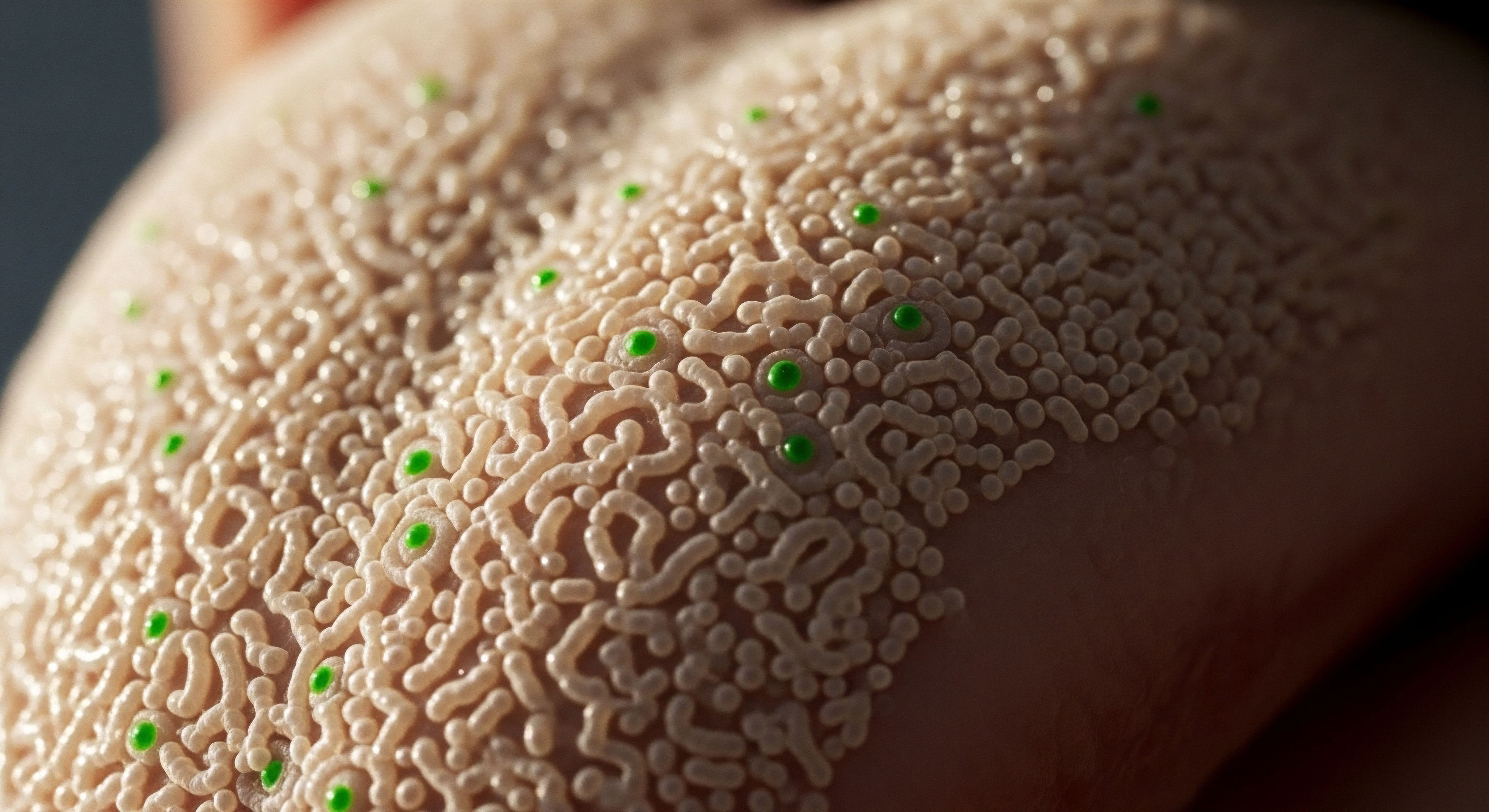
How Does a Cell Normally Hear Insulins Message?
The process of insulin signaling is a beautifully coordinated sequence of events. When this system operates correctly, your body maintains a state of metabolic harmony. Each step is a critical link in a chain of communication that translates a meal into cellular energy.
The sequence unfolds with remarkable speed and accuracy:
- Signal Reception ∞ Insulin, the key, docks with its specific receptor on the outer membrane of the cell. This connection is the very first step in the communication process.
- Internal Activation ∞ The binding of insulin activates the receptor’s internal component, a process known as autophosphorylation. This event initiates a chain reaction inside the cell, activating a series of proteins called insulin receptor substrates (IRS).
- Signal Amplification ∞ The activated IRS proteins function as docking stations and amplifiers, relaying the message to numerous downstream pathways. One of the most important is the PI3K/Akt pathway, which is central to glucose metabolism.
- Glucose Transporter Mobilization ∞ The activation of the Akt protein is the final command. It directs vesicles containing GLUT4 transporters to move to the cell membrane and fuse with it. This action places the GLUT4 “doors” on the cell surface.
- Glucose Uptake ∞ With the GLUT4 transporters in place, glucose can now move from the bloodstream into the cell, effectively lowering blood sugar levels and providing the cell with the fuel it needs to perform its functions.
This entire sequence is a testament to the body’s innate intelligence, a system designed for efficient energy management. When we feel vibrant and energetic, it is because this cellular conversation is happening seamlessly, millions of times over, throughout our bodies. The disruption of this conversation is what we experience as insulin resistance.
The sensation of metabolic slowdown begins when our cells lose their ability to properly respond to the hormonal messenger insulin.
The genesis of insulin resistance often lies in a state of chronic energy surplus. A consistent influx of energy, particularly from diets rich in refined carbohydrates and certain types of fats, places an immense demand on this signaling system. The pancreas works harder, producing more insulin to try and clear glucose from the blood.
Over time, the cells, overwhelmed by the constant barrage of insulin’s signal and the sheer volume of fuel, begin to protect themselves. They turn down the volume of the conversation. This protective adaptation is the beginning of insulin resistance. The cell becomes progressively “deaf” to insulin’s message.
The key is still present, often in greater numbers than ever before, but the lock becomes harder to turn. The result is that glucose remains in the bloodstream, and the cells, despite being surrounded by fuel, begin to experience a functional state of starvation. This is the central paradox of insulin resistance ∞ a state of energy excess in the blood coexisting with an energy deficit within the cell.


Intermediate
To truly grasp the progression from a healthy metabolic state to one of insulin resistance, we must move our examination from the systemic overview into the cell itself. The phenomenon is rooted in specific molecular disruptions that interfere with the elegant insulin signaling cascade.
These disruptions are frequently initiated by an accumulation of specific types of fat molecules within cells that were never designed for significant lipid storage, such as muscle and liver cells. This process, known as cellular lipotoxicity, represents a foundational mechanism through which dietary choices directly impair metabolic function.

The Lipotoxicity Hypothesis a Closer Look
When dietary energy intake, particularly from fats and refined carbohydrates, consistently exceeds the body’s capacity to burn or safely store it, fat begins to accumulate in non-adipose tissues. This ectopic fat storage is a primary driver of insulin resistance. Inside the cell, these excess fatty acids are converted into signaling molecules that actively disrupt the insulin pathway.

Diacylglycerol the Signal Jammer
One of the most significant of these molecules is diacylglycerol (DAG). In a healthy cell, DAG is a transient intermediate in the synthesis of triglycerides, the safe storage form of fat. In a state of energy overload, DAG levels build up within the cell.
This accumulation has a direct consequence ∞ the activation of a family of enzymes called protein kinase C (PKC), with a specific isoform, PKC-theta (PKC-θ) in muscle and PKC-epsilon (PKC-ε) in the liver, being particularly implicated.
The activation of these PKC isoforms is a critical misstep. Once active, PKC targets the insulin receptor substrate (IRS-1), the crucial docking protein in the insulin cascade. It phosphorylates IRS-1 on a serine residue. This action is akin to putting glue in the lock.
The normal function of IRS-1 requires it to be phosphorylated on tyrosine residues by the insulin receptor. Serine phosphorylation by PKC prevents this from happening effectively, blocking the signal from being passed down to Akt and, consequently, preventing the GLUT4 transporters from moving to the cell surface. The insulin key is in the lock, but the internal mechanism is jammed.

The Role of Ceramides
Alongside DAG, another class of lipid molecules called ceramides contributes to the development of insulin resistance. Ceramide accumulation is linked to both excess dietary saturated fat and the body’s own inflammatory responses. These lipids exert their effects through several mechanisms, including the activation of protein phosphatases that can dephosphorylate and inactivate Akt, further downstream in the insulin signaling pathway.
Ceramides are also potent inducers of cellular stress and apoptosis (programmed cell death), contributing to the overall dysfunction of the cell and promoting a low-grade inflammatory state that exacerbates insulin resistance.
| Lipid Intermediate | Primary Tissue of Action | Mechanism of Interference | Primary Downstream Effect |
|---|---|---|---|
| Diacylglycerol (DAG) | Skeletal Muscle, Liver |
Activates novel protein kinase C (PKC) isoforms (PKC-θ and PKC-ε). |
Inhibits Insulin Receptor Substrate (IRS-1) via serine phosphorylation, blocking the signal at an early stage. |
| Ceramides | Skeletal Muscle, Liver, Adipose Tissue |
Activates protein phosphatases (like PP2A) and promotes inflammatory signaling. |
Inhibits Akt directly and promotes cellular stress, blocking the signal further down the cascade. |

What Happens When the Cells Powerhouse Is Overwhelmed?
The accumulation of these disruptive lipid molecules is a direct result of another cellular process reaching its limit ∞ mitochondrial function. Mitochondria are the cell’s powerhouses, responsible for oxidizing fatty acids for energy in a process called beta-oxidation. A diet high in energy-dense foods creates a tidal wave of fatty acids arriving at the mitochondria.
The mitochondria, like a factory with finite production capacity, become overwhelmed. They cannot keep up with the demand to process all the incoming fuel.
Insulin resistance deepens as the cell’s energy-burning organelles, the mitochondria, are overwhelmed by excessive fuel from dietary sources.
This state of mitochondrial overload leads to what is known as incomplete fatty acid oxidation. Instead of being fully burned to produce ATP, the fatty acids are only partially processed, leading to a buildup of metabolic intermediates, including the very acyl-CoAs that are used to synthesize DAG and ceramides.
This metabolic traffic jam ensures a steady supply of the lipid molecules that interfere with insulin signaling. This establishes a vicious cycle ∞ excess dietary fat leads to mitochondrial overload, which causes the accumulation of lipotoxic intermediates, which in turn causes insulin resistance. The resulting high insulin levels further promote fat storage, perpetuating the entire cycle. Understanding this connection makes it clear that insulin resistance is a physiological response to a chronic state of cellular energy overload.


Academic
A comprehensive understanding of diet-induced insulin resistance requires a systems-biology perspective, viewing the cell as an integrated network where metabolic, inflammatory, and organelle-specific stress pathways converge. The accumulation of lipotoxic intermediates is a foundational event, yet it precipitates a cascade of other stress responses that solidify and amplify the insulin-resistant state.
Two of the most critical secondary mechanisms are endoplasmic reticulum stress and the activation of cellular inflammation, which are deeply intertwined with mitochondrial function and the overarching control of metabolic sensor proteins like sirtuins.

Endoplasmic Reticulum Stress the Protein Folding Factory under Duress
The endoplasmic reticulum (ER) is a vast network within the cell responsible for synthesizing and folding a large proportion of the cell’s proteins, including the insulin receptor itself. A chronic influx of nutrients, particularly glucose and saturated fatty acids, places an enormous burden on the ER’s protein-folding capacity.
This overload leads to an accumulation of unfolded or misfolded proteins, a condition known as ER stress. In response, the cell activates a sophisticated defense program called the Unfolded Protein Response (UPR).
The UPR has three main branches, each initiated by a specific sensor protein (IRE1α, PERK, and ATF6). While the UPR is initially adaptive, aiming to restore homeostasis, its chronic activation in the context of metabolic overload directly antagonizes insulin action. The IRE1α branch is particularly relevant, as it activates the c-Jun N-terminal kinase (JNK) pathway.
JNK is a stress-activated kinase that, once activated, directly phosphorylates the IRS-1 protein on inhibitory serine residues, the same mechanism employed by PKC in lipotoxicity. This provides a second, independent pathway through which cellular stress impairs the initial steps of insulin signaling.
Furthermore, prolonged ER stress can inhibit the proper processing and transport of newly synthesized insulin pro-receptors from the ER to the cell surface, effectively reducing the number of functional receptors available to bind insulin. The cell becomes less sensitive because it literally has fewer “ears” to hear insulin’s message.

How Do Cellular Stress Responses Drive Insulin Resistance?
The convergence of lipotoxicity and ER stress creates a pro-inflammatory environment within the cell. This low-grade, chronic inflammation is a core feature of insulin resistance. It is mediated by the activation of key inflammatory signaling hubs, most notably the inhibitor of nuclear factor kappa-B kinase subunit beta (IKK-β) and the NLRP3 inflammasome.
- IKK-β and NF-κB ∞ Both excess fatty acids and ER stress can activate IKK-β. This kinase, in turn, activates the transcription factor NF-κB, which orchestrates the expression of a wide array of pro-inflammatory cytokines like TNF-α and IL-6. These cytokines can then act in an autocrine (on the same cell) or paracrine (on nearby cells) fashion to induce insulin resistance, often by activating JNK and other serine kinases.
- The NLRP3 Inflammasome ∞ This intracellular protein complex functions as a sensor for cellular danger signals, including excess ceramides and reactive oxygen species (ROS) spilling from overloaded mitochondria. Upon activation, the NLRP3 inflammasome triggers the production of the potent inflammatory cytokine interleukin-1β (IL-1β), which is a powerful driver of insulin resistance in tissues like the liver and pancreas.
| Stress Pathway | Primary Trigger | Key Kinase/Mediator Activated | Mechanism of Insulin Signal Inhibition |
|---|---|---|---|
| Lipotoxicity |
Excess intracellular diacylglycerol (DAG) and ceramides. |
Protein Kinase C (PKC-θ/ε) |
Inhibitory serine phosphorylation of IRS-1. |
| ER Stress (UPR) |
Accumulation of unfolded proteins due to nutrient overload. |
c-Jun N-terminal Kinase (JNK) |
Inhibitory serine phosphorylation of IRS-1; reduced insulin receptor processing. |
| Inflammatory Signaling |
Activation by lipids, ER stress, and ROS. |
IKK-β, NLRP3 Inflammasome |
Production of inflammatory cytokines (TNF-α, IL-1β) that further activate JNK/IKK-β. |
| Oxidative Stress |
Electron leakage from overloaded mitochondria. |
Reactive Oxygen Species (ROS) |
Direct damage to proteins in the insulin signaling pathway and activation of JNK/IKK-β. |

Sirtuins the Master Metabolic Regulators
Overseeing this complex network of stress responses are sirtuins, a family of NAD+-dependent protein deacetylases that function as critical energy sensors. Sirtuin 1 (SIRT1), in particular, plays a protective role in metabolic health. Its activity is dependent on the cellular level of NAD+, a coenzyme whose levels increase during states of energy deficit (like fasting or exercise). When active, SIRT1 acts to improve insulin sensitivity and mitochondrial function.
SIRT1 accomplishes this through several mechanisms:
- Deacetylation of PGC-1α ∞ It activates PGC-1α, a master regulator of mitochondrial biogenesis, promoting the creation of new, efficient mitochondria to handle fatty acid oxidation.
- Inhibition of NF-κB ∞ SIRT1 can deacetylate and inhibit the RelA/p65 subunit of NF-κB, directly suppressing the inflammatory signaling that drives insulin resistance.
- Regulation of Hepatic Glucose Production ∞ In the liver, SIRT1 modulates the activity of transcription factors like FOXO1, helping to regulate glucose output in response to fasting and feeding.
A diet characterized by chronic energy surplus leads to a decrease in the NAD+/NADH ratio, which reduces SIRT1 activity. This impairment of a key protective system allows the feed-forward cycles of lipotoxicity, ER stress, and inflammation to proceed unchecked, cementing the insulin-resistant state.
This reveals that insulin resistance is a multifaceted cellular condition driven by the simultaneous failure of fuel disposal systems and the chronic activation of interconnected stress pathways, all under the supervision of nutrient-sensing regulators that are themselves compromised by the dietary environment.

References
- Shulman, G. I. “Cellular mechanisms of insulin resistance.” The Journal of clinical investigation 106.2 (2000) ∞ 171-176.
- Samuel, V. T. and G. I. Shulman. “Mechanisms for insulin resistance ∞ common threads and pathways.” Cell 148.5 (2012) ∞ 852-871.
- Hotamisligil, G. S. “Endoplasmic reticulum stress and the inflammatory basis of metabolic disease.” Cell 140.6 (2010) ∞ 900-917.
- Perry, R. J. et al. “The role of hepatic lipids in hepatic insulin resistance and type 2 diabetes.” Nature 510.7503 (2014) ∞ 84-91.
- Summers, S. A. “Ceramides in insulin resistance and metabolic disease.” Nature Reviews Endocrinology 16.1 (2020) ∞ 47-60.
- Petersen, M. C. and G. I. Shulman. “Mechanisms of insulin action and insulin resistance.” Physiological reviews 98.4 (2018) ∞ 2133-2223.
- Ozcan, U. et al. “Endoplasmic reticulum stress links obesity, insulin action, and type 2 diabetes.” Science 306.5695 (2004) ∞ 457-461.
- Schenk, S. et al. “Skeletal muscle triglycerides and insulin resistance.” Current cardiology reports 10.4 (2008) ∞ 307-314.
- Nakatani, Y. et al. “Involvement of the c-Jun N-terminal kinase pathway in insulin resistance.” Journal of Biological Chemistry 279.44 (2004) ∞ 45803-45809.
- Li, Y. et al. “Sirtuins in insulin resistance ∞ an updated review.” Oxidative Medicine and Cellular Longevity 2018 (2018).

Reflection
The journey through the cellular landscape of insulin resistance reveals a system of profound intelligence responding logically to the signals it receives. The fatigue, the metabolic slowdown, the persistent struggle ∞ these are not signs of a broken body. They are the predictable consequences of a system under siege from an environment of energy overload.
The knowledge of these mechanisms, from the jammed receptor at the cell’s surface to the stressed organelles within, shifts the perspective from one of passive suffering to one of active participation. Your body is in a constant dialogue with your choices.
Understanding the language of that dialogue ∞ the molecular signals of DAG, the stress calls from the ER, the protective actions of sirtuins ∞ is the first and most critical step. This scientific foundation provides the ‘why’ behind the lived experience. The path forward involves learning how to change the conversation, to send signals that promote sensitivity, efficiency, and vitality. This knowledge is your starting point for a more personalized and informed approach to reclaiming your own biological potential.

Glossary

insulin receptor

insulin signaling

insulin resistance

lipotoxicity

fatty acids

diacylglycerol

protein kinase c

insulin receptor substrate

serine phosphorylation
ceramides

cellular stress

inflammatory signaling

endoplasmic reticulum stress
cellular inflammation

c-jun n-terminal kinase
nlrp3 inflammasome

nf-κb

sirtuins
