


Fundamentals
You may have observed subtle shifts in your skin’s texture and resilience over time. A fine line that appears more defined, a loss of the plumpness you once took for granted, or a newfound dryness that seems unrelated to the weather. These experiences are valid and often point to a complex, internal symphony of biochemical signals.
Your body operates through an intricate communication network, and its messengers, hormones, direct cellular activities that manifest in your physical appearance. Understanding the roles of two primary female sex hormones, estrogen and progesterone, provides a direct insight into the biological processes that govern skin health. Their balance is fundamental to the skin’s structural integrity and youthful function.
Viewing these hormones as powerful regulators of cellular behavior allows us to appreciate their influence. They are the conductors of an orchestra, and the skin cells are the musicians. When the conductors are in sync and their signals are clear, the performance is seamless. When their signals become erratic or diminished, as they do during perimenopause and menopause, the harmony is disrupted, and the visible results are changes in skin quality.

The Architect of Skin Structure Estrogen
Estrogen, specifically estradiol (E2), functions as a primary architect for the skin’s foundation. Its presence signals to key cells within the dermis, the skin’s thickest layer, to perform their vital functions. Dermal fibroblasts, the cells responsible for producing structural proteins, are highly responsive to estrogen.
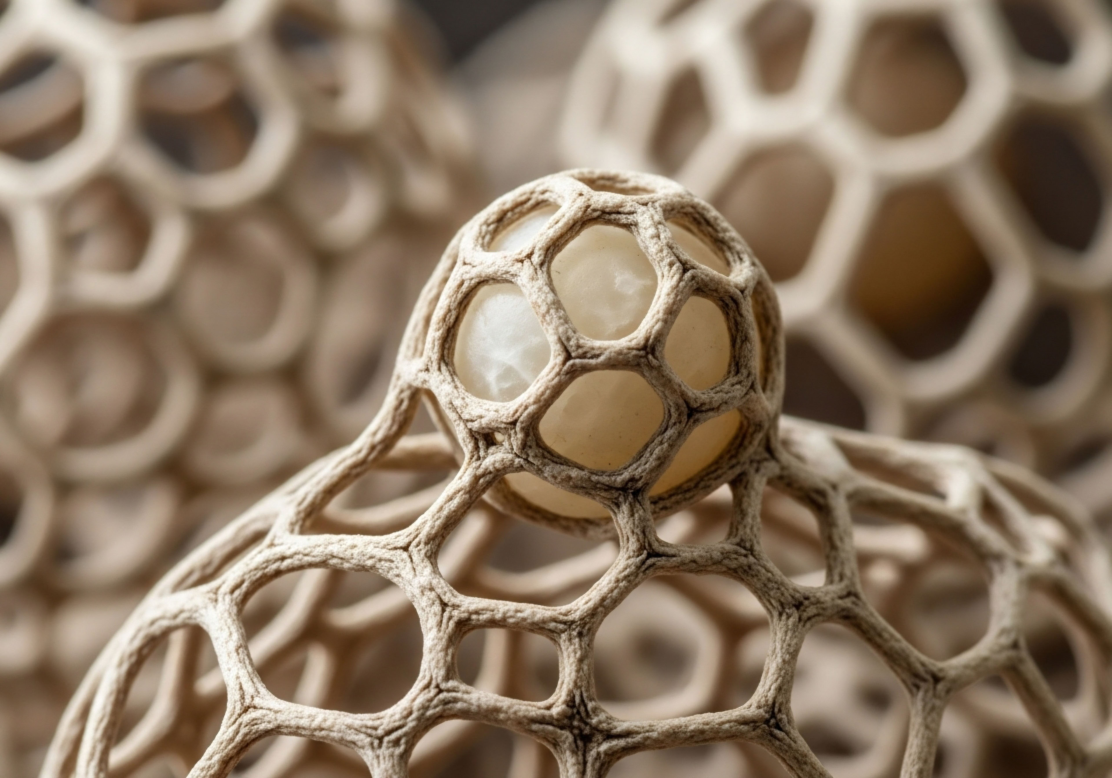
When estrogen binds to its receptors on these fibroblasts, it initiates a cascade of events that leads to the synthesis of collagen and elastin. Collagen provides the skin with its firmness and strength, acting as the scaffolding that holds everything together. Elastin, as its name suggests, gives the skin its ability to stretch and recoil. Optimal estrogen levels maintain a robust production line for these proteins, preserving the skin’s dense, supple structure.
Beyond structural proteins, estrogen also governs the skin’s hydration levels from within. It stimulates the production of hyaluronic acid, a molecule with a remarkable capacity to bind and hold water molecules. This substance fills the spaces between collagen and elastin fibers, creating a hydrated, gel-like matrix that supports the cells and gives the skin a plump, smooth appearance.
A well-hydrated dermis translates to a luminous and dewy complexion. Consequently, a decline in estrogen leads directly to a reduction in hyaluronic acid, causing internal dehydration that manifests as surface dryness and fine lines.

The Calibrator of Skin Function Progesterone
Progesterone works in concert with estrogen, acting as a sophisticated calibrator of skin function. While estrogen builds the primary architecture, progesterone refines and modulates the environment. One of its significant contributions is to skin elasticity. Studies have shown that progesterone can increase the skin’s elastic properties, contributing to its firmness and ability to snap back into place. This action complements estrogen’s role in building the skin’s structural framework.
The interplay between estrogen and progesterone is a dynamic system where each hormone’s actions influence the other to maintain dermal health.
Progesterone also influences sebum production. Sebum is the natural oil produced by the sebaceous glands in the skin, and it forms a protective barrier called the acid mantle. This barrier helps to lock in moisture and protect the skin from external irritants.
Progesterone helps to regulate sebum production, ensuring the skin remains adequately moisturized without becoming overly oily. When progesterone levels decline, this regulation can be disrupted, often contributing to skin dryness and a compromised barrier function. The coordinated action of both estrogen and progesterone is what creates a resilient, hydrated, and structurally sound integumentary system.


Intermediate
Progressing from a foundational awareness of hormonal roles to an intermediate understanding requires examining the precise mechanisms through which estrogen and progesterone exert their influence on skin cells. The aesthetic benefits observed are the direct result of these hormones binding to specific receptors within the skin, thereby activating or deactivating genetic pathways that control cellular growth, protein synthesis, and inflammation.
The concept of balance becomes particularly meaningful here, as the ratio of estrogen to progesterone, not just their absolute quantities, dictates the overall physiological outcome. Disruptions in this ratio, common during the menopausal transition, lead to accelerated changes in skin architecture.

Cellular Targets and Receptor-Mediated Actions
The skin is a major endocrine organ, replete with receptors for sex hormones. Both estrogen receptors (ERα and ERβ) and progesterone receptors (PR) are present in keratinocytes (the primary cells of the epidermis), dermal fibroblasts, and sebaceous glands. Estrogen’s profound effect on collagen is a direct receptor-mediated process.
When estradiol binds to ERα and ERβ on dermal fibroblasts, it promotes the transcription of genes for type I and type III collagen, the two most abundant types of collagen in the skin. Simultaneously, estrogen downregulates the expression of matrix metalloproteinases (MMPs), a family of enzymes responsible for degrading collagen and elastin. This dual action both stimulates new collagen production and protects existing collagen from breakdown, resulting in a net increase in dermal density and thickness.
Progesterone’s influence, while less extensively documented, appears to be similarly receptor-driven. Topical application of progesterone has been clinically demonstrated to improve skin firmness and elasticity in postmenopausal women. This suggests that progesterone, upon binding to its receptors in the skin, may modulate the expression of elastin or other components of the extracellular matrix that contribute to the skin’s pliable yet resilient nature. It also appears to have a calming effect on the skin, potentially by modulating local inflammatory responses.

How Do Clinical Protocols Address Hormonal Imbalance?
Understanding these mechanisms provides the rationale for clinical interventions designed to restore hormonal equilibrium. For women experiencing symptoms associated with perimenopause or post-menopause, such as skin thinning, increased wrinkling, and dryness, hormonal optimization protocols are designed to replenish diminished hormone levels and re-establish a more favorable physiological balance. These are not one-size-fits-all approaches; they are tailored based on an individual’s symptoms and comprehensive lab testing.
- Testosterone Cypionate for Women ∞ While often considered a male hormone, testosterone plays a role in female health, including maintaining skin thickness and collagen production. Low-dose weekly subcutaneous injections (typically 10 ∞ 20 units) can be prescribed to support the structural integrity of the skin and improve overall vitality.
- Progesterone Supplementation ∞ The form and timing of progesterone administration depend on a woman’s menopausal status. For peri-menopausal women with irregular cycles, cyclical progesterone can help regulate the cycle and balance estrogen’s proliferative effects. For post-menopausal women, continuous low-dose progesterone is often used in conjunction with estrogen to provide steady-state benefits, including support for skin elasticity and sleep quality, which itself is vital for skin repair.
- Pellet Therapy ∞ For some individuals, long-acting subcutaneous pellets containing testosterone (and sometimes estradiol) offer a convenient delivery method. These pellets release a steady, low dose of hormones over several months, providing consistent support for skin and other tissues. When testosterone is administered, an aromatase inhibitor like Anastrozole may be included in the protocol to manage the conversion of testosterone to estrogen, ensuring the hormonal ratio remains within the desired therapeutic range.
Hormonal optimization protocols aim to re-establish the biochemical signals that direct cells to maintain youthful function and structure.

Comparing Hormonal States and Their Cutaneous Effects
The visible difference between skin influenced by optimal hormonal balance and skin affected by hormonal decline is stark. The following table outlines the specific impacts on key aesthetic parameters.
| Skin Parameter | Effect of Optimal Estrogen & Progesterone Balance | Effect of Low Estrogen & Progesterone Levels |
|---|---|---|
| Collagen Density | Robust synthesis of Type I and III collagen; inhibition of collagen-degrading enzymes (MMPs). Skin is firm and structurally supported. | Reduced collagen synthesis and increased MMP activity. Skin becomes thinner, less resilient, and more prone to wrinkling. |
| Hydration & Volume | High levels of hyaluronic acid production, leading to excellent water retention in the dermis. Skin appears plump, dewy, and smooth. | Decreased hyaluronic acid levels, resulting in dermal dehydration. Skin looks dry, crepey, and fine lines become more apparent. |
| Elasticity | Healthy elastin fiber function, supported by both hormones. Skin readily returns to its original shape after being stretched. | Elastin fibers become fragmented and lose function. Skin begins to sag and loses its taught quality, a condition known as elastosis. |
| Epidermal Thickness | Maintained keratinocyte proliferation and a healthy epidermal layer. The skin’s barrier function is strong. | The rate of cell turnover slows, leading to a thinning of the epidermis. The skin becomes more fragile and translucent. |
| Wound Healing | Efficient and organized repair processes, with balanced inflammation and rapid tissue regeneration. | Delayed and impaired wound healing. The inflammatory response may be prolonged, and tissue repair is less efficient. |


Academic
An academic exploration of the cutaneous benefits of hormonal equilibrium necessitates a deep analysis of the upstream regulatory systems and the downstream molecular biology within the dermal microenvironment. The aesthetic qualities of skin are emergent properties of complex, interconnected biological pathways. The primary axis governing the production of estrogen and progesterone is the Hypothalamic-Pituitary-Gonadal (HPG) axis.
Its age-related dysregulation is the central event that precipitates the cascade of changes observed in menopausal skin. Understanding this system provides a complete picture, from central nervous system signaling to protein expression in a single fibroblast.

The HPG Axis and the Chronology of Steroid Hormone Decline
The HPG axis functions as a classic endocrine feedback loop. The hypothalamus secretes Gonadotropin-Releasing Hormone (GnRH) in a pulsatile manner, which signals the anterior pituitary gland to release Luteinizing Hormone (LH) and Follicle-Stimulating Hormone (FSH). These gonadotropins travel through the bloodstream to the ovaries, where FSH stimulates follicular growth and LH triggers ovulation and subsequent formation of the corpus luteum.
The developing follicles produce estradiol, while the corpus luteum produces both estradiol and progesterone. These ovarian steroids, in turn, exert negative feedback on both the hypothalamus and the pituitary, suppressing GnRH, LH, and FSH release to maintain homeostasis.
With advancing reproductive age, the pool of ovarian follicles diminishes, and their responsiveness to FSH declines. This leads to lower inhibin B production (a protein that normally suppresses FSH) and subsequently, elevated FSH levels, a hallmark of perimenopause. Estradiol levels become erratic, characterized by periods of both high and low output, while progesterone production wanes due to anovulatory cycles.
Ultimately, with follicular exhaustion at menopause, estradiol and progesterone production plummets, and the loss of negative feedback results in persistently high levels of LH and FSH. This sustained state of hypoestrogenism is the primary driver of accelerated cutaneous aging.
What Are the Molecular Implications in the Dermis?
The loss of circulating estradiol has profound consequences at the molecular level within the dermis. Dermal fibroblasts express both ERα and ERβ, and their activation by estradiol directly influences the expression of genes critical for the extracellular matrix (ECM).
Research has shown that estradiol treatment increases the mRNA levels for COL1A1 and COL3A1, the genes encoding type I and type III procollagen, respectively. Furthermore, estrogen signaling suppresses the transcription of MMP-1 (collagenase) and MMP-3 (stromelysin), enzymes that catabolize the ECM. The net effect of optimal estrogen levels is therefore powerfully anabolic and anti-catabolic for the dermal matrix.
The decline in steroid hormones removes the primary signaling that maintains the skin’s extracellular matrix and structural integrity.
The functionality of Selective Estrogen Receptor Modulators (SERMs) highlights the complexity of this signaling. SERMs like tamoxifen and raloxifene bind to estrogen receptors but induce different conformational changes in the receptor protein. This altered shape affects the recruitment of co-activator and co-repressor proteins, leading to tissue-specific agonist or antagonist effects.
For example, some studies suggest raloxifene may have a stimulatory effect on collagen biosynthesis in skin fibroblasts, demonstrating that it is possible to selectively activate certain beneficial pathways of the estrogen receptor in specific tissues. This field of research underscores the potential for developing highly targeted therapies for skin aging.

Clinical Evidence for Hormonal Intervention on Skin Parameters
A body of clinical research substantiates the connection between hormone replacement therapy (HRT) and the preservation of skin quality in postmenopausal women. These studies provide quantitative evidence for the effects described at the molecular level.
| Study Focus | Intervention | Key Findings and Observations | Source Indication |
|---|---|---|---|
| Skin Collagen Content | Oral conjugated equine estrogens and cyproterone acetate for 6 months. | Demonstrated a statistically significant increase of 6.49% in skin collagen fiber content compared to a non-treated control group. This provides direct evidence of HRT’s anabolic effect on the dermal matrix. | |
| Skin Thickness and Hydration | Topical estradiol cream applied to the face of postmenopausal women. | Resulted in increased epidermal thickness and improved skin hydration. The study highlighted that topical application can exert local benefits, potentially minimizing systemic exposure. | |
| Skin Elasticity and Firmness | Topical 2% progesterone cream applied by peri- and postmenopausal women for 16 weeks. | Showed a significant increase in skin elasticity and firmness compared to a vehicle-controlled group. Wrinkle depth and count were also reduced, isolating the specific benefits of progesterone. | |
| Wrinkling and Rigidity | Long-term oral estrogen therapy in women at least 5 years post-menopause. | The hormone-treated group had significantly lower average wrinkle scores and reduced skin rigidity compared to a group that had never used HRT. This points to the long-term preventative effects of sustained hormonal support. |
These clinical findings, grounded in the principles of HPG axis function and molecular endocrinology, create a cohesive and evidence-based understanding of how maintaining a balanced hormonal environment directly translates to tangible aesthetic benefits. The visible youthfulness of the skin is a reflection of its underlying cellular health, which is meticulously orchestrated by estrogen and progesterone.

References
- Stevenson, Susan, and Julie Thornton. “Effect of estrogens on skin aging and the potential role of SERMs.” Clinical Interventions in Aging, vol. 2, no. 3, 2007, pp. 283-97.
- Gasser, S. et al. “Impact of progesterone on skin and hair in menopause ∞ a comprehensive review.” Gynecological Endocrinology, vol. 37, no. 2, 2021, pp. 1-7.
- Holzer, G. et al. “Effects and side effects of 2%-progesterone cream on the skin of peri- and postmenopausal women ∞ Results from a double blind, vehicle-controlled, randomized study.” British Journal of Dermatology, vol. 153, no. 3, 2005, pp. 626-34.
- Rzepecki, Alexandra K. et al. “Estrogen-deficient skin ∞ The role of topical therapy.” International Journal of Women’s Dermatology, vol. 5, no. 2, 2019, pp. 85-90.
- Singh, Priyali. “Collagen and Estrogen ∞ How Hormones Affect Skin, Joints, and Aging in Women.” Meto Blog, 3 July 2025.

Reflection

Your Biological Narrative
The information presented here offers a map of the intricate biological landscape that shapes your skin’s health. It translates the abstract language of endocrinology into a tangible understanding of your own physical experience. This knowledge is a powerful tool.
It allows you to reframe the changes you observe not as inevitable decline, but as predictable outcomes of shifts in your internal signaling. Your body is communicating its needs through these visible signs. Recognizing the connection between how you feel, how you look, and your underlying hormonal status is the first, most significant step toward proactive stewardship of your own well-being.

A Foundation for Informed Action
This deep dive into the science of hormonal influence is designed to be a foundation, preparing you for a more productive and collaborative dialogue with a clinical expert. A personalized wellness protocol is built upon this type of evidence-based understanding, combined with specific diagnostic data from your own body.
Armed with this clarity, you can ask more precise questions, better understand the rationale behind specific therapeutic recommendations, and become an active participant in the process of calibrating your own health. The potential for vitality and function is not something to be reclaimed from the past; it is something to be built for the future, based on a sophisticated understanding of your unique biology.