
Fundamentals
Embarking on a journey of hormonal optimization is a deeply personal decision, often born from a feeling that your body’s vitality is slipping away. You might notice a persistent fatigue, a decline in strength, or a general sense of diminished well-being.
When you consider testosterone replacement therapy (TRT), you are looking for a way to reclaim that function. A central question that arises in this process is how introducing testosterone from an external source affects the body’s own production centers, specifically the testes. Understanding this relationship is fundamental to making informed decisions about your long-term health.
The body’s hormonal system operates on a sophisticated feedback mechanism, much like a thermostat regulating a room’s temperature. This network, known as the Hypothalamic-Pituitary-Gonadal (HPG) axis, constantly monitors and adjusts hormone levels to maintain a state of balance, or homeostasis.
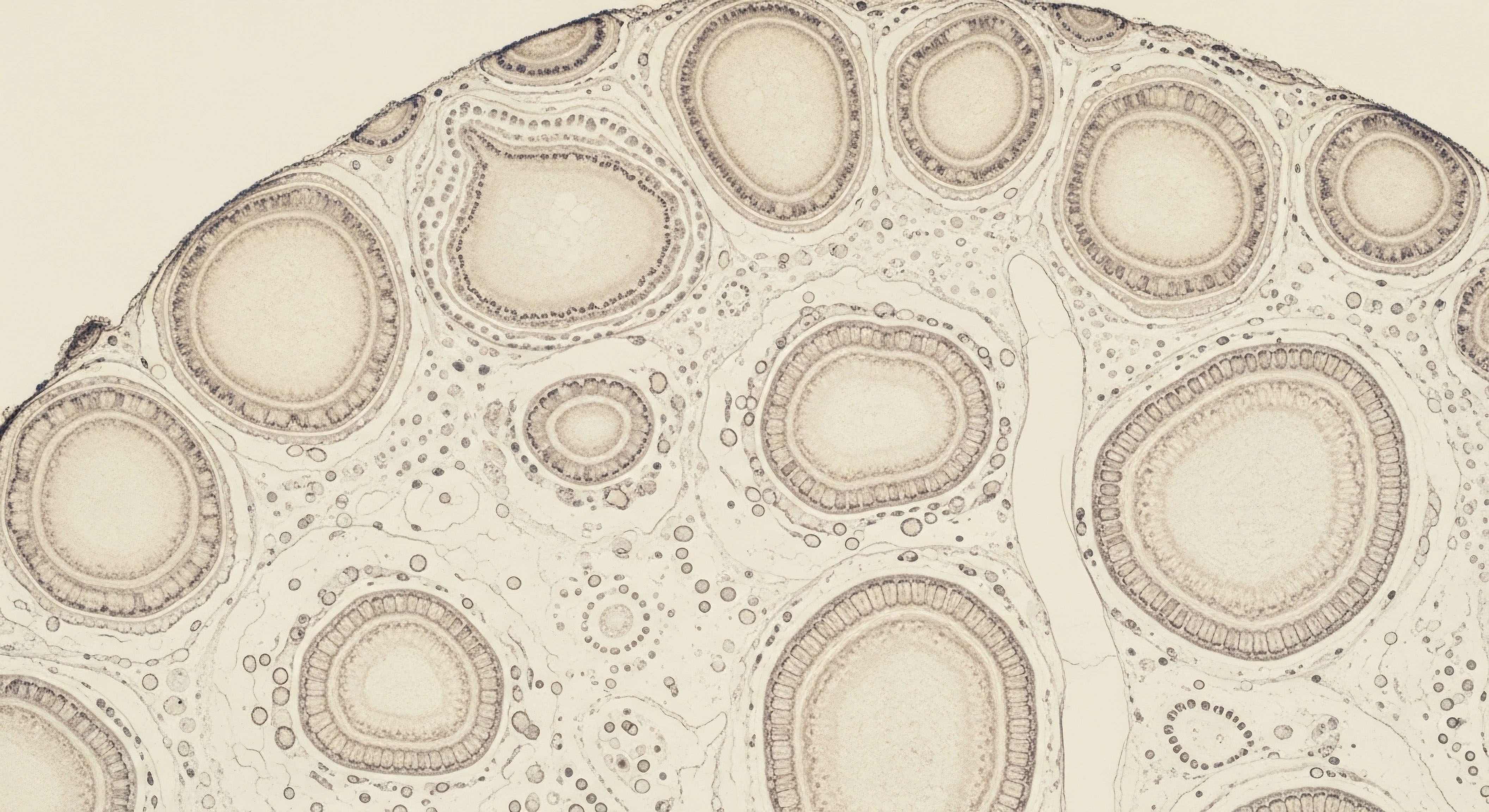
The hypothalamus in the brain releases Gonadotropin-Releasing Hormone (GnRH), which signals the pituitary gland to produce two key hormones ∞ Luteinizing Hormone (LH) and Follicle-Stimulating Hormone (FSH). LH travels to the testes and instructs specialized cells, the Leydig cells, to produce testosterone. FSH, in parallel, acts on another set of testicular cells, the Sertoli cells, to facilitate sperm production (spermatogenesis).
Introducing external testosterone signals the body’s control center to halt its own production, leading to a predictable suppression of testicular function.
When you begin a TRT protocol, you introduce testosterone into your bloodstream from an external source. Your brain’s intricate monitoring system detects these elevated levels. In response, the hypothalamus reduces its GnRH signals, believing the body has more than enough testosterone.
This action has a direct cascading effect ∞ the pituitary gland slows, or even stops, its release of LH and FSH. Without the stimulating signals from LH and FSH, the testes are no longer instructed to perform their primary functions. The Leydig cells cease producing endogenous testosterone, and the Sertoli cells halt the process of spermatogenesis.
This systematic shutdown is a predictable and normal physiological response. It is the body’s way of conserving resources when it perceives an abundance of a particular hormone. The direct consequence for the testicular environment is a significant decrease in intratesticular testosterone levels, which are crucial for sperm maturation.
This leads to a state of temporary infertility and a noticeable reduction in testicular size, often referred to as testicular atrophy. These effects are a direct and expected outcome of long-term TRT, highlighting the profound influence exogenous hormones have on the body’s natural endocrine rhythms.


Intermediate
For individuals undertaking hormonal optimization, moving beyond the foundational understanding of testicular suppression opens the door to a more sophisticated appreciation of clinical protocols. These strategies are designed to manage the physiological consequences of long-term TRT. The primary objective is to support the testicular environment even as the HPG axis is being intentionally suppressed by external testosterone. This requires a nuanced approach that addresses the specific hormonal signals that are diminished during therapy.
Maintaining Testicular Integrity during Therapy
The shutdown of the HPG axis by exogenous testosterone creates a communication breakdown. The testes no longer receive the LH and FSH signals from the pituitary gland that are essential for their function. To counteract this, clinicians can introduce agents that mimic these natural signals, thereby preserving testicular activity and size. The two primary compounds used for this purpose are Human Chorionic Gonadotropin (hCG) and Gonadorelin.
Human Chorionic Gonadotropin (hCG) is a hormone that structurally resembles LH. When administered, it binds to the LH receptors on the Leydig cells in the testes, directly stimulating them to produce testosterone and maintain their volume. This process effectively bypasses the suppressed HPG axis, providing the stimulation the testes need to remain active. By keeping the Leydig cells engaged, hCG helps prevent the significant testicular atrophy and decline in natural testosterone production that would otherwise occur.
Gonadorelin is a synthetic version of GnRH, the hormone released by the hypothalamus. Its mechanism is different from hCG. Instead of directly stimulating the testes, Gonadorelin works upstream by stimulating the pituitary gland to release its own LH and FSH. This approach helps maintain the natural signaling pathway from the pituitary to the testes. It is often prescribed to prevent testicular shrinkage and preserve some level of endogenous hormone production during TRT.
Clinical protocols utilize agents like hCG or Gonadorelin to mimic natural hormonal signals, thereby preserving testicular function during TRT.
The choice between these agents depends on individual goals, such as fertility preservation or maintaining testicular volume for psychological well-being. Protocols often involve administering these compounds alongside testosterone cypionate injections to create a more balanced hormonal environment. For example, a standard protocol might pair weekly testosterone injections with twice-weekly subcutaneous injections of Gonadorelin or hCG.

What Is the Role of Aromatase Inhibitors?
A secondary, yet important, consideration in long-term TRT is the management of estrogen. Testosterone can be converted into estradiol, a form of estrogen, through a process called aromatization. While some estrogen is necessary for male health, elevated levels can lead to side effects such as water retention and gynecomastia.
Aromatase inhibitors, like Anastrozole, are medications that block this conversion process, helping to maintain a healthy testosterone-to-estrogen ratio. Their inclusion in a protocol is based on an individual’s lab results and clinical presentation.
| Compound | Mechanism of Action | Primary Goal in TRT | Administration |
|---|---|---|---|
| hCG | Mimics LH, directly stimulating Leydig cells in the testes. | Prevents testicular atrophy, maintains endogenous testosterone production, and supports fertility. | Subcutaneous injection, typically 2-3 times per week. |
| Gonadorelin | Mimics GnRH, stimulating the pituitary to release LH and FSH. | Maintains testicular size and function by preserving the pituitary-testicular signaling pathway. | Subcutaneous injection, frequency varies based on protocol. |
| Anastrozole | Blocks the aromatase enzyme, preventing the conversion of testosterone to estrogen. | Manages estrogen levels to mitigate potential side effects. | Oral tablet, typically taken a couple of times per week. |

Post-Therapy Recovery and Fertility
For men who wish to discontinue TRT, particularly for fertility reasons, a specific protocol is required to restart the HPG axis. The goal is to encourage the body to resume its natural production of GnRH, LH, and FSH. This often involves a combination of medications designed to stimulate the endocrine system at different points.
- Clomiphene Citrate (Clomid) and Tamoxifen ∞ These are Selective Estrogen Receptor Modulators (SERMs). They work by blocking estrogen receptors in the hypothalamus, which tricks the brain into thinking estrogen levels are low. This stimulates the release of GnRH, and subsequently LH and FSH, kickstarting testicular function.
- Gonadorelin ∞ As in on-cycle therapy, Gonadorelin can be used to directly stimulate the pituitary gland.
The timeline for recovery of spermatogenesis after stopping long-term TRT varies significantly among individuals. Factors such as the duration of therapy, the dosage used, and the individual’s age all play a role. While many men can recover normal sperm production, it can take several months to over a year. Some studies indicate that with proper management, a significant majority of men can expect sperm counts to return to fertile levels within 6 to 12 months.

Academic
A sophisticated analysis of long-term testosterone replacement therapy’s influence on testicular function extends beyond the systemic suppression of the HPG axis to the cellular and molecular dynamics within the testicular microenvironment. The administration of exogenous androgens initiates a profound shift in the paracrine signaling and metabolic interplay between Leydig and Sertoli cells, the two cornerstone cell types governing testicular homeostasis.
Understanding this disruption at a cellular level provides a more complete picture of the resulting testicular quiescence and the challenges associated with its reversal.

Cellular Mechanisms of Testicular Suppression
The primary mechanism of TRT-induced testicular suppression is the cessation of gonadotropin support. Luteinizing Hormone (LH) is the principal trophic factor for Leydig cells, stimulating not only testosterone synthesis but also ensuring their long-term survival and functional integrity. Follicle-Stimulating Hormone (FSH) provides critical support to Sertoli cells, which in turn orchestrate the complex process of spermatogenesis.
When exogenous testosterone administration suppresses pituitary LH and FSH secretion, the testes are deprived of these vital signals. The Leydig cells, lacking LH stimulation, downregulate the expression of key steroidogenic enzymes, including the crucial rate-limiting enzyme, Cholesterol side-chain cleavage enzyme (CYP11A1), and 17β-hydroxysteroid dehydrogenase (HSD17B3), which converts androstenedione to testosterone.
This leads to a dramatic fall in intratesticular testosterone concentrations, which are normally maintained at levels 50-100 times higher than in peripheral circulation. This high local concentration is indispensable for the progression of meiosis and the maturation of spermatids.
The absence of gonadotropin signaling during TRT disrupts the intricate cellular communication between Leydig and Sertoli cells, leading to a profound suppression of steroidogenesis and spermatogenesis.
Simultaneously, the absence of FSH signaling to Sertoli cells compromises their ability to support developing germ cells. Sertoli cells are responsible for providing structural support, nutrients, and regulatory factors to the germ cells. The reduction in FSH, coupled with the collapse of intratesticular testosterone, leads to a breakdown of this supportive architecture, resulting in increased germ cell apoptosis and a halt in spermatogenesis. This process manifests clinically as azoospermia or severe oligozoospermia.

What Are the Long-Term Consequences for Cellular Function?
Prolonged withdrawal of gonadotropin support can lead to more persistent changes in testicular cell populations. While much of the initial testicular atrophy is due to the loss of germ cells and a reduction in seminiferous tubule diameter, there can also be changes to the Leydig and Sertoli cells themselves. Some evidence suggests that long-term suppression may lead to Leydig cell dedifferentiation or apoptosis, potentially complicating the recovery of endogenous testosterone production after TRT cessation.
The recovery of spermatogenesis is contingent on the successful re-establishment of the HPG axis and the restoration of intratesticular testosterone. Studies examining recovery post-TRT show a wide variability in timelines, influenced by the duration of androgen exposure and patient age.
Data extrapolated from male contraceptive studies, which use testosterone to suppress fertility, indicate that while recovery is likely for most, it can be a protracted process. Probability estimates suggest recovery of spermatogenesis in 90% of men at 12 months and 100% at 24 months after discontinuing exogenous testosterone.
| Cell Type | Primary Gonadotropin Signal | Effect of TRT-Induced Suppression | Functional Consequence |
|---|---|---|---|
| Leydig Cells | Luteinizing Hormone (LH) | Downregulation of steroidogenic enzymes (e.g. CYP11A1, HSD17B3). Potential for cellular dedifferentiation over time. | Drastic reduction in intratesticular testosterone production. |
| Sertoli Cells | Follicle-Stimulating Hormone (FSH) | Reduced supportive function for germ cells. Compromised structural integrity of seminiferous tubules. | Increased germ cell apoptosis and cessation of spermatogenesis. |

How Does Adjunctive Therapy Influence Cellular Dynamics?
The use of adjunctive therapies like hCG and SERMs (e.g. Clomiphene) directly targets these cellular mechanisms. hCG acts as an LH analog, providing the necessary trophic support to Leydig cells to maintain steroidogenesis and intratesticular testosterone levels, thereby preserving the conditions required for spermatogenesis.
This intervention helps mitigate the profound cellular quiescence induced by TRT. SERMs, used in post-cycle therapy, work at the hypothalamic level to restore the endogenous production of LH and FSH, initiating the cascade that reactivates both Leydig and Sertoli cell function. The success of these recovery protocols underscores the plasticity of the testicular environment, although the potential for incomplete recovery in cases of very long-term use remains a critical area of clinical consideration.

References
- Ramasamy, R. et al. “Recovery of spermatogenesis following testosterone replacement therapy or anabolic-androgenic steroid use.” Fertility and Sterility, vol. 105, no. 3, 2016, pp. 569-77.
- Bhattacharya, T. et al. “The effects of long-term testosterone treatment on endocrine parameters in hypogonadal men ∞ 12-year data from a prospective controlled registry study.” The Aging Male, vol. 25, no. 1, 2022, pp. 185-191.
- McColl, E.M. et al. “The effects of testosterone replacement therapy on the prostate ∞ a clinical perspective.” Urology, vol. 79, no. 4, 2012, pp. 763-70.
- Liu, P. Y. et al. “The rate, extent, and modifiers of spermatogenic recovery after hormonal male contraception ∞ an integrated analysis.” The Lancet, vol. 367, no. 9520, 2006, pp. 1412-20.
- O’Donnell, L. et al. “Testosterone and spermatogenesis.” Endocrine Reviews, vol. 22, no. 3, 2001, pp. 289-318.
- Matthew W. Retzloff, et al. “Age and Duration of Testosterone Therapy Predict Time to Return of Sperm Count after hCG Therapy.” Fertility and Sterility, vol. 104, no. 3, 2015, E249.
- Payne, A. H. and G. L. Youngblood. “Regulation of expression of steroidogenic enzymes in Leydig cells.” Biology of Reproduction, vol. 52, no. 2, 1995, pp. 217-25.
- Walker, W. H. “Testosterone signaling and the regulation of spermatogenesis.” Spermatogenesis, vol. 1, no. 2, 2011, pp. 116-20.
- Chen, H. et al. “Intercellular communication between Sertoli cells and Leydig cells in the absence of Follicle-Stimulating Hormone-Receptor signaling.” Biology of Reproduction, vol. 73, no. 4, 2005, pp. 655-61.
- Saad, F. et al. “Effects of testosterone on metabolic syndrome components.” The Journal of Steroid Biochemistry and Molecular Biology, vol. 116, no. 3-5, 2009, pp. 162-67.

Reflection
The journey toward hormonal balance is a path of profound self-discovery. The information presented here illuminates the intricate biological systems at play, translating the clinical science of testosterone therapy into a more tangible understanding of your own body. This knowledge is the first, most critical step. It transforms abstract feelings of fatigue or decline into a clear map of physiological processes, allowing you to see the connections between symptoms and systems.

Your Personal Health Blueprint
As you move forward, consider this knowledge a foundation upon which to build a personalized health strategy. Your body has its own unique history and its own distinct needs. The decision to engage with hormonal optimization protocols is one that should be made in partnership with a clinical expert who can interpret your specific biological data and align it with your personal goals.
The ultimate aim is to create a protocol that restores function and vitality in a way that is sustainable and true to your individual blueprint. The power to reclaim your well-being begins with this deeper understanding of the elegant, complex machinery within.

Glossary

hormonal optimization

testosterone replacement therapy

follicle-stimulating hormone

luteinizing hormone

endogenous testosterone

pituitary gland

intratesticular testosterone

testicular atrophy

long-term trt

hpg axis

human chorionic gonadotropin

gonadorelin

testosterone production
leydig cells

testicular function

clomiphene citrate

spermatogenesis

testosterone replacement

sertoli cells

increased germ cell apoptosis


