
Fundamentals
You may feel a pervasive sense of fatigue, a subtle loss of vitality, or notice changes in your body composition that seem disconnected from your lifestyle. These experiences are valid and often point toward a complex internal conversation your body is having, a conversation where the hormones insulin and testosterone are central figures.
Understanding how insulin resistance directly affects testosterone levels is the first step in decoding these signals and reclaiming your body’s intended function. The connection is a two-way street, a reciprocal relationship where the function of one profoundly influences the other, creating a feedback loop that can either support or undermine your overall well-being.

The Cellular Dialogue between Insulin and Testosterone
At its core, your body operates through a series of intricate signaling systems. Think of hormones as messengers carrying specific instructions to cells. Insulin’s primary role is to instruct cells to absorb glucose from the bloodstream for energy.
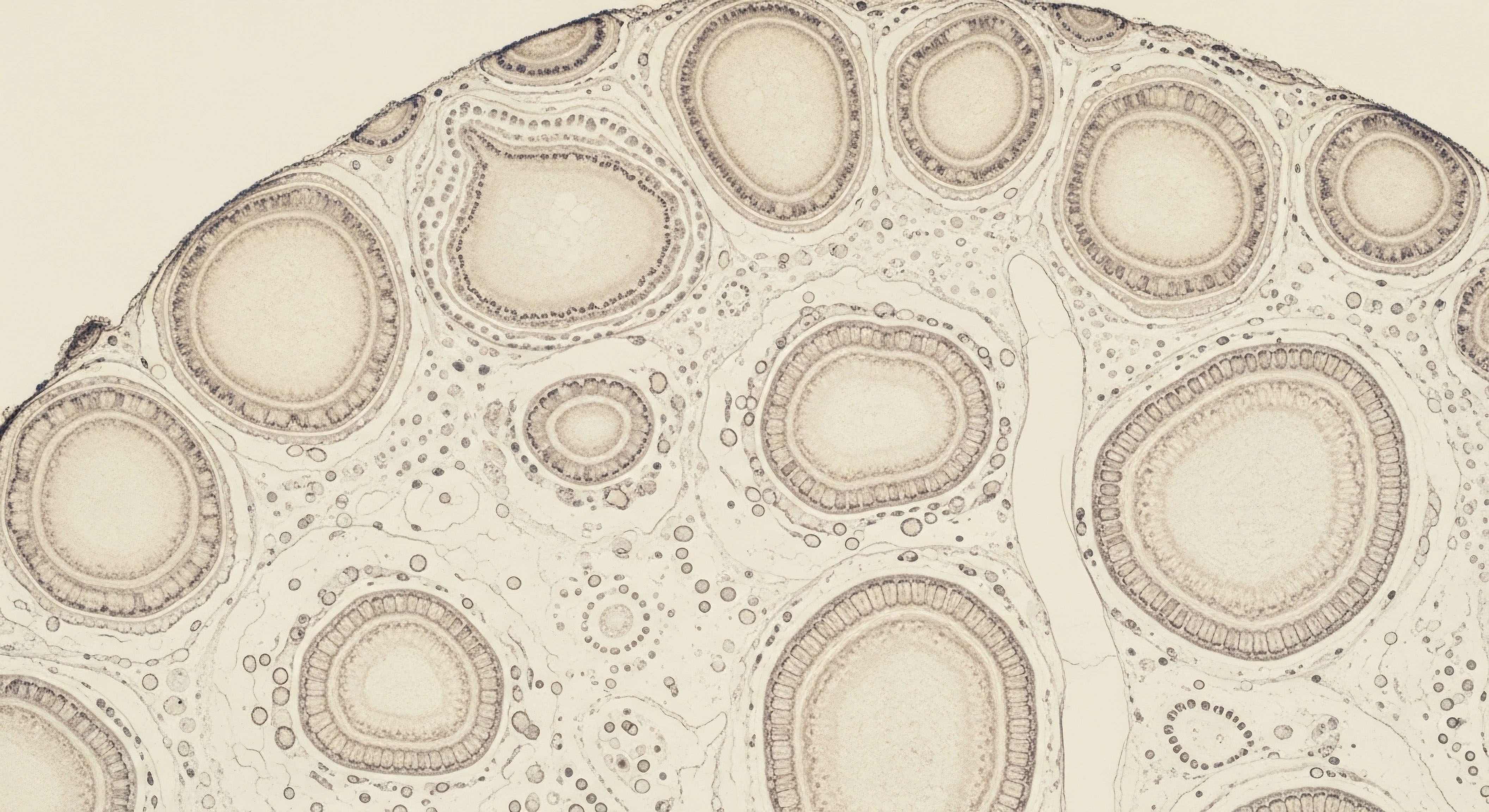
When cells become resistant to this message, the pancreas compensates by producing more insulin, leading to a state of high insulin levels, or hyperinsulinemia. This elevated insulin level is where the direct impact on testosterone begins. The Leydig cells in the testes, the primary sites of testosterone production in men, are one of the many cell types that listen to insulin’s signals.
Research indicates that a state of insulin resistance is associated with a decrease in the ability of these Leydig cells to secrete testosterone. This suggests a direct impairment at the very source of testosterone synthesis.
Insulin resistance creates a state of high circulating insulin, which directly impairs the testosterone-producing cells in the testes.
This biological crosstalk means that the metabolic state of your body has a direct and measurable effect on your hormonal state. The fatigue, difficulty in building muscle, and changes in mood you might be experiencing are not isolated symptoms; they are downstream consequences of this cellular miscommunication. The process is a cascade. Insulin resistance promotes conditions that further suppress testosterone, and low testosterone can, in turn, worsen insulin sensitivity, creating a self-perpetuating cycle.

The Role of Body Composition
Insulin resistance is closely linked to an increase in visceral adipose tissue, the fat stored deep within the abdominal cavity. This type of fat is metabolically active and functions almost like an endocrine organ itself. It releases inflammatory substances and contributes to the conversion of testosterone into estrogen via an enzyme called aromatase.
This process does two things ∞ it lowers the available amount of active testosterone and increases estrogen levels, which then signals the brain to reduce its stimulation for testosterone production. It is a compounding effect where the consequence of insulin resistance (increased visceral fat) becomes a cause of lower testosterone. Addressing insulin resistance, therefore, becomes a foundational strategy for restoring hormonal balance. It is about recalibrating the body’s internal environment to support its natural, optimal function.


Intermediate
For those already familiar with the basic connection between metabolic health and hormonal balance, the next step is to understand the specific mechanisms through which insulin resistance systematically dismantles healthy testosterone levels. This involves looking beyond the direct effects on the testes and examining the broader endocrine system, including transport proteins and the central command center in the brain.
The relationship is a complex interplay of signals and feedback loops, where a disruption in one area creates ripple effects throughout the system. A clinically informed perspective reveals a few key pathways that are profoundly impacted.

Sex Hormone-Binding Globulin a Key Regulator
Testosterone circulates in the bloodstream in two primary states ∞ bound and unbound (or free). The majority of testosterone is bound to proteins, primarily Sex Hormone-Binding Globulin (SHBG). Only unbound testosterone is biologically active and available for your cells to use. The liver produces SHBG, and its production is directly suppressed by insulin.
In a state of insulin resistance, the resulting high insulin levels send a strong signal to the liver to decrease SHBG production. This leads to lower total SHBG levels in the blood. While this might intuitively seem to increase free testosterone, the body’s regulatory systems compensate.
The lower SHBG levels mean that testosterone is cleared from the bloodstream more quickly, and the overall pool of total testosterone often decreases. Low SHBG is a well-established marker for insulin resistance and is strongly associated with a higher likelihood of metabolic syndrome.
Elevated insulin levels directly suppress the liver’s production of SHBG, the primary transport protein for testosterone, leading to lower total testosterone levels and disrupting hormonal regulation.
This is a critical point in understanding lab results. A man might have a “normal” free testosterone level but a low total testosterone and low SHBG, a pattern that points directly toward underlying insulin resistance as a primary driver of the hormonal imbalance. Therapeutic approaches that focus solely on increasing testosterone without addressing the root cause of insulin resistance often fail to achieve optimal outcomes because they do not correct the underlying systemic dysfunction.

How Does SHBG Influence Testosterone Bioavailability?
The concentration of SHBG effectively determines the distribution of testosterone between its bound and free states. Think of SHBG as a reservoir that holds testosterone, protecting it from rapid degradation and ensuring a stable supply. When SHBG levels are healthy, this reservoir is well-maintained. When insulin resistance drives SHBG down, the reservoir shrinks, leading to a more volatile and often deficient supply of this vital hormone.
To illustrate the clinical relevance, consider the following table comparing typical hormonal profiles:
| Hormonal Marker | Healthy Metabolic State | Insulin-Resistant State |
|---|---|---|
| Fasting Insulin | Low to Normal | High |
| SHBG | Normal to High | Low |
| Total Testosterone | Optimal | Low to Borderline |
| Free Testosterone | Optimal | Variable, often low-normal |

The Hypothalamic-Pituitary-Gonadal Axis Disruption
The production of testosterone is regulated by a sophisticated feedback system known as the Hypothalamic-Pituitary-Gonadal (HPG) axis. The hypothalamus in the brain releases Gonadotropin-Releasing Hormone (GnRH) in pulses. This signals the pituitary gland to release Luteinizing Hormone (LH), which then travels to the testes and stimulates the Leydig cells to produce testosterone. The frequency and amplitude of these GnRH pulses are critical for maintaining healthy LH levels and, consequently, testosterone production.
Insulin resistance and the associated metabolic changes can disrupt this central pulse generator. Several factors are at play:
- Leptin ResistanceObesity and insulin resistance are often accompanied by high levels of the hormone leptin, which is produced by fat cells. While leptin is normally a key signal for the brain to regulate energy balance and support reproductive function, chronic high levels can lead to leptin resistance in the hypothalamus. This resistance can interfere with the proper functioning of the GnRH pulse generator, leading to disordered LH release and inadequate stimulation of the testes.
- Inflammatory SignalsInsulin resistance is fundamentally an inflammatory state. The increased visceral fat produces inflammatory cytokines like TNF-α and IL-6. These cytokines can cross the blood-brain barrier and directly suppress hypothalamic function, further dampening the GnRH pulses.
The result is a state of secondary hypogonadism, where the problem originates not in the testes themselves, but in the signaling from the brain. This is why simply administering testosterone may not be a complete solution. Protocols that include agents like Gonadorelin, which mimics GnRH, or Enclomiphene, which stimulates the pituitary, are designed to address this central suppression and support the entire HPG axis.


Academic
A sophisticated analysis of the relationship between insulin resistance and testosterone requires a systems-biology perspective, examining the intricate molecular and cellular mechanisms that link metabolic dysregulation to endocrine failure. The connection extends beyond simple hormonal suppression and involves a complex network of inflammatory pathways, cellular stress responses, and genetic expression changes.
At this level, we see how insulin resistance acts as a systemic stressor that directly compromises the function of the Leydig cells, the hypothalamic GnRH pulse generator, and the bioavailability of androgens.

Direct Leydig Cell Inhibition via Inflammatory Cytokines
Insulin resistance and the associated visceral adiposity create a chronic, low-grade inflammatory environment. This state is characterized by elevated circulating levels of pro-inflammatory cytokines, such as Tumor Necrosis Factor-alpha (TNF-α), Interleukin-1 beta (IL-1β), and Interleukin-6 (IL-6).
These cytokines have been shown in vitro and in vivo to exert a direct inhibitory effect on Leydig cell steroidogenesis. The mechanism is multifactorial. These cytokines can suppress the expression of key steroidogenic enzymes, including Steroidogenic Acute Regulatory (StAR) protein, which is the rate-limiting step in transporting cholesterol into the mitochondria for conversion into pregnenolone.
They also downregulate the expression of enzymes further down the cascade, such as CYP17A1 and HSD3B1, which are essential for the synthesis of testosterone from its precursors.
The chronic inflammation associated with insulin resistance directly suppresses testosterone synthesis by inhibiting the expression of critical enzymes within the Leydig cells of the testes.
This inflammatory-mediated suppression provides a direct molecular link between metabolic disease and hypogonadism. It explains why conditions associated with high levels of inflammation, even outside of obesity, can lead to reduced testosterone levels. The Leydig cells are, in effect, caught in the crossfire of a systemic inflammatory response, leading to a measurable decline in their primary function.

How Does Inflammation Specifically Impair Testicular Function?
The impact of inflammatory mediators on testicular function is a critical area of research. The following table outlines the specific actions of key cytokines on the testosterone production pathway.
| Cytokine | Mechanism of Action on Leydig Cells | Resulting Effect on Steroidogenesis |
|---|---|---|
| TNF-α | Inhibits the expression of StAR protein and key steroidogenic enzymes like CYP17A1. | Significant reduction in testosterone production. |
| IL-6 | Can block the differentiation of stem Leydig cells into mature, testosterone-producing cells. | Reduced capacity for Leydig cell regeneration and long-term testosterone synthesis. |
| IL-1β | Decreases testosterone synthesis in a dose-dependent manner. | Direct suppression of acute testosterone output. |

The Central Disruption of the GnRH Pulse Generator
The hypothalamic GnRH pulse generator, now understood to be driven by kisspeptin neurons in the arcuate nucleus, is highly sensitive to metabolic cues. In a state of insulin resistance, this central regulator is bombarded with disruptive signals. The hyperinsulinemia itself can have a direct effect.
While acute insulin administration can stimulate GnRH release, the chronic hyperinsulinemia and subsequent insulin resistance at the neuronal level seen in obesity appears to disrupt the delicate pulsatility required for proper LH secretion. Studies in animal models show that mice with specific deletion of the insulin receptor on GnRH neurons have altered reproductive function in the context of diet-induced obesity, highlighting the direct role of insulin signaling in these neurons.
Furthermore, the associated hyperleptinemia and subsequent leptin resistance are critical factors. Leptin is a permissive signal for reproduction, indicating sufficient energy stores. When the hypothalamus becomes resistant to leptin’s signal, it interprets this as a state of energy deficit, even in the presence of excess body fat.
This perceived energy crisis leads to a downregulation of the GnRH pulse generator as a protective mechanism to conserve resources, further suppressing the HPG axis. This creates a paradoxical situation where metabolic excess leads to a hormonal state that mimics starvation.
The therapeutic implications are significant. Addressing the central component of hypogonadism in insulin-resistant individuals requires strategies that go beyond simple testosterone replacement. Interventions may need to focus on improving insulin and leptin sensitivity in the brain, potentially through metabolic therapies or targeted peptide protocols designed to restore hypothalamic function. This systems-based approach recognizes that the hormonal deficiency is a symptom of a much broader metabolic and neuroendocrine dysregulation.

References
- Pitteloud, N. et al. “Increasing Insulin Resistance Is Associated with a Decrease in Leydig Cell Testosterone Secretion in Men.” The Journal of Clinical Endocrinology & Metabolism, vol. 90, no. 5, 2005, pp. 2636-41.
- Kelly, D. M. and T. H. Jones. “Testosterone and Glucose Metabolism in Men ∞ Current Concepts and Controversies.” Journal of Endocrinology, vol. 217, no. 3, 2013, R25-45.
- Ding, E. L. et al. “Sex Hormone ∞ Binding Globulin and Risks of Type 2 Diabetes in Women and Men.” New England Journal of Medicine, vol. 361, no. 12, 2009, pp. 1152-63.
- Traish, A. M. et al. “The Dark Side of Testosterone Deficiency ∞ I. Metabolic Syndrome and Erectile Dysfunction.” Journal of Andrology, vol. 30, no. 1, 2009, pp. 10-22.
- Herbison, A. E. “The Gonadotropin-Releasing Hormone Pulse Generator.” Endocrinology, vol. 159, no. 9, 2018, pp. 3249-56.
- Hales, D. B. and A. H. Payne. “Glucocorticoid-mediated repression of P450scc mRNA and protein in cultured mouse Leydig cells.” Endocrinology, vol. 124, no. 5, 1989, pp. 2099-104.
- Laaksonen, D. E. et al. “Associations of Total Testosterone and Sex Hormone ∞ Binding Globulin Levels With Insulin Sensitivity in Middle-Aged Finnish Men.” Diabetes Care, vol. 30, no. 4, 2007, pp. 902-04.
- Vikan, T. et al. “The association between serum testosterone and insulin resistance ∞ a longitudinal study.” BMC Endocrine Disorders, vol. 18, no. 1, 2018, p. 89.
- Zitzmann, M. “Testosterone deficiency, insulin resistance and the metabolic syndrome.” Nature Reviews Endocrinology, vol. 5, no. 12, 2009, pp. 673-81.
- Chen, C. et al. “The in vitro modulation of steroidogenesis by inflammatory cytokines and insulin in TM3 Leydig cells.” Reproductive Biology and Endocrinology, vol. 16, no. 1, 2018, p. 26.

Reflection
The information presented here provides a map of the biological territory where your metabolic and hormonal health intersect. Understanding these complex connections is a profound act of self-awareness. It shifts the perspective from viewing symptoms as isolated problems to seeing them as communications from a deeply interconnected system.
This knowledge is the foundation. The next step in your personal health journey is to consider how this map applies to your unique physiology. What are the signals your body is sending? How might your metabolic health be influencing your vitality? This is where the path becomes personalized, moving from general understanding to specific, actionable insight guided by a comprehensive evaluation of your individual biology.


