

Fundamentals
You feel it in the morning, a stiffness in your knees or a subtle ache in your hands that wasn’t there a decade ago. It’s a physical sensation that speaks to a deeper biological truth, a gradual shift in your body’s internal environment. This experience is a valid and important signal.
Your body is communicating a change in its capacity for maintenance and repair. The question of how hormonal protocols protect your joints for the long term is not about masking a symptom; it is about understanding and addressing the root cause of this very real, physical experience.
It begins with acknowledging that your joints are living, dynamic tissues, constantly engaged in a process of breakdown and renewal, a process orchestrated by a complex network of biochemical messengers, with your sex hormones acting as the master conductors.
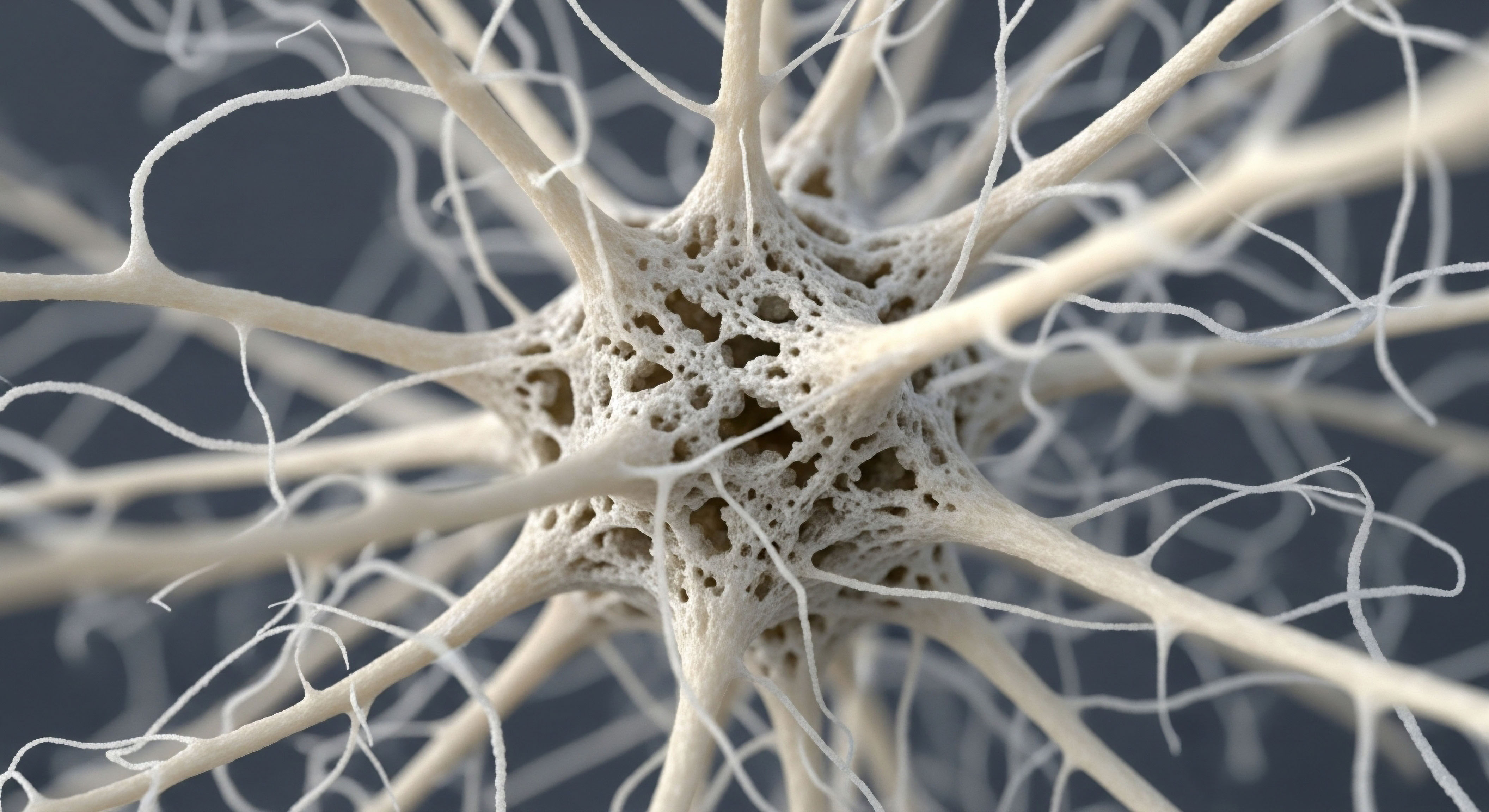
To truly grasp how hormonal optimization guards your joints, we must first look at the architecture of the joint itself. Imagine a joint, like your knee or hip, as a sophisticated biological hinge. The ends of the bones are capped with a smooth, resilient material called articular cartilage.
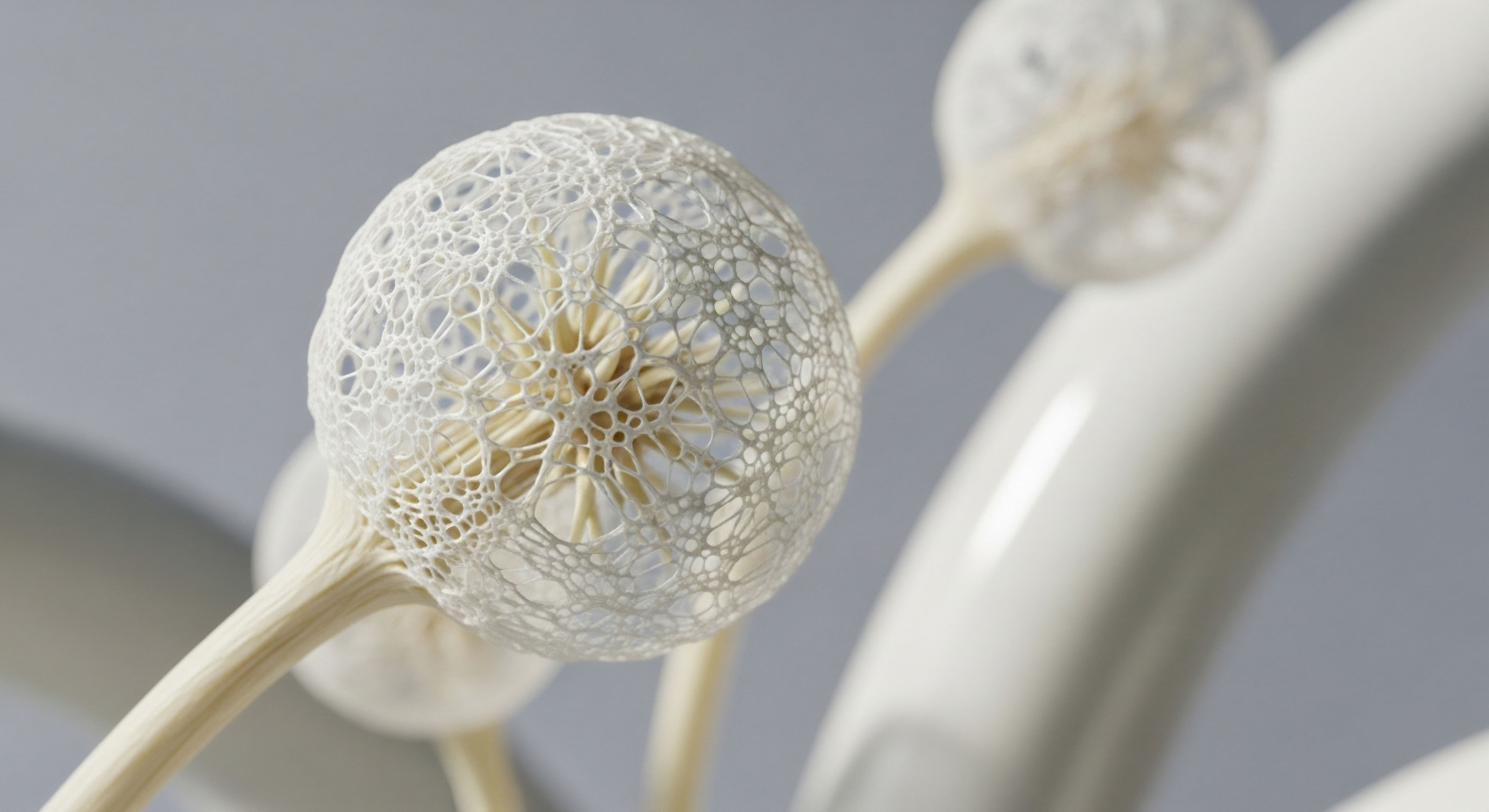
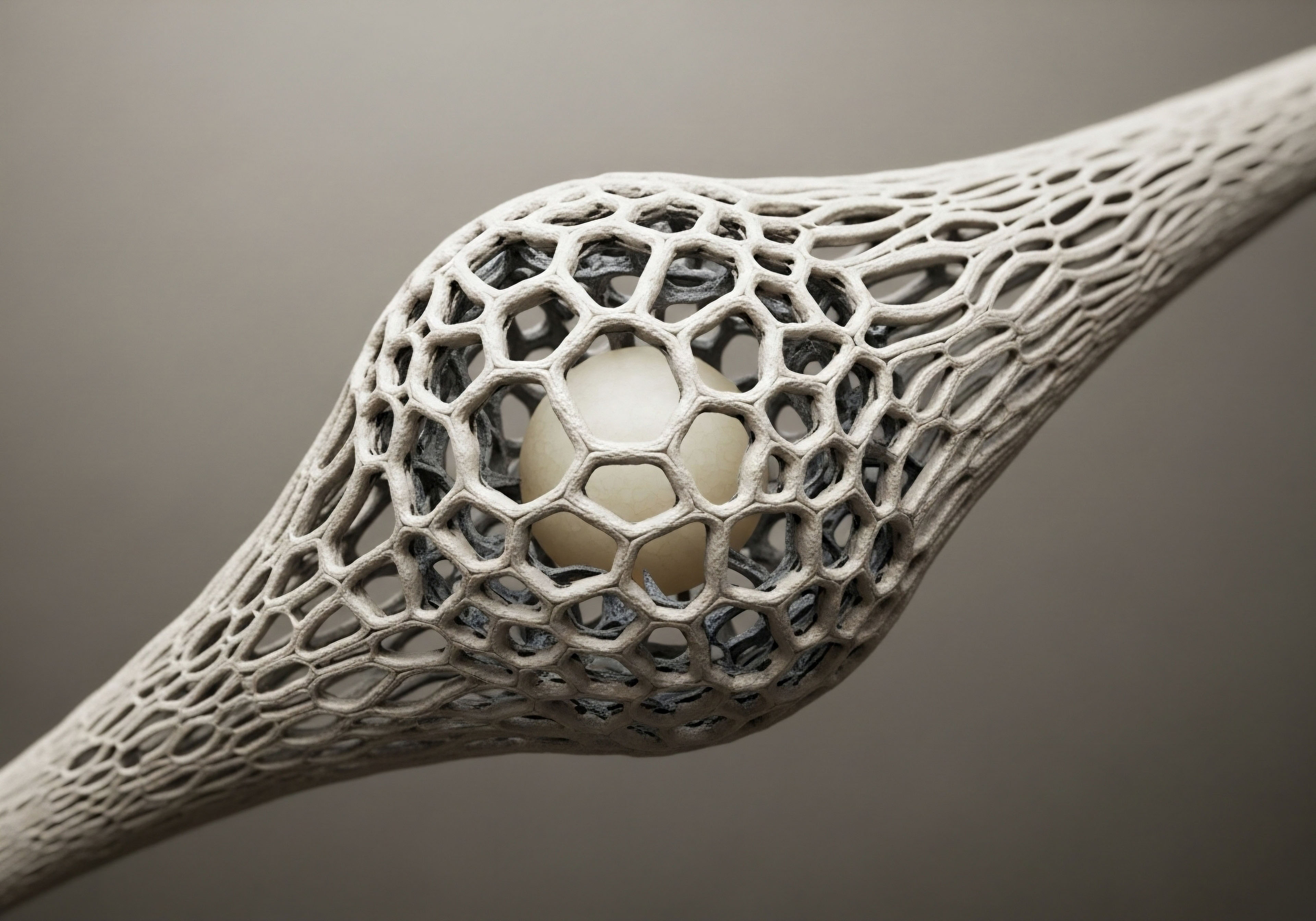
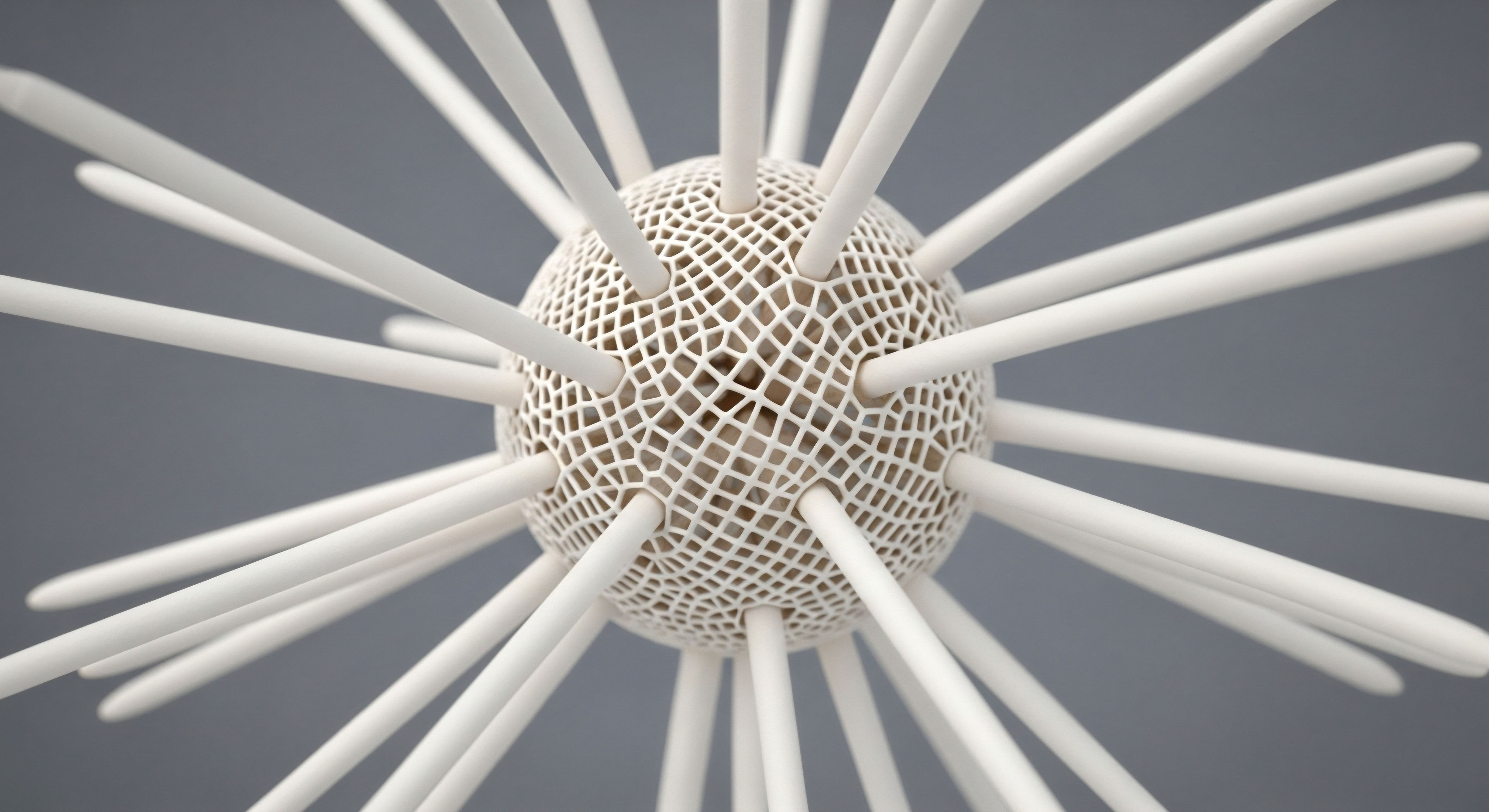
This cartilage is the critical component for frictionless movement, acting as a cushion that absorbs shock and distributes load. It is a living tissue, composed of specialized cells called chondrocytes, which are embedded within a complex scaffold known as the extracellular matrix (ECM).
This matrix, primarily made of collagen and proteoglycans, is what gives cartilage its strength and compressive resistance. The health of your joints depends entirely on the ability of your chondrocytes to maintain this matrix. They are the resident mechanics, tirelessly working to repair microscopic damage and replenish the structural proteins that wear down with daily use.

The Cellular Conductors
Your body’s endocrine system functions as a sophisticated communication network, and hormones are its primary messengers. Estrogen and testosterone, often narrowly viewed through the lens of reproduction, are in fact powerful systemic hormones that influence a vast array of tissues, including your bones, muscles, brain, and critically, the components of your joints.
Both chondrocytes and the cells in the synovial membrane, the tissue that lines the joint capsule, are equipped with specific receptors for these hormones. These receptors act like docking stations. When a hormone like estrogen or testosterone binds to its receptor, it initiates a cascade of signals inside the cell, directly influencing its behavior.
This signaling can instruct a chondrocyte to increase its production of collagen or command an immune cell to stand down, reducing inflammation. For much of your life, robust levels of these hormones ensure that the balance of power within the joint favors repair and maintenance.

When the Signals Fade
The process of aging is accompanied by a natural and progressive decline in the production of sex hormones. In women, this change is relatively abrupt with menopause, leading to a significant drop in estrogen levels. In men, the decline of testosterone is more gradual, a slow tapering that occurs over decades.
This hormonal shift changes the internal environment of the joint. The “build and repair” signals become weaker and less frequent, while the signals for breakdown and inflammation become more dominant. Without adequate hormonal stimulation, chondrocytes become less efficient at replenishing the ECM.
The collagen network can become disorganized, and the cartilage can begin to thin and lose its resilience. Simultaneously, the synovial membrane may become more prone to inflammation, creating a hostile environment that further accelerates cartilage degradation. This is the biological reality behind the increased prevalence of osteoarthritis, a condition of degenerative joint disease, in postmenopausal women and older men.
Hormonal optimization protocols are designed to restore these essential signals, re-establishing a biological environment that favors preservation and longevity for your joints.
The integrity of your joints is actively managed by hormonal signals that direct cellular repair and control inflammation.
Understanding this fundamental connection is the first step. The discomfort you may feel is not a sign of inevitable decay but a direct consequence of a change in your body’s internal chemistry. By addressing this change, we can influence the function and future of these vital tissues. The goal is to provide your cells with the instructions they need to perform their duties, protecting the intricate architecture of your joints so they can serve you for a lifetime.


Intermediate
Moving beyond the foundational understanding that hormones influence joint health, we can now examine the specific mechanisms through which this protection is conferred. Hormonal optimization is a clinical strategy that recalibrates the body’s internal signaling to favor anabolic, or tissue-building, processes over catabolic, or breakdown, processes.
This is achieved by restoring key hormones to levels that support cellular function, directly impacting the chondrocytes and synovial environment within the joint. The process is akin to restoring the proper operating system for your cells, allowing them to execute their maintenance and repair protocols with renewed efficiency.

Estrogen the Master Regulator of Chondrocyte Survival
The dramatic increase in osteoarthritis rates in women following menopause points directly to the critical role of estrogen in joint preservation. Estrogen’s influence is multifaceted, operating through several distinct cellular pathways to protect cartilage. Its receptors are found on chondrocytes, and their activation triggers a series of protective events.

Modulating Cellular Autophagy and Stress
One of the most significant ways estrogen protects cartilage is by promoting a process called autophagy in chondrocytes. Autophagy is essentially the cell’s quality control and recycling system. It allows the cell to break down and remove damaged or dysfunctional components, like misfolded proteins or worn-out organelles.
This process is essential for maintaining cellular health and preventing the accumulation of toxic waste products that can trigger cell death. Research has shown that estrogen stimulates autophagy in chondrocytes by influencing the ERK-mTOR signaling pathway. By keeping this pathway in check, estrogen ensures that the cellular cleanup crews remain active, preserving the health and functionality of the chondrocytes.
Furthermore, estrogen helps to mitigate endoplasmic reticulum (ER) stress. The ER is the cellular factory responsible for producing many of the proteins that make up the extracellular matrix, including collagen. When this factory is overworked or disrupted, it can lead to an accumulation of unfolded proteins, a condition known as ER stress.
If this stress is not resolved, it triggers apoptosis, or programmed cell death. Studies have demonstrated that estradiol can reduce ER stress-induced apoptosis in chondrocytes, effectively protecting these essential cells from self-destruction and preserving the cellularity of the cartilage.

Testosterone the Architect of the Joint Matrix
While estrogen is a key regulator of cell survival, testosterone plays a more direct structural role, acting as a primary architect of the joint’s connective tissues. Its influence extends to cartilage, ligaments, and tendons, all of which are critical for joint stability and function. The decline in testosterone is linked to an increased risk of arthritis, underscoring its protective function.
Stimulating Collagen and Matrix Synthesis
Testosterone’s primary contribution to joint health is its powerful effect on collagen synthesis. Collagen is the most abundant protein in the body and the main structural component of the extracellular matrix in cartilage, tendons, and ligaments. Testosterone directly stimulates chondrocytes and fibroblasts (the cells in tendons and ligaments) to produce more collagen.
Specifically, it has been shown to increase the production of Type II collagen, which is the specific type that forms the resilient framework of articular cartilage. By promoting robust collagen production, testosterone ensures that these tissues remain strong, dense, and better able to withstand mechanical stress. This results in thicker, more resilient cartilage and stronger, more stable ligaments and tendons, reducing the risk of injury and degenerative changes.
Hormonal therapies function by directly instructing joint cells to enhance their natural repair processes and suppress inflammatory signals.
Moreover, testosterone influences the production of other essential matrix components, such as glycosaminoglycans, which help the cartilage retain water and resist compression. It also plays a vital role in maintaining the health of the subchondral bone, the layer of bone directly beneath the cartilage. A strong and healthy subchondral bone provides a stable foundation for the cartilage, and testosterone’s ability to increase bone mineral density contributes to the overall integrity of the joint structure.

How Do Clinical Protocols Support Joint Health?
Understanding these mechanisms allows us to see how specific clinical protocols for hormonal optimization directly translate to joint protection. The goal of these therapies is to restore the essential signaling that has diminished with age, thereby reactivating the body’s innate repair and maintenance systems.
For men experiencing the symptoms of andropause, a protocol involving Testosterone Cypionate is designed to restore serum testosterone to a healthy, youthful range. This directly addresses the decline in collagen synthesis and bone density. By providing a consistent and stable level of testosterone, the therapy ensures that the cells within the joints are continuously receiving the signal to build and repair the extracellular matrix.
The inclusion of Gonadorelin helps maintain the body’s own testosterone production pathways, while a carefully managed dose of Anastrozole prevents the excessive conversion of testosterone to estrogen, maintaining a balanced hormonal profile.
For women, particularly those in perimenopause or post-menopause, hormonal therapy is critical for mitigating the rapid decline in joint health. Protocols often involve estrogen replacement to directly counteract the increased risk of osteoarthritis by protecting chondrocytes from apoptosis and inflammation.
The addition of a small, physiologic dose of Testosterone Cypionate can further support joint integrity by stimulating collagen production, addressing a component of joint health that estrogen alone does not fully cover. Progesterone is also a key component, particularly for women with an intact uterus, and it has its own anti-inflammatory properties that contribute to the overall protective effect.
The following tables illustrate the distinct yet complementary roles of these hormones and a sample protocol structure.
| Joint Component | Primary Effect of Estrogen | Primary Effect of Testosterone |
|---|---|---|
| Chondrocytes (Cartilage Cells) | Reduces apoptosis (cell death), promotes survival, and decreases inflammation. | Stimulates production of Type II collagen and other matrix proteins. |
| Extracellular Matrix (ECM) | Inhibits enzymes that degrade the matrix (MMPs). | Directly promotes the synthesis of new collagen fibers, increasing matrix density. |
| Synovial Membrane | Exerts a powerful anti-inflammatory effect, reducing swelling and pain signals. | Contributes to systemic anti-inflammatory effects. |
| Subchondral Bone | Helps maintain bone density, preventing resorption. | Significantly increases bone mineral density, providing a stronger foundation for cartilage. |
| Patient Group | Core Therapy Component | Supporting Component | Clinical Rationale for Joint Health |
|---|---|---|---|
| Male (Andropause) | Testosterone Cypionate (weekly injection) | Anastrozole (as needed to manage estrogen) | Restores systemic signals for collagen synthesis in cartilage and ligaments, and increases bone mineral density, strengthening the entire joint unit. |
| Female (Post-Menopause) | Estradiol (transdermal or oral) | Testosterone Cypionate (low-dose weekly injection) & Progesterone | Provides powerful chondrocyte protection, reduces joint inflammation, and slows cartilage degradation, while low-dose testosterone supports the structural integrity of the collagen matrix. |
| Both Groups | Peptide Therapy (e.g. Ipamorelin/CJC-1295) | N/A | Stimulates the body’s own production of growth hormone, which promotes systemic tissue repair and can enhance the healing of connective tissues, complementing the effects of HRT. |


Academic
A sophisticated analysis of hormonal influence on joint longevity requires a systems-biology perspective, viewing the joint not as an isolated mechanical structure but as a complex biological system where the endocrine, immune, and musculoskeletal systems are deeply intertwined.
The protective effects of hormonal replacement therapy (HRT) are a direct consequence of its ability to modulate the intricate crosstalk between these systems at a cellular and molecular level. The primary locus of this activity is the regulation of the inflammatory state and the metabolic phenotype of the resident cells of the joint, particularly the chondrocytes and synoviocytes.

The Immuno-Endocrine Axis in Synovial Homeostasis
The synovium, a thin membrane lining the joint capsule, is a critical battleground in joint health. It is rich in immune cells and is the primary source of synovial fluid, which lubricates the joint and nourishes the avascular cartilage. In a state of hormonal decline, this environment shifts towards a pro-inflammatory phenotype. This is where the immuno-endocrine axis becomes paramount. Both estrogen and testosterone exert profound immunomodulatory effects.
Estrogen, for instance, has a complex, concentration-dependent effect on the immune system. At physiological levels seen in premenopausal women, it tends to suppress the activity of pro-inflammatory cytokines such as Tumor Necrosis Factor-alpha (TNF-α) and Interleukin-1 beta (IL-1β), both of which are potent drivers of cartilage degradation.
It achieves this by acting on estrogen receptors (ERs), specifically ERα and ERβ, which are expressed on synovial macrophages and fibroblasts. The binding of estradiol to these receptors can inhibit the activation of transcription factors like NF-κB, a master regulator of the inflammatory response. This prevents the transcription of genes that code for inflammatory mediators and matrix-degrading enzymes like matrix metalloproteinases (MMPs).
Androgens, including testosterone, generally exert an anti-inflammatory effect. They have been shown to suppress the production of TNF-α, IL-1, and IL-6 by monocytes and macrophages. Furthermore, testosterone can promote the production of the anti-inflammatory cytokine Interleukin-10 (IL-10). This hormonal regulation of the local immune environment is a key mechanism by which HRT protects the joint.
By restoring hormonal levels, these therapies re-establish an anti-inflammatory tone within the synovium, protecting the nearby cartilage from a constant barrage of destructive inflammatory signals.

What Is the Molecular Basis of Chondroprotection?
The long-term survival and function of articular cartilage depend on the health of a relatively small population of chondrocytes. These cells are responsible for maintaining a large volume of extracellular matrix. The decline in sex hormones makes these cells vulnerable to apoptosis, leading to a hypocellular cartilage that is unable to repair itself. Hormonal optimization directly counters this process at the molecular level.
Estrogen’s chondroprotective effects are mediated, in part, through its interaction with intracellular signaling pathways that regulate cell fate. As noted, its ability to inhibit ER stress-induced apoptosis is critical. ER stress activates the Unfolded Protein Response (UPR), a signaling network designed to restore homeostasis.
However, if the stress is chronic, the UPR shifts from a pro-survival to a pro-apoptotic signal. Estradiol signaling through ERα can bolster the adaptive capacity of the UPR, preventing this fatal shift and thereby preserving the chondrocyte population. Similarly, its modulation of the ERK-mTOR pathway to promote basal autophagy is a vital homeostatic mechanism, ensuring the efficient removal of cellular debris that could otherwise trigger inflammation or cell death.
Androgen Receptor (AR) activation in chondrocytes also contributes to a pro-survival and pro-anabolic phenotype. Overexpression of the AR in chondrocytes has been shown to promote the expression of key chondrogenic transcription factors like SOX9, and subsequently, increase the synthesis of aggrecan and Type II collagen, the defining components of healthy articular cartilage.
Concurrently, AR activation can downregulate the expression of catabolic enzymes like MMP-13, which is a primary driver of collagen degradation in osteoarthritis. This dual action, simultaneously promoting matrix synthesis and inhibiting its breakdown, places androgens as a critical factor in maintaining the structural integrity of cartilage.
Hormonal therapies work by recalibrating the complex interplay between the immune and endocrine systems within the joint’s microenvironment.
The Cartilage-Bone Unit and Systemic Influences
The health of articular cartilage cannot be divorced from the health of the underlying subchondral bone. The two tissues form a functional unit, and pathological changes in one inevitably affect the other. The age-related decline in sex hormones is a well-established driver of osteoporosis, a condition of reduced bone mineral density.
This weakening of the subchondral bone can alter the mechanical forces experienced by the overlying cartilage, increasing its susceptibility to damage. HRT, particularly therapies involving testosterone, directly addresses this by promoting bone formation. Testosterone’s ability to increase bone mineral density strengthens the subchondral plate, providing a more stable foundation for the articular cartilage and protecting it from mechanical failure.
Finally, it is important to consider the systemic effects of hormonal and peptide therapies. Protocols that include growth hormone secretagogues, such as Sermorelin or a combination of Ipamorelin and CJC-1295, introduce another layer of support for joint health. These peptides stimulate the pulsatile release of endogenous growth hormone (GH) from the pituitary gland.
GH and its primary mediator, Insulin-like Growth Factor 1 (IGF-1), are powerful anabolic agents that promote tissue growth and repair throughout the body. In the context of joint health, IGF-1 is known to stimulate chondrocyte proliferation and matrix synthesis. By enhancing the body’s natural regenerative capacity, these peptide therapies can work synergistically with HRT, amplifying the repair signals and promoting the healing of connective tissues, further contributing to long-term joint durability.

Can HRT Reverse Existing Joint Damage?
A crucial question for individuals already experiencing joint degeneration is whether hormonal optimization can reverse existing damage. While articular cartilage has a very limited capacity for regeneration, the primary goal of HRT in this context is to halt the progression of further degradation and optimize the function of the remaining tissue.
By reducing inflammation, preventing further chondrocyte death, and stimulating the existing cells to produce new matrix components, these therapies can significantly improve symptoms and preserve the joint for a longer period. The evidence suggests that early intervention yields the most significant protective benefits. Restoring the body’s hormonal milieu creates an environment where the joint’s own maintenance systems can function optimally, slowing the degenerative process and preserving mobility and function for the long term.
- Hormonal Receptors ∞ The presence of Androgen Receptors and Estrogen Receptors within chondrocytes and synovial cells is the direct molecular gateway through which HRT exerts its influence on joint tissue.
- Signaling Cascades ∞ Hormonal binding initiates complex intracellular signaling, such as the modulation of the NF-κB, ERK-mTOR, and UPR pathways, which collectively govern the cell’s inflammatory state, metabolic activity, and survival.
- Gene Transcription ∞ Ultimately, these signals alter gene transcription, upregulating anabolic genes (e.g. COL2A1 for Type II collagen, SOX9) and downregulating catabolic genes (e.g. MMP13, ADAMTS5).

References
- Cutolo, M. et al. “Sex hormones and rheumatoid arthritis.” Autoimmunity Reviews, vol. 7, no. 5, 2008, pp. 376-82.
- Li, Ke, et al. “Estrogen prevents articular cartilage destruction in a mouse model of AMPK deficiency via ERK-mTOR pathway.” Journal of Orthopaedic Translation, vol. 18, 2019, pp. 87-96.
- O’Brien, M. et al. “Estrogen receptor Alpha in human knee articular cartilage of healthy and osteoarthritic females.” Journal of Orthopaedic Research, vol. 35, no. 1, 2017, pp. 1-8.
- Cheng, L. and S. Wang. “Lower serum testosterone is associated with increased likelihood of arthritis.” Scientific Reports, vol. 13, no. 1, 2023, p. 19448.
- Klose, S. et al. “Estradiol Inhibits ER Stress-Induced Apoptosis in Chondrocytes and Contributes to a Reduced Osteoarthritic Cartilage Degeneration in Female Mice.” Frontiers in Cell and Developmental Biology, vol. 10, 2022, p. 880404.
- Cutolo, M. and R. H. Straub. “Sex hormones and osteoarthritis.” Menopause, vol. 26, no. 1, 2019, pp. 1-3.
- Ko, M. J. et al. “Hormone therapy and risk of knee osteoarthritis in postmenopausal women ∞ a nationwide study.” Menopause, vol. 26, no. 4, 2019, pp. 366-373.
- Daigle, D. S. et al. “The effects of androgen receptor overexpression on chondrogenic ability of rabbit articular chondrocytes.” IUBMB Life, vol. 73, no. 9, 2021, pp. 1156-1166.
- Hansen, M. and M. Kjaer. “Sex Hormones and Tendon.” Advances in Experimental Medicine and Biology, vol. 920, 2016, pp. 131-41.
- Janssen, I. et al. “Testosterone and muscle atrophy in aging men.” Journal of Clinical Endocrinology & Metabolism, vol. 89, no. 9, 2004, pp. 4434-4439.

Reflection
The information presented here provides a map of the biological pathways that connect your internal chemistry to the physical experience of joint health. This knowledge is a powerful tool, shifting the perspective from one of passive endurance to one of proactive engagement. The feelings of stiffness, the aches, the concerns about future mobility ∞ these are not endpoints.
They are data points, signals from a complex and intelligent system that is responding to a changing internal environment. Your personal health journey is unique, written in the language of your own biology and experiences.

What Is Your Body Communicating?
Consider the narrative your body has been telling. Are there patterns? Do the changes you feel correspond to other shifts in your energy, your vitality, your overall sense of well-being? The science of endocrinology teaches us that these systems are interconnected.
The same hormonal shifts that influence your joints are also shaping your metabolic health, your cognitive function, and your resilience to stress. Understanding these connections allows you to see your health not as a collection of isolated symptoms, but as a unified system.
This clinical knowledge serves as a foundation for a more informed dialogue, a starting point for a deeper inquiry into your own health. The path forward involves translating this understanding into a personalized strategy, a protocol that acknowledges your unique biochemistry and goals. The potential for preserving your vitality and function is immense when you begin to work with your body’s own systems, providing the resources and signals they need to thrive for the long term.

Glossary

sex hormones

hormonal optimization

articular cartilage

extracellular matrix

osteoarthritis

joint health

collagen synthesis
increase bone mineral density

subchondral bone

testosterone cypionate

immuno-endocrine axis

matrix metalloproteinases

androgen receptor

bone mineral density