
Fundamentals
Embarking on a protocol of hormonal optimization is a significant step in reclaiming your vitality. You may have started this process to address persistent fatigue, a decline in physical strength, or a general sense that your internal systems were no longer operating with their youthful efficiency.
The decision to begin testosterone therapy was likely born from a desire to feel like yourself again. As you experience the benefits ∞ the return of energy, mental clarity, and physical capacity ∞ you also become more attuned to your body’s intricate internal communications.
It is within this heightened awareness that you might notice other changes, such as a reduction in testicular volume. This observation is a direct and expected consequence of the body’s sophisticated feedback mechanisms. It is a sign that your endocrine system has recognized the presence of external testosterone and has adjusted its own production accordingly. Understanding this process is the first step toward managing it effectively.
Your body operates under the direction of a magnificent command-and-control system known as the Hypothalamic-Pituitary-Gonadal (HPG) axis. This network is the primary regulator of your reproductive and hormonal health. At the top of this hierarchy sits the hypothalamus, a small but powerful region in your brain that constantly monitors your body’s internal environment.
When the hypothalamus detects a need for more testosterone, it releases a signaling molecule called Gonadotropin-Releasing Hormone (GnRH). This is the initial message, the top-level directive that sets the entire chain of events in motion. Think of the hypothalamus as the system’s central thermostat, constantly sensing the ambient temperature and deciding when to turn the heat on.
The GnRH message travels a short distance to the pituitary gland, another critical structure at the base of the brain. The pituitary acts as the mid-level manager, receiving the directive from the hypothalamus and translating it into specific instructions for the downstream workforce.
In response to GnRH, the pituitary gland produces and releases two key hormones into the bloodstream ∞ Luteinizing Hormone (LH) and Follicle-Stimulating Hormone (FSH). These gonadotropins are the specific messengers that travel throughout the body to deliver their instructions directly to the testes.
LH is the primary signal for testosterone production, while FSH is central to the process of spermatogenesis, or sperm production. The coordinated release of these two hormones ensures that the testes perform their dual functions of producing both androgens and sperm.
When external testosterone is introduced, the body’s natural signaling cascade from the brain to the testes is suppressed, leading to a decrease in testicular activity.
When you begin testosterone replacement therapy (TRT), you are introducing testosterone from an external source. Your hypothalamus and pituitary gland, ever vigilant, detect these elevated levels of testosterone in the bloodstream. Interpreting this as a sign that production is more than adequate, the hypothalamus reduces its release of GnRH.
This, in turn, causes the pituitary to dramatically decrease its output of LH and FSH. The communication channel effectively goes quiet. Without the stimulating signals of LH, the Leydig cells within the testes cease their production of endogenous testosterone. Without FSH, the Sertoli cells receive no instruction to support sperm maturation.
The result is that the testes, deprived of their hormonal cues to function, enter a state of dormancy. This leads to a reduction in their size, a condition known as testicular atrophy, and a halt in sperm production. This is a natural and predictable biological response, a testament to the efficiency of your body’s regulatory systems.
This is where Human Chorionic Gonadotropin (HCG) enters the clinical picture. HCG is a hormone that possesses a remarkable molecular resemblance to LH. Its structure is so similar that it can bind to and activate the LH receptors on the Leydig cells within the testes.
When administered during testosterone therapy, HCG effectively acts as a surrogate for the suppressed LH signal. It bypasses the dormant HPG axis and directly stimulates the testes, instructing them to resume their critical functions. By providing this direct stimulation, HCG prompts the Leydig cells to produce intratesticular testosterone, the testosterone made within the testes themselves.
This internal production is what maintains testicular volume and cellular health. The renewed presence of intratesticular testosterone also provides essential support for the adjacent Sertoli cells, helping to preserve the machinery of spermatogenesis. In essence, HCG keeps the testicular engine running, even while the brain’s signals are temporarily offline due to the presence of therapeutic testosterone.


Intermediate
Understanding the fundamental principles of the HPG axis and the effect of exogenous testosterone provides the ‘what’ and ‘why’ of testicular atrophy during therapy. Now, we can advance to the clinical ‘how’ ∞ the specific mechanisms and protocols that utilize Human Chorionic Gonadotropin to preserve testicular form and function.
The use of HCG in a hormonal optimization protocol is a sophisticated strategy that acknowledges the interconnectedness of the endocrine system. It represents a shift from simple replacement to intelligent system management, ensuring that while serum testosterone levels are optimized, the health and activity of the gonadal tissues are not compromised.

The Molecular Mimicry of Luteinizing Hormone
The efficacy of HCG lies in its ability to function as an analogue of Luteinizing Hormone (LH). Both LH and HCG are glycoproteins, complex molecules composed of two subunits ∞ an alpha subunit and a beta subunit. The alpha subunit is virtually identical across several pituitary hormones, including LH, FSH, and Thyroid-Stimulating Hormone (TSH).
The beta subunit is unique to each hormone and confers its specific biological activity. The beta subunit of HCG shares significant structural homology with the beta subunit of LH, which allows it to bind to the same receptor, the LHCGR, located on the surface of the Leydig cells in the testes.
There is a key pharmacological difference between the two hormones. Endogenous LH is released by the pituitary gland in a pulsatile manner and has a relatively short half-life of about 20-30 minutes. This pulsatile signaling is crucial for normal physiological function. HCG, conversely, has a much longer half-life, estimated to be around 24-36 hours.
This extended duration of action means that HCG provides a more sustained, non-pulsatile stimulation of the Leydig cells. This is why HCG can be administered through subcutaneous injections just two or three times per week, providing a continuous maintenance signal to the testes that prevents them from becoming dormant during prolonged testosterone therapy. This sustained action effectively keeps the local testosterone production online, preserving both the physical volume of the testes and their capacity for sperm production.
HCG acts as a direct and sustained stimulus to the testes by mimicking the body’s natural Luteinizing Hormone, thereby maintaining intratesticular testosterone production and testicular volume.

Leydig Cells and Sertoli Cells a Partnership in Function
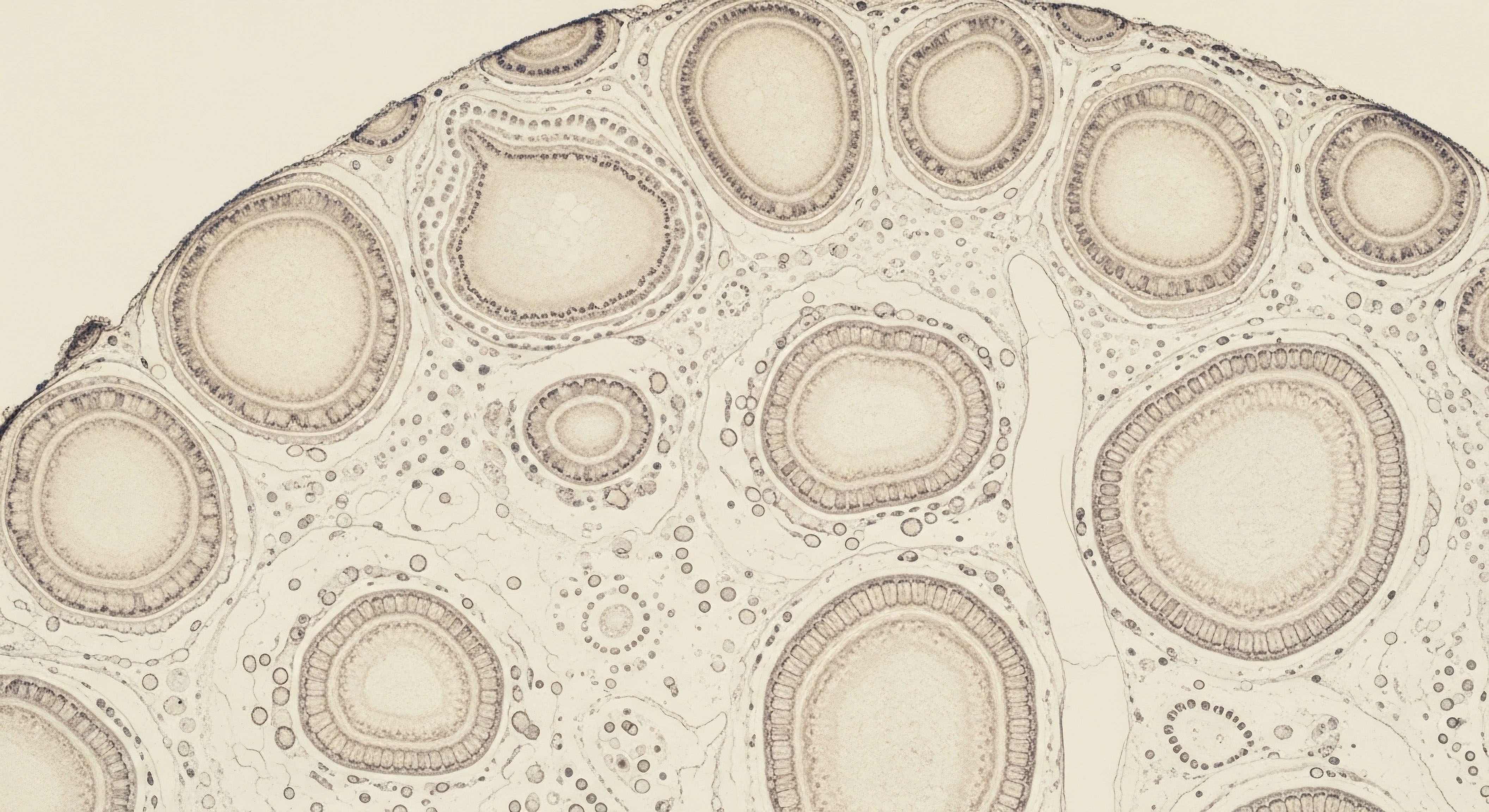
The testes are comprised of a complex architecture of tissues, with two cell types being particularly important for male reproductive function ∞ the Leydig cells and the Sertoli cells. These cells work in a synergistic partnership, and understanding their distinct roles clarifies the impact of HCG.
- Leydig Cells ∞ Located in the interstitial tissue between the seminiferous tubules, Leydig cells are the primary producers of testosterone in the male body. Their function is almost exclusively regulated by LH (or, in a therapeutic context, HCG). When LH binds to the LHCGR on the Leydig cell surface, it triggers a cascade of intracellular signals that results in the conversion of cholesterol into testosterone. This locally produced testosterone is known as intratesticular testosterone, and its concentration within the testes is many times higher than the concentration of testosterone found in the bloodstream.
- Sertoli Cells ∞ Situated within the walls of the seminiferous tubules, Sertoli cells are the “nurse” cells for spermatogenesis. Their primary function is to support the development and maturation of sperm cells, from their earliest germ cell stage to mature spermatozoa. This process is directly stimulated by FSH from the pituitary gland. Critically, the function of Sertoli cells is also highly dependent on the extremely high concentrations of intratesticular testosterone produced by the neighboring Leydig cells.
During TRT, the suppression of the HPG axis shuts down both LH and FSH. The lack of LH is what causes the Leydig cells to stop producing testosterone. The lack of FSH directly removes the primary stimulus for the Sertoli cells. The concurrent crash in intratesticular testosterone further cripples Sertoli cell function.
HCG administration directly addresses the LH deficiency, reactivating the Leydig cells. This restores the vital production of intratesticular testosterone, which in turn provides the necessary paracrine support to the Sertoli cells, helping to maintain the intricate process of spermatogenesis even in the absence of FSH. While HCG cannot replace the function of FSH entirely, preserving high levels of intratodesterone is a powerful mechanism for maintaining fertility potential during TRT.

Clinical Protocols and Dosing Strategies
The goal of incorporating HCG into a TRT protocol is to use the minimum effective dose to maintain testicular function without causing unwanted side effects. Overstimulation of the Leydig cells with excessively high doses of HCG can lead to testicular desensitization and an overproduction of estrogen, as some testosterone is converted to estradiol via the aromatase enzyme, which is also present in the testes. Standard clinical practice has therefore gravitated towards low-dose, high-frequency protocols.
A typical protocol for a man on weekly testosterone cypionate injections might involve the subcutaneous administration of HCG two times per week, on the day before and the day of the testosterone injection, for instance. This timing helps to ensure that testicular stimulation is consistent throughout the week.
The table below compares the expected outcomes of TRT with and without the inclusion of HCG.
| Parameter | TRT Monotherapy | TRT with Concomitant HCG |
|---|---|---|
| Serum Testosterone | Maintained in optimal range via exogenous source. | Maintained in optimal range, with a small contribution from endogenous production. |
| LH / FSH Levels | Suppressed to near-zero levels. | Suppressed to near-zero levels. |
| Testicular Volume | Significant reduction (atrophy) over time. | Maintained at or near baseline size. |
| Spermatogenesis | Severely impaired or completely halted, leading to infertility. | Largely preserved due to maintenance of intratesticular testosterone. |
| Libido & Well-Being | Generally improved due to optimized serum testosterone. | Often reported as further enhanced, potentially due to the preservation of other testicular hormones and steroids. |
A sample weekly protocol demonstrates how these medications are integrated. This is a representative example, and individual protocols must be tailored by a qualified physician based on lab work and clinical response.
| Day of Week | Medication Protocol |
|---|---|
| Monday | Administer 500 IU HCG (subcutaneous injection) |
| Tuesday | Administer 100mg Testosterone Cypionate (intramuscular injection) |
| Wednesday | Administer 0.5mg Anastrozole (oral tablet, if required for estrogen management) |
| Thursday | – |
| Friday | Administer 500 IU HCG (subcutaneous injection) |
| Saturday | Administer 100mg Testosterone Cypionate (intramuscular injection) |
| Sunday | Administer 0.5mg Anastrozole (oral tablet, if required) |

Academic
A sophisticated clinical understanding of Human Chorionic Gonadotropin’s role in androgen replacement requires an appreciation of its molecular pharmacology and the intricate intracellular signaling it initiates. Moving beyond the systemic overview, an academic exploration focuses on the precise biochemical events that occur at the cellular level within the testis.
This perspective illuminates the quantitative and qualitative differences between LH and HCG signaling, the downstream consequences for steroidogenesis, and the critical importance of intratesticular androgen concentrations for maintaining the complex ecosystem of the seminiferous tubules. The decision to use HCG is grounded in this deep physiological science, aiming to replicate biological fidelity as closely as possible when the natural regulatory axis is intentionally suppressed.

The LHCGR and Downstream Signal Transduction
The biological actions of both LH and HCG are mediated by a single transmembrane receptor ∞ the Luteinizing Hormone/Chorionic Gonadotropin Receptor (LHCGR). This receptor is a member of the G protein-coupled receptor (GPCR) superfamily, the largest and most diverse group of membrane receptors in eukaryotes.
When HCG binds to the extracellular domain of the LHCGR on a Leydig cell, it induces a conformational change in the receptor. This change is transmitted to the intracellular portion of the receptor, causing it to activate a heterotrimeric G protein, specifically the Gs (stimulatory) alpha subunit.
Activation of Gs leads to its dissociation from the beta-gamma subunits and its subsequent interaction with the enzyme adenylyl cyclase. Adenylyl cyclase then catalyzes the conversion of ATP into cyclic adenosine monophosphate (cAMP), a ubiquitous second messenger. The accumulation of intracellular cAMP is the central event in the canonical signaling pathway.
cAMP proceeds to activate Protein Kinase A (PKA) by binding to its regulatory subunits, causing them to release the catalytic subunits. The now-active PKA catalytic subunits phosphorylate a host of downstream protein targets within the cell.
Key PKA targets include transcription factors like the cAMP Response Element-Binding protein (CREB), which, upon phosphorylation, translocates to the nucleus and binds to cAMP response elements (CREs) on the DNA. This binding event initiates the transcription of genes essential for steroidogenesis. The most critical of these is the gene for Steroidogenic Acute Regulatory Protein (StAR).
The StAR protein facilitates the rate-limiting step in steroid hormone production ∞ the transport of cholesterol from the outer mitochondrial membrane to the inner mitochondrial membrane, where the P450scc enzyme (cholesterol side-chain cleavage enzyme) awaits to convert it into pregnenolone. From pregnenolone, a series of enzymatic steps within the mitochondria and smooth endoplasmic reticulum leads to the synthesis of testosterone. HCG’s primary molecular function is to keep this entire cAMP-PKA-StAR pathway active.
HCG preserves testicular function during testosterone therapy by activating the same intracellular signaling pathways as luteinizing hormone, ensuring the continued transcription of genes vital for testosterone synthesis.

What Are the Molecular Consequences of HCG Administration on Leydig Cell Steroidogenesis?
While HCG and LH activate the same primary signaling pathway, subtle differences in their interaction with the LHCGR lead to distinct downstream effects, which have important clinical implications. Research suggests that HCG is a more potent agonist at the LHCGR than LH, inducing a greater accumulation of cAMP for a given receptor occupancy. Furthermore, its significantly longer half-life (approximately 24-36 hours versus 20-30 minutes for LH) results in a more prolonged and intense activation of the signaling cascade.
This has two major consequences. First, it makes HCG a highly efficient therapeutic agent for maintaining steroidogenesis. A low-dose injection can sustain Leydig cell stimulation for several days. Second, it introduces the risk of receptor desensitization and downregulation.
Continuous, high-intensity stimulation of a GPCR can lead to its phosphorylation by GPCR kinases (GRKs), which promotes the binding of arrestin proteins. Arrestin binding uncouples the receptor from the G protein, effectively turning off the signal, and targets the receptor for internalization and subsequent degradation.
This is a protective mechanism to prevent cellular overstimulation. Clinically, this means that excessive HCG dosing can, paradoxically, lead to a reduction in Leydig cell responsiveness over time. This underpins the clinical rationale for using minimal effective doses (e.g. 250-500 IU two to three times per week) to maintain function without inducing significant desensitization.

The Indispensable Role of Intratesticular Testosterone
The ultimate goal of HCG co-administration is the maintenance of intratesticular testosterone (ITT). The concentration of testosterone inside the testes is approximately 100-fold higher than in the peripheral circulation. This incredibly high local androgen concentration is absolutely essential for spermatogenesis. Exogenous testosterone therapy, while normalizing serum levels, obliterates ITT levels by suppressing LH.
A landmark study by Coviello et al. (2012) demonstrated that TRT alone reduced ITT by 94%. However, the co-administration of low-dose HCG (500 IU every other day) with TRT completely prevented this drop and, in fact, increased ITT by 26% from baseline. This preservation of the intratesticular androgen environment is the direct mechanism by which HCG maintains fertility.
The high ITT bathes the Sertoli cells, providing the powerful androgenic signal required to drive the differentiation of spermatogonia into mature spermatozoa. Even with suppressed FSH levels, the presence of robust ITT can sustain spermatogenesis, a finding that has been repeatedly confirmed in clinical practice. This makes HCG a cornerstone of fertility preservation for men requiring long-term androgen replacement.
Furthermore, the maintenance of the entire steroidogenic pathway ensures the continued production of other important steroid intermediates, such as DHEA and pregnenolone, within the testes. While the systemic impact of these locally produced steroids is still an area of active research, some clinicians theorize that their preservation may contribute to the enhanced sense of well-being and libido that many patients report with combined TRT and HCG therapy, compared to TRT alone. It represents a more holistic approach to hormonal support, acknowledging that the testes produce more than just testosterone.
- Signal Initiation ∞ HCG binds to the LHCGR on Leydig cells, activating the Gs alpha subunit of the associated G protein.
- Second Messenger Production ∞ The activated Gs protein stimulates adenylyl cyclase to convert ATP to cAMP, rapidly increasing intracellular cAMP levels.
- Kinase Activation ∞ cAMP binds to the regulatory subunits of PKA, releasing the active catalytic subunits.
- Protein Phosphorylation ∞ PKA phosphorylates key proteins, including the transcription factor CREB.
- Gene Transcription ∞ Phosphorylated CREB moves to the nucleus and promotes the transcription of steroidogenic genes, most notably the gene for the StAR protein.
- Cholesterol Transport ∞ The StAR protein facilitates the transport of cholesterol into the mitochondria, the rate-limiting step of the entire process.
- Testosterone Synthesis ∞ A series of enzymatic reactions within the mitochondria and endoplasmic reticulum convert cholesterol into testosterone, maintaining high intratesticular concentrations.
References
- Bhasin, Shalender, et al. “Testosterone therapy in men with hypogonadism ∞ an Endocrine Society clinical practice guideline.” The Journal of Clinical Endocrinology & Metabolism 103.5 (2018) ∞ 1715-1744.
- Coviello, Andrea D. et al. “Effects of combined testosterone and HCG on testicular function in men on testosterone replacement therapy.” Journal of Andrology 33.3 (2012) ∞ 416-422.
- Habous, Mohamad, et al. “Human chorionic gonadotropin monotherapy for the treatment of hypogonadal symptoms in men with total testosterone > 300 ng/dL.” Urology Annals 10.4 (2018) ∞ 414.
- Hsieh, T. Mike, and Larry I. Lipshultz. “Indications for the use of human chorionic gonadotropic hormone for the management of infertility in hypogonadal men.” Translational Andrology and Urology 4.4 (2015) ∞ 558.
- Payne, Anita H. and Matthew P. Hardy. “Leydig cells ∞ formation, function, and regulation.” Biology of Reproduction 76.6 (2007) ∞ 920-928.
- Ricci, E. et al. “Human LH and hCG stimulate differently the early signalling pathways but result in equal testosterone synthesis in mouse Leydig cells in vitro.” Molecular and Cellular Endocrinology 440 (2017) ∞ 64-72.
- Wenker, Evan P. et al. “The use of HCG-based combination therapy for recovery of spermatogenesis after testosterone use.” Journal of Sexual Medicine 12.6 (2015) ∞ 1334-1337.

Reflection
The journey into hormonal optimization is deeply personal. The information presented here serves as a map, detailing the intricate biological terrain you are navigating. Understanding the interplay between your body’s natural signaling systems and the therapeutic protocols you undertake is a powerful act of self-advocacy.
The science of the HPG axis, the function of Leydig cells, and the role of HCG provides a framework for interpreting your own experience and for engaging in a more informed dialogue with your clinical provider. This knowledge transforms you from a passive recipient of care into an active participant in your own wellness.
Your unique physiology and personal health goals are the most important variables in this equation. The path forward is one of partnership, where this clinical understanding is paired with expert guidance to calibrate a protocol that restores function, preserves health, and allows you to operate at your fullest potential.
