


Fundamentals
You may feel a profound sense of disconnect when your body seems to operate by a set of rules you were never taught. The fatigue that settles deep in your bones, the unpredictable shifts in mood, or the subtle but persistent sense of being unwell are all valid experiences.
These feelings are frequently the first indication that the intricate communication network within your body requires attention. This network, your endocrine system, uses hormones as its chemical messengers, and the control center for this entire operation resides within your brain. The way you feel is deeply connected to how well your brain is receiving these hormonal signals. The food you consume provides the raw materials and the critical instructions that dictate the quality of this communication.
Your brain, specifically a region called the hypothalamus, acts as the body’s primary command center for hormonal regulation. It is constantly monitoring your internal environment, taking in information about your energy status, stress levels, and safety.
Based on this information, it sends out precise instructions to the pituitary gland, which in turn directs the function of your thyroid, adrenal glands, and gonads (testes or ovaries). This entire system is known as the Hypothalamic-Pituitary-Gonadal (HPG) axis, and its smooth operation is the foundation of metabolic and reproductive health. The macronutrients you eat ∞ proteins, fats, and carbohydrates ∞ are the primary external factors that inform the hypothalamus about your body’s condition.
Your diet directly informs the brain’s hormonal control centers, shaping your body’s entire endocrine response.
Each macronutrient provides a distinct type of signal. Carbohydrates are rapidly converted to glucose, causing a release of insulin from the pancreas. Insulin’s job is to shuttle glucose into cells for energy, and the brain reads insulin levels as a signal of immediate energy availability.
Fats, on the other hand, influence the release of leptin from your adipose tissue. Leptin acts as a long-term indicator of your energy reserves, essentially telling your brain how much fuel you have stored. Protein provides amino acids, which are the building blocks for tissues and neurotransmitters, signaling to the brain that resources are available for growth and repair.
The ratio of these macronutrients in your diet creates a unique metabolic signature that the hypothalamus interprets to make critical decisions about your body’s operations, including energy expenditure, reproductive readiness, and stress response.

How Does Your Brain Listen to Food?
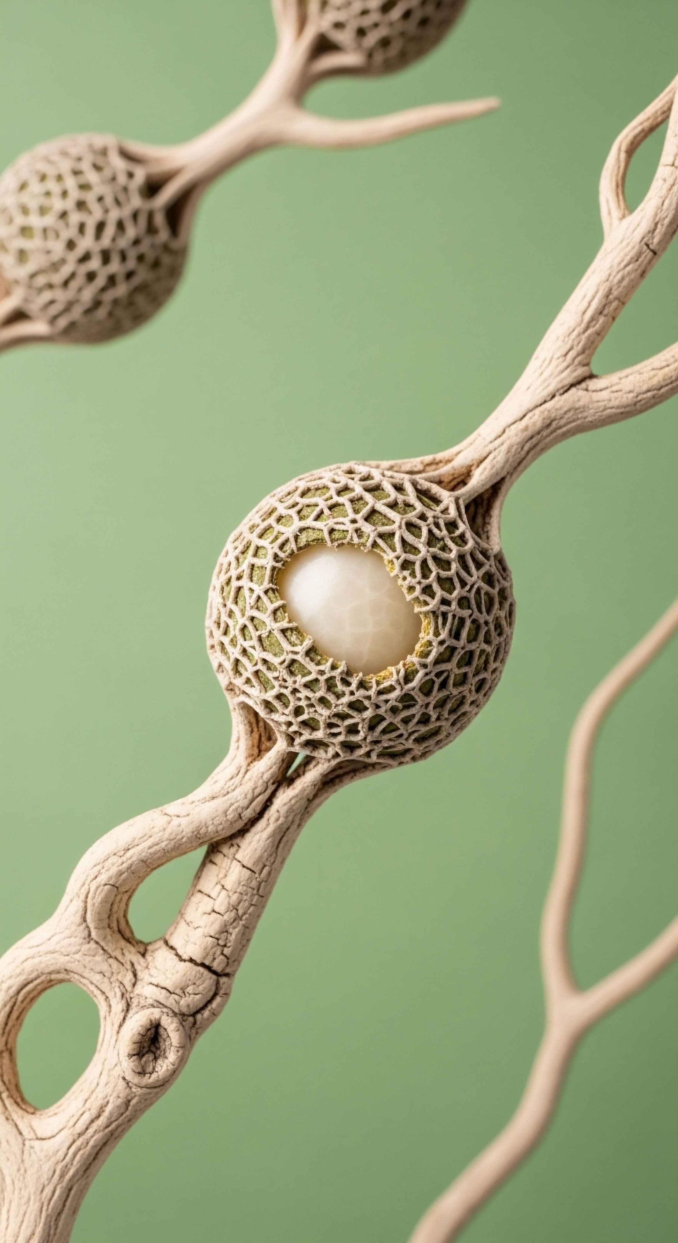
The brain’s ability to receive hormonal messages depends on cellular structures called receptors. Think of a hormone as a key and a receptor as a specifically shaped lock. For a hormonal message to be delivered, the key must fit perfectly into the lock.
The composition of your diet directly affects the integrity and sensitivity of these locks. A diet rich in certain types of fats, like omega-3s, helps build flexible and responsive cell membranes, making it easier for hormone keys to fit their locks.
Conversely, a diet high in processed components can lead to cellular inflammation and stiffness, effectively ‘rusting’ the locks and making them difficult to open. This concept of receptor sensitivity is central to understanding why two individuals can have similar hormone levels on a lab report but experience vastly different symptoms. One person’s brain may be “listening” effectively, while the other’s is functionally deaf to the signals being sent.


Intermediate
To understand how macronutrient ratios affect brain hormone reception, we must examine the specific biochemical dialogues they initiate. The brain does not just register calories; it deciphers the metabolic language spoken by insulin, leptin, and other signaling molecules.
The proportion of carbohydrates, fats, and proteins in your diet determines the volume and clarity of these signals, which in turn modulates the function of the Hypothalamic-Pituitary-Gonadal (HPG) axis. An imbalance in these dietary signals can lead to a cascade of hormonal disruptions that manifest as tangible symptoms.

The Carbohydrate Signal and Insulin’s Role
Diets dominated by refined carbohydrates create a state of chronically elevated insulin. Insulin is a powerful anabolic hormone, and while necessary, its constant presence is a disruptive signal to the hypothalamus. In a state of high insulin, the brain’s insulin receptors can become desensitized, a condition known as insulin resistance.
This resistance has profound consequences for hormonal health. The hypothalamus contains neurons that produce Gonadotropin-Releasing Hormone (GnRH), the master pulse generator for the entire reproductive system. These GnRH neurons have insulin receptors. In an insulin-resistant state, the normal pulsatile release of GnRH is disrupted.
Studies in female mice have shown that obesity-associated hyperinsulinemia directly stimulates GnRH neurons, leading to an increased pulse amplitude and contributing to reproductive dysfunction. This mechanism is a key factor in the pathophysiology of Polycystic Ovary Syndrome (PCOS) in women and can contribute to suppressed testosterone production in men by altering the downstream signals of Luteinizing Hormone (LH) and Follicle-Stimulating Hormone (FSH).
Chronic exposure to high insulin levels from carbohydrate-heavy diets can alter the brain’s master reproductive clock.

The Fat Signal and Membrane Integrity
The type of dietary fat consumed has a direct structural and functional impact on brain cells. Neuronal membranes, which house the all-important hormone receptors, are composed of a lipid bilayer. The fluidity and integrity of this membrane are dictated by the fatty acids available from your diet.
- Omega-3 Fatty Acids ∞ Found in sources like fatty fish, these polyunsaturated fats are incorporated into neuronal membranes, increasing their fluidity. This flexibility enhances the ability of receptors to change shape and bind effectively with their corresponding hormones, improving signal transmission.
- Saturated and Trans Fats ∞ Diets high in these fats can lead to more rigid and less functional cell membranes. This structural change can impair receptor function, making it harder for hormones to dock and deliver their messages. High saturated fat intake is also associated with increased neuroinflammation, which further degrades the brain’s ability to process hormonal signals correctly.
The quality of dietary fat therefore directly translates into the quality of hormonal communication at a cellular level. It is the difference between a well-oiled machine and one clogged with rust.

Macronutrient Impact on Key Hormonal Signals
The table below outlines how different dietary patterns influence the primary hormones that communicate with the brain’s regulatory centers.
| Dietary Pattern | Primary Insulin Response | Primary Leptin Signal | Effect on GnRH Pulsatility | Potential Clinical Outcome |
|---|---|---|---|---|
| High Refined Carbohydrate | Chronically High / Spiking | Often Elevated (Leptin Resistance) | Irregular / High Amplitude Pulses | Insulin Resistance, PCOS, Hypogonadism |
| High Healthy Fat / Low Carbohydrate | Low and Stable | Normalized Sensitivity | Stabilized / Regular Pulses | Improved Insulin Sensitivity, Hormonal Regulation |
| Balanced Macronutrients | Moderate and Meal-Dependent | Stable and Responsive | Generally Regular Pulses | Stable Metabolic and Hormonal Function |

What Is the Role of Dietary Protein?
Protein’s influence is multifaceted. Adequate protein intake is necessary for the synthesis of peptide hormones and neurotransmitters. Beyond this foundational role, specific amino acids act as direct signaling molecules within the hypothalamus. They are sensed by specialized cells that communicate with GnRH neurons, providing another layer of information about the body’s nutritional status.
A sufficient supply of amino acids signals that the body has the resources for complex processes like reproduction and tissue repair, thus supporting a healthy HPG axis function. Nutritional deficiencies, including inadequate protein, consistently impair the function of the HPG axis by reducing the secretion of LH and FSH.


Academic
The regulation of hormonal homeostasis by diet is a highly sophisticated process mediated by specialized cells within the mediobasal hypothalamus. A deep analysis reveals that the brain’s reception of hormonal signals is actively managed by a population of glial cells known as tanycytes.
These cells function as the primary nutrient sensors of the central nervous system, directly linking the metabolic state, as determined by macronutrient intake, to the functional output of the entire neuroendocrine system. Their unique anatomical position and molecular machinery place them at the critical interface between the circulation, the cerebrospinal fluid (CSF), and key hypothalamic neuronal populations, including the GnRH neurons that govern the reproductive axis.
Tanycytes as the Hypothalamic Nutrient Gatekeepers
Tanycytes are radial glial-like cells that line the floor and ventrolateral walls of the third ventricle. They are broadly classified into alpha (α) and beta (β) subtypes, each with distinct projections and functions.
Their cell bodies are in direct contact with the CSF, allowing them to sample its nutrient content, while their long basal processes extend deep into the hypothalamic parenchyma, synapsing with neurons of the arcuate (ARC) and ventromedial (VMN) nuclei, or terminating on the fenestrated capillaries of the median eminence. This architecture allows them to act as a dynamic barrier and transport system, selectively controlling the passage of metabolic signals from the periphery into the brain’s control centers.

The Molecular Sensing Apparatus of Tanycytes
Tanycytes are equipped with a diverse array of transporters and receptors that enable them to detect the concentration of all three macronutrient classes. This molecular toolkit allows them to provide an integrated reading of the body’s metabolic status to the hypothalamus.
| Macronutrient Class | Sensing Mechanism in Tanycytes | Signaling Pathway and Consequence |
|---|---|---|
| Carbohydrates | Glucose transporters (GLUTs), Glucokinase (GK), Sweet Taste Receptors (Tas1r2/Tas1r3) | Glucose uptake and metabolism within the tanycyte triggers calcium signaling and can lead to the release of signaling molecules like lactate, which then influences adjacent neuronal activity. |
| Fats | Fatty acid transporters (e.g. CD36), enzymes for lipid metabolism | Tanycytes can take up free fatty acids from the CSF and circulation. This process influences intracellular signaling cascades and is linked to the regulation of local inflammation and neuronal lipid sensing. |
| Proteins | Umami Taste Receptors (Tas1r1/Tas1r3), various amino acid transporters | Detection of specific amino acids like glutamate and leucine triggers calcium mobilization in tanycytes, directly signaling nutrient availability to the hypothalamic network. |

How Do Tanycytes Modulate GnRH Secretion?
One of the most elegant mechanisms of hormonal control is the physical regulation of GnRH neuron terminals by tanycytes. This process, often termed “GnRH gating,” involves structural plasticity of the tanycytic end-feet that surround the axon terminals of GnRH neurons in the median eminence.
In a state of nutritional abundance, signaled by a balanced influx of macronutrients, the tanycytic processes may retract. This retraction uncovers the GnRH terminals, allowing them to secrete their peptides into the portal blood supply, which carries the signal to the pituitary gland.
Conversely, during periods of nutritional stress or significant metabolic imbalance, these tanycytic processes can enshroud the GnRH terminals, physically blocking their access to the circulation and thereby suppressing the reproductive axis. This physical gating is a direct, mechanistic link between what we eat and the brain’s decision to promote or inhibit reproductive function.
Tanycytes act as physical gates, controlling hormone release based on the nutrient information they sense from your diet.
This system illustrates that the brain’s reception of hormonal signals is an active, regulated process. The sensitivity of the HPG axis is therefore dependent on the health and function of these glial intermediaries. A diet that leads to chronic inflammation or metabolic dysfunction, such as one high in saturated fats and refined sugars, can impair tanycyte function.
This impairment disrupts their ability to accurately sense nutrients and appropriately gate GnRH release, leading to the dysregulation of the entire HPG axis. The pathologies we observe at the systemic level, such as infertility and hypogonadism, have their origins in this microscopic, diet-driven dialogue between nutrients, glial cells, and neurons.

References
- Di Gregorio, M. et al. “Insulin Receptor Signaling in the GnRH Neuron Plays a Role in the Abnormal GnRH Pulsatility of Obese Female Mice.” PLoS ONE, vol. 10, no. 3, 2015, e0121093.
- Badger, Thomas. “Nutrition and the Hypothalamic-Pituitary-Gonadal Axis.” Grantome, 1985.
- Vaz, J. C. et al. “Effect of Nutritional Stress on the Hypothalamo-Pituitary-Gonadal Axis in the Growing Male Rat.” Hormone Research in Paediatrics, vol. 57, no. 1-2, 2002, pp. 49-56.
- Ciofi, Philippe, et al. “Tanycytes As Regulators of Seasonal Cycles in Neuroendocrine Function.” Frontiers in Neuroendocrinology, vol. 57, 2020, 100829.
- Bolborea, M. and F. J. P. Ebling. “Role of hypothalamic tanycytes in nutrient sensing and energy balance.” Journal of Neuroendocrinology, vol. 30, no. 11, 2018, e12651.
- Augusto, T. V. et al. “Impact of Dietary Fats on Brain Functions.” International Journal of Molecular Sciences, vol. 21, no. 23, 2020, p. 9242.
- Shanmugam, H. et al. “Autoimmune activation of the GnRH receptor induces insulin resistance independent of obesity in a female rat model.” American Journal of Physiology-Endocrinology and Metabolism, vol. 316, no. 5, 2019, pp. E776-E787.
- Gómez-Apo, E. et al. “Nutrient Sensing by Hypothalamic Tanycytes.” Frontiers in Cellular Neuroscience, vol. 13, 2019, p. 165.
- Gómez, Fernando, and Soledad de la. “Brain foods ∞ the effects of nutrients on brain function.” Nature Reviews Neuroscience, vol. 9, no. 7, 2008, pp. 568-578.
- Cleveland Clinic. “Hormones ∞ What They Are, Function & Types.” Cleveland Clinic, 2022.

Reflection

A Personal Biological Blueprint
Understanding the intricate science of how your dietary choices are translated into hormonal commands by your brain is a profound step. The information presented here provides a map of the underlying biological territory. Your own body, however, has a unique history and a specific context.
The symptoms you experience are the expression of these complex systems operating within your individual life. This knowledge is designed to be a tool for introspection, a lens through which you can view your own patterns of eating, energy, and well-being.
The path toward recalibrating your internal systems begins with recognizing that you have the ability to influence the conversation between your diet and your brain. This journey is deeply personal, and the next step involves considering how these principles apply to the unique architecture of your own health.

Glossary

amino acids

metabolic signature

insulin resistance

gonadotropin-releasing hormone

gnrh neurons

omega-3 fatty acids

hpg axis