


Fundamentals
You may feel it as a deep ache after a strenuous day, a sense of fragility that wasn’t there a decade ago, or simply a curiosity about why your body feels different. This perception of your skeletal structure changing over time is a tangible reflection of a profound biological reality.
Your bones are not static, inert scaffolding. They are a dynamic, living tissue, a vibrant endocrine organ that is constantly remodeling itself, responding to every signal your body sends. Understanding this continuous conversation within your own physiology is the first step toward actively participating in your long-term wellness.
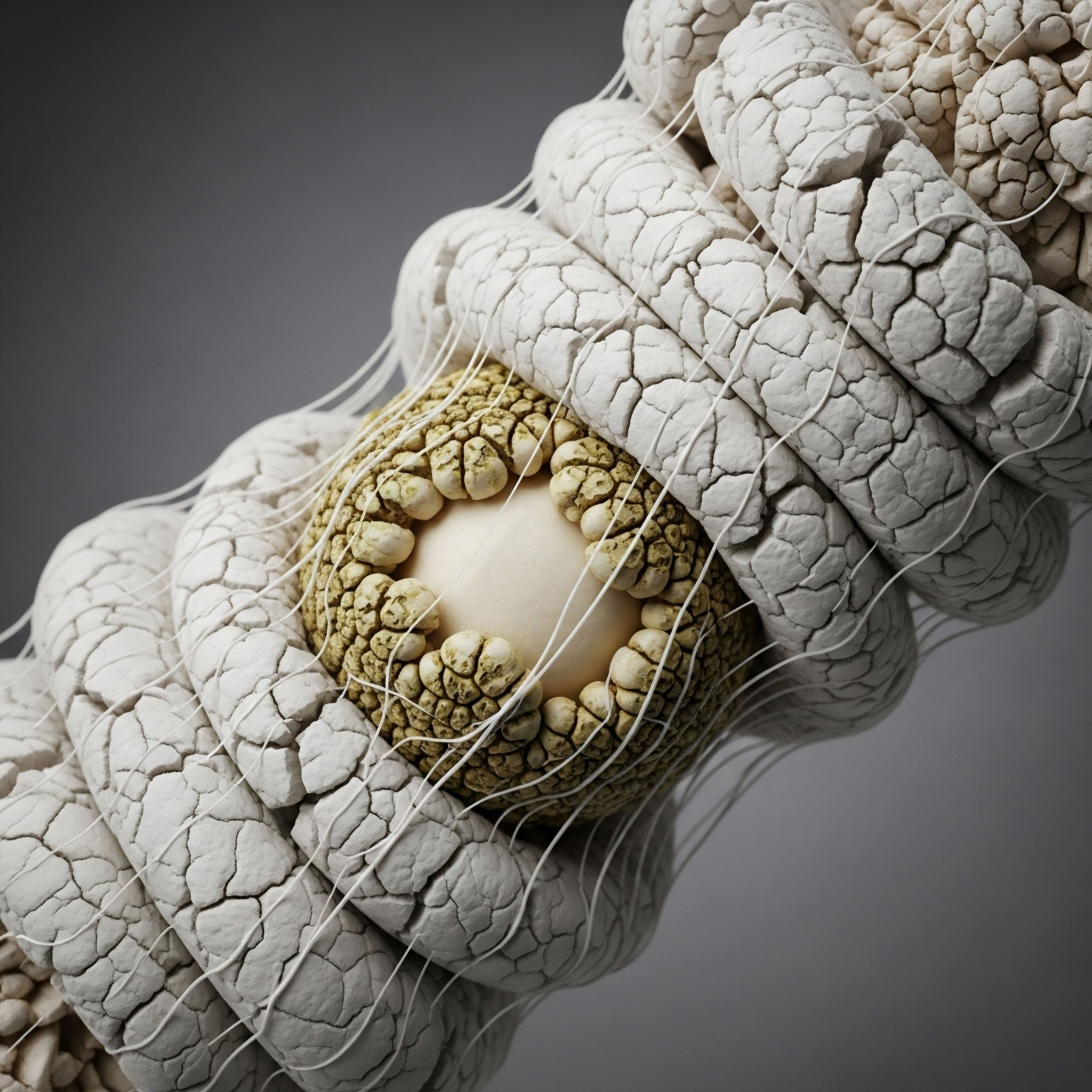
This process, known as bone remodeling, is a beautifully balanced cycle of breakdown and rebuilding, essential for repairing microscopic damage and adapting to mechanical stress. Two primary cell types orchestrate this process ∞ osteoclasts, which resorb old or damaged bone tissue, and osteoblasts, which synthesize new bone matrix to replace it. Your body’s ability to maintain skeletal integrity depends entirely on the sophisticated communication that governs the activity of these two cell populations.

The Core Regulatory System
At the heart of this communication lies a critical signaling triad known as the RANKL/RANK/OPG pathway. Think of it as the primary command system for bone resorption. Osteoblasts, the bone-building cells, produce a protein called Receptor Activator of Nuclear Factor-κB Ligand (RANKL). When RANKL binds to its receptor, RANK, on the surface of osteoclast precursor cells, it triggers their maturation and activation. This activation is the “go” signal for osteoclasts to begin breaking down bone tissue.
To ensure this process remains in check, osteoblasts also secrete a protective protein called osteoprotegerin (OPG). OPG functions as a decoy receptor, intercepting RANKL before it can bind to RANK on osteoclasts. By binding to RANKL, OPG effectively neutralizes the resorption signal, acting as the “stop” signal.
The relative balance, or ratio, between RANKL and OPG is a critical determinant of your bone mass. A higher RANKL-to-OPG ratio favors bone resorption and can lead to a decrease in bone density, while a lower ratio favors bone stability and formation.

The Bone Formation Pathway
Complementing the RANKL/OPG system is another essential signaling cascade called the Wnt pathway. The canonical Wnt/β-catenin pathway is a powerful promoter of bone formation. When Wnt proteins bind to receptors on the surface of mesenchymal stem cells, they initiate a cascade that leads to the differentiation of these precursor cells into mature, bone-building osteoblasts.
This pathway directly stimulates the creation of the cellular machinery required for synthesizing new bone. Moreover, enhanced Wnt signaling also encourages osteoblasts to produce more OPG, which indirectly suppresses bone resorption. This dual effect ∞ promoting formation while inhibiting breakdown ∞ makes the Wnt pathway a central player in achieving a net increase in bone mass.
Bone remodeling is a dynamic process where osteoclast cells resorb old tissue and osteoblast cells form new tissue, governed by precise molecular signals.

Hormonal Oversight of the System
These intricate cellular pathways do not operate in isolation. They are profoundly influenced by the broader endocrine environment, particularly by sex hormones like testosterone and estrogen. Both men and women produce these hormones, which are essential for maintaining skeletal health throughout life.
Estrogen, for instance, is a potent suppressor of bone resorption, primarily by increasing the production of OPG and limiting the lifespan of osteoclasts. Testosterone contributes to bone health by stimulating the proliferation of osteoblasts, the cells responsible for bone formation.
The age-related decline in these hormones is a primary contributor to the shift in remodeling balance that leads to bone loss. Peptides introduce a new layer of communication, offering a way to directly influence these foundational cellular processes to support and restore skeletal integrity.


Intermediate
Understanding the foundational balance of bone remodeling opens the door to a more targeted question ∞ How can we therapeutically influence this system to favor bone formation and repair? This is where specific peptide protocols become relevant. Peptides are short chains of amino acids that act as precise signaling molecules. Within the context of bone health, they function as sophisticated biological messengers that can modulate the core pathways of skeletal regeneration, offering a more nuanced approach to wellness.

Growth Hormone Peptides the Anabolic Signal
A key strategy for enhancing bone formation involves augmenting the body’s own anabolic systems. Growth hormone (GH) is a primary driver of tissue growth and repair, and its influence extends directly to bone. Growth Hormone Secretagogues (GHSs) are a class of peptides designed to stimulate the pituitary gland to release its own endogenous GH. This approach works with the body’s natural pulsatile rhythm of GH secretion. Two of the most well-studied GHSs used in combination are Ipamorelin and CJC-1295.

How Do Growth Hormone Secretagogues Work Together?
Ipamorelin is a selective growth hormone-releasing peptide (GHRP). It mimics the action of ghrelin, binding to the GHSR in the pituitary to trigger a clean, potent pulse of GH release. CJC-1295 is a synthetic analogue of Growth Hormone-Releasing Hormone (GHRH).
It works on a different receptor in the pituitary to increase the overall quantity of GH that is stored and released. When used together, they create a powerful synergistic effect. CJC-1295 amplifies the baseline level and storage of GH, and Ipamorelin triggers a strong, immediate release of this amplified supply.
This dual-action stimulation results in a significant, yet physiologically patterned, increase in circulating GH and, subsequently, Insulin-like Growth Factor 1 (IGF-1), which is a key mediator of GH’s anabolic effects on bone tissue. This elevation in GH and IGF-1 directly stimulates osteoblast activity, leading to increased bone formation and, over time, improvements in bone mineral content and density.
| Peptide | Mechanism of Action | Primary Effect on GH Release | Contribution to Synergy |
|---|---|---|---|
| Ipamorelin |
Selective GHRP; mimics ghrelin to bind to the GHSR on the pituitary gland. |
Induces a strong, pulsatile release of Growth Hormone. |
Triggers the release of the GH pool amplified by CJC-1295. |
| CJC-1295 |
GHRH analogue; stimulates the pituitary to produce and store more Growth Hormone. |
Increases the baseline and total amount of GH available for release. |
Provides a larger reservoir of GH for Ipamorelin to act upon. |

BPC-157 the Local Repair Operator
While GHSs provide a systemic anabolic signal, other peptides work at a more localized level to facilitate the mechanics of tissue repair. BPC-157, a peptide derived from a protein found in gastric juice, is renowned for its potent cytoprotective and healing properties. Its influence on bone remodeling stems from its ability to accelerate the biological processes required for repair.
BPC-157 has been shown to significantly promote angiogenesis, the formation of new blood vessels. A robust blood supply is absolutely essential for healing, as it delivers the oxygen, nutrients, and precursor cells needed to regenerate tissue at the site of injury or microfracture.
Furthermore, BPC-157 enhances the migration and proliferation of fibroblasts, the cells that produce collagen, which forms the structural matrix of bone and connective tissues. Perhaps one of its most sophisticated mechanisms is its ability to upregulate the expression of growth hormone receptors on local tissues, such as tendon fibroblasts.
This action makes the target tissue more sensitive to the circulating GH that the body is already producing, or that is being amplified by GHSs like Ipamorelin and CJC-1295. In essence, BPC-157 prepares the construction site, while GHSs call in the construction crew.
Peptides such as Ipamorelin and BPC-157 act as precise biological messengers to enhance the body’s natural systems for bone growth and repair.

The Hormonal Foundation
It is important to place these peptide therapies in their proper context. They are modulators and amplifiers of existing biological systems. Their efficacy is maximized when the body’s foundational hormonal environment is balanced. Since sex hormones like testosterone and estrogen are primary regulators of the RANKL/OPG ratio and osteoblast function, ensuring their optimal levels is a prerequisite for any advanced protocol.
Hormonal optimization creates a permissive environment where bone resorption is controlled and bone formation is encouraged. Peptide therapies then act as targeted agents to further enhance the formation side of the equation, leading to more robust and efficient bone remodeling.
- Testosterone ∞ Directly stimulates the proliferation of osteoblast precursor cells, providing the cellular building blocks for new bone. It is essential for maintaining the structural integrity of the musculoskeletal system.
- Estrogen ∞ Acts as the primary brake on bone resorption by suppressing the activity and lifespan of osteoclasts. It is the dominant hormone for preventing excessive bone breakdown in both men and women.
- Growth Hormone Peptides ∞ Function as systemic anabolic signals, increasing the overall drive for tissue growth and directly stimulating osteoblast activity to enhance bone mineral density.
- Repair Peptides (BPC-157) ∞ Work locally to improve the infrastructure for healing, promoting blood flow and increasing cellular sensitivity to growth signals, thereby accelerating repair at specific sites.


Academic
A sophisticated analysis of peptide influence on bone remodeling requires a deep examination of the molecular crosstalk between the principal signaling pathways. The skeletal system’s dynamic equilibrium is maintained through a complex interplay of systemic hormonal signals and local paracrine factors.
Peptides intervene in this system as highly specific modulators, capable of altering pathway sensitivity and function to shift the homeostatic set point towards anabolism and repair. The interaction between the Wnt/β-catenin pathway and the OPG/RANKL/RANK axis represents a central node of this regulation, and it is here that the influence of peptide therapy can be most clearly understood from a systems-biology perspective.

Molecular Crosstalk Wnt and RANKL OPG Signaling
The canonical Wnt/β-catenin signaling pathway and the OPG/RANKL/RANK system are intricately linked. The activation of Wnt signaling in osteoblasts and their precursors does more than just promote their differentiation into bone-forming cells. A key downstream effect of β-catenin stabilization is the transcriptional upregulation of the gene encoding for OPG (TNFRSF11B).
This action directly increases the OPG/RANKL ratio, thereby suppressing osteoclastogenesis and bone resorption. This mechanism reveals the elegant efficiency of the system ∞ the primary bone-building pathway actively inhibits the primary bone-resorbing pathway. Pathologies like osteoporosis often involve a disruption in this crosstalk.
For instance, antagonists of the Wnt pathway, such as sclerostin (produced by osteocytes) and Dickkopf-1 (Dkk-1), bind to Wnt co-receptors, preventing signaling. This inhibition reduces osteoblast differentiation and simultaneously decreases OPG production, tipping the remodeling balance towards net bone loss.
The intricate communication between the Wnt and RANKL/OPG signaling pathways forms the central regulatory axis of bone metabolism.

What Is the Role of Endocrine Disruption in Bone Pathology?
Age-related hormonal decline directly impacts this delicate molecular balance. Estrogen deficiency, a hallmark of menopause, leads to an increase in the expression of RANKL and a decrease in the expression of OPG, directly promoting osteoclast activity. In men, both declining estrogen and testosterone levels contribute to bone loss.
Estrogen remains the dominant regulator of resorption, while testosterone is critical for sustaining bone formation. This endocrine shift creates a systemic environment that favors bone catabolism. Therapeutic peptides must operate within this context, acting as targeted interventions to counteract these systemic drifts.
- Systemic Anabolic Stimulation ∞ Growth Hormone Secretagogues like Ipamorelin/CJC-1295 function by elevating systemic levels of GH and IGF-1. IGF-1 is known to promote the survival of osteoblasts and stimulate their synthesis of type I collagen. Furthermore, evidence suggests that the GH/IGF-1 axis can positively influence Wnt signaling, creating a powerful pro-anabolic effect that supports both osteoblast proliferation and function. By increasing the upstream anabolic drive, these peptides help to restore the signaling environment necessary for positive bone remodeling.
- Local Healing Potentiation ∞ Regenerative peptides like BPC-157 exert their effects through distinct, localized mechanisms. BPC-157’s pro-angiogenic effect, mediated partly through the upregulation of Vascular Endothelial Growth Factor (VEGF), is critical. Angiogenesis is coupled with osteogenesis; the invasion of blood vessels into cartilage is a required step for endochondral ossification and fracture healing. Moreover, the finding that BPC-157 upregulates GH receptor expression in fibroblasts suggests a mechanism of local sensitization. This makes the damaged tissue more responsive to the systemic anabolic signals provided by endogenous or peptide-stimulated GH, thereby amplifying the healing response precisely where it is needed.
| Molecule | Primary Function | Cellular Source | Influenced By |
|---|---|---|---|
| RANKL |
Activates osteoclast differentiation and bone resorption. |
Osteoblasts, Osteocytes |
Increased by low estrogen; Decreased by OPG. |
| OPG |
Inhibits RANKL, thus preventing bone resorption. |
Osteoblasts, Osteocytes |
Increased by Wnt signaling and estrogen. |
| Wnt Proteins |
Promote osteoblast differentiation and bone formation. |
Osteoblasts, Bone Marrow Cells |
Inhibited by Sclerostin and Dkk-1. |
| Sclerostin/Dkk-1 |
Antagonize Wnt signaling, inhibiting bone formation. |
Osteocytes (Sclerostin) |
Mechanical loading suppresses sclerostin. |
| Growth Hormone/IGF-1 |
Systemic anabolic signal; stimulates osteoblast activity. |
Pituitary (GH), Liver/Local (IGF-1) |
Increased by GHS peptides (Ipamorelin/CJC-1295). |

A Systems-Biology Synthesis
From a systems-biology perspective, peptides do not simply “build bone.” They strategically modulate key informational nodes within the complex regulatory network of skeletal homeostasis. The foundational hormonal state, dictated by the HPG axis, sets the systemic tone. Age-related decline shifts this tone towards catabolism.
Peptide therapies then offer a multi-pronged intervention. GHSs restore a potent, systemic anabolic signal (GH/IGF-1) that positively influences the entire network. Simultaneously, regenerative peptides like BPC-157 optimize the local microenvironment for repair, enhancing vascularity and increasing receptor sensitivity. This integrated approach, which combines foundational hormonal optimization with targeted peptide modulation, represents a sophisticated clinical strategy for influencing bone remodeling dynamics to support long-term skeletal health and function.

References
- Chang, Chih-Hao, et al. “Pentadecapeptide BPC 157 Enhances the Growth Hormone Receptor Expression in Tendon Fibroblasts.” Molecules, vol. 22, no. 11, 2017, p. 1905.
- Finkelstein, Joel S. et al. “Relative Contributions of Testosterone and Estrogen in Regulating Bone Resorption and Formation in Normal Elderly Men.” The Journal of Clinical Endocrinology & Metabolism, vol. 83, no. 10, 1998, pp. 3489-94.
- Hegedüs, B. et al. “Regulatory Effects and Interactions of the Wnt and OPG-RANKL-RANK Signaling at the Bone-Cartilage Interface in Osteoarthritis.” International Journal of Molecular Sciences, vol. 22, no. 16, 2021, p. 8642.
- Svensson, Johan, et al. “The GH Secretagogues Ipamorelin and GH-Releasing Peptide-6 Increase Bone Mineral Content in Adult Female Rats.” Journal of Endocrinology, vol. 165, no. 3, 2000, pp. 569-77.
- Teitelbaum, Steven L. and M. Neale Weinstock. “The Role of Wnt Signaling in Bone Formation and Osteoporosis.” The Journal of Clinical Investigation, vol. 124, no. 5, 2014, pp. 1836-41.
- Mohan, Subburaman, and John C. Jennings. “The Role of Testosterone in Bone Metabolism.” Journal of Clinical Endocrinology & Metabolism, vol. 89, no. 8, 2004, pp. 3661-70.
- Raun, K. et al. “Ipamorelin, the First Selective Growth Hormone Secretagogue.” European Journal of Endocrinology, vol. 139, no. 5, 1998, pp. 552-61.
- Keramati, Mohammad, et al. “The Role of BPC 157 in Management of Bone and Joint Disorders.” Journal of Orthopaedic Surgery and Research, vol. 16, no. 1, 2021, p. 645.

Reflection
The information presented here offers a map of the complex biological landscape that governs your skeletal health. It details the cellular conversations, the signaling pathways, and the molecular messengers that determine the strength and vitality of your bones. This knowledge is a powerful tool, transforming abstract feelings of physical change into a clear understanding of your body’s inner workings.
The next step in your personal health journey involves using this map not as a destination, but as a guide for a deeper, more informed conversation with a clinical professional who can help translate this science into a personalized protocol that aligns with your unique physiology and wellness goals. Your body is communicating constantly; learning its language is the key to reclaiming your vitality.

Glossary

bone remodeling

bone resorption

osteoclast

bone formation

wnt signaling

testosterone

estrogen

growth hormone secretagogues

growth hormone

ipamorelin

cjc-1295

osteoblast

systemic anabolic signal

bpc-157

rankl/opg ratio

bone mineral density

regenerative peptides like bpc-157
