


Fundamentals
You have put in the work. The sessions are intense, the diet is structured, and the commitment is absolute. Yet, the progress you expect feels distant. The soreness lingers longer than it should, strength gains are slow to materialize, and a persistent sense of fatigue clouds your efforts.
This experience, this disconnect between effort and outcome, is a deeply personal and often frustrating one. It is a biological conversation, and it begins with understanding that your body might be speaking a language you haven’t yet learned to interpret. The process of muscle recovery is a complex and elegant biological orchestration.
It is a cycle of controlled damage followed by a meticulously managed repair and reinforcement process. When you subject your muscles to strenuous work, you create microscopic tears in the fibers. This is the necessary stimulus. The subsequent recovery and growth depend entirely on the quality of the internal environment and the clarity of the biochemical signals that manage the reconstruction project.
Think of your endocrine system as the project management team for this reconstruction. It dispatches chemical messengers, hormones, that dictate the pace, efficiency, and quality of the repair. These hormones operate in a delicate balance, with specific teams assigned to either building up (anabolism) or breaking down (catabolism).
When this management team is functioning optimally, the response to the stimulus of exercise is efficient and effective. The microscopic tears are repaired, and the muscle fibers are rebuilt stronger and more resilient than before. This is the foundation of adaptation and progress.
The endocrine system acts as the body’s internal communication network, using hormones to direct the complex process of muscle repair and growth after exercise.

The Anabolic Construction Crew
On one side of this equation are the anabolic hormones, the primary drivers of growth and repair. These are the signals that tell your body to build. The two most significant members of this construction crew are Testosterone and the Growth Hormone/Insulin-Like Growth Factor-1 (GH/IGF-1) axis.
Testosterone is a principal architect of muscle tissue. It directly signals muscle cells to increase protein synthesis, the process of creating new proteins to repair the damage. More profoundly, testosterone activates a special population of muscle stem cells known as satellite cells.
These cells lie dormant on the surface of muscle fibers and are the true agents of repair and growth. When activated by signals like testosterone, they multiply and fuse with existing muscle fibers, donating their nuclei and enhancing the fiber’s capacity for protein synthesis. This process, myonuclear accretion, is fundamental to long-term muscle hypertrophy.
The GH/IGF-1 axis functions as a coordinated growth-promoting system. The brain signals the pituitary gland to release Growth Hormone (GH), particularly during deep sleep and in response to intense exercise. GH then travels to the liver and other tissues, stimulating the production of Insulin-Like Growth Factor-1 (IGF-1).
IGF-1 is a potent anabolic factor that works in concert with testosterone to promote muscle protein synthesis and support the activation and proliferation of satellite cells. Together, this axis provides a powerful, systemic signal for the body to enter a state of repair and growth.

The Catabolic Demolition Crew
On the other side of the balance is the primary catabolic hormone, cortisol. Released from the adrenal glands in response to stress ∞ be it psychological, physiological, or from the physical act of training itself ∞ cortisol’s role is to mobilize energy.
It does this by breaking down tissues, including muscle protein, to release amino acids that can be converted into glucose for immediate energy. In short bursts, this is a necessary and healthy survival mechanism. The physical stress of a workout elevates cortisol, which is a normal part of the exercise response.
The issue arises when cortisol levels become chronically elevated. A state of sustained high cortisol, often resulting from a combination of life stress, poor sleep, and inadequate recovery, creates a persistently catabolic internal environment. In this state, the “demolition crew” is working overtime.
Chronically high cortisol directly inhibits muscle protein synthesis, effectively blocking the “build” signals from testosterone and IGF-1. It also accelerates muscle protein breakdown, meaning your body is actively deconstructing muscle tissue at a rate that outpaces repair. This creates a scenario where, despite your best efforts in the gym, you may be in a state of net muscle loss, leading to prolonged soreness, stagnant progress, and an increased risk of injury.


Intermediate
Understanding the fundamental roles of anabolic and catabolic hormones provides the ‘what’; exploring their specific mechanisms of action reveals the ‘how’. The efficiency of your muscle recovery is determined at the cellular level, dictated by the hormonal signals that reach your muscle fibers and their resident stem cells.
An imbalance in these signals creates a cascade of downstream effects that can either accelerate your progress or bring it to a complete halt. This deeper layer of physiology is where we can begin to connect symptoms to systems and understand the rationale behind targeted clinical interventions.
Hormonal imbalances directly alter the cellular machinery of muscle repair, affecting protein synthesis rates, satellite cell activity, and inflammatory responses.

The Cellular Mechanics of Testosterone and Recovery
Testosterone’s influence on muscle recovery extends far beyond a simple signal to “grow.” Its primary mechanism involves binding to androgen receptors (AR) located within muscle cells and, critically, on satellite cells. This binding initiates a cascade of genomic events, activating specific genes that drive the machinery of muscle protein synthesis.
Studies show that testosterone administration increases the number of myonuclei and the size of muscle fibers, a direct result of enhanced satellite cell activity. These satellite cells, once activated by testosterone, proliferate and fuse with damaged muscle fibers, donating their nuclei. This increases the muscle fiber’s capacity for producing contractile proteins, leading to hypertrophy and enhanced strength.
Furthermore, testosterone has a moderating effect on the inflammatory response to exercise-induced muscle damage. While some inflammation is a necessary signal for repair, excessive or prolonged inflammation can impede recovery. Testosterone appears to help create a more favorable environment for satellite cells to perform their regenerative functions by mitigating excessive inflammation. A deficiency in testosterone, therefore, results in a multi-faceted impairment of the recovery process ∞ reduced protein synthesis, diminished satellite cell activation, and a potentially dysregulated inflammatory response.
| Recovery Metric | Optimal Testosterone Environment | Low Testosterone (Hypogonadal) Environment |
|---|---|---|
| Muscle Protein Synthesis (MPS) | Significantly upregulated post-exercise. | Blunted or suppressed response to training stimulus. |
| Satellite Cell Activation | Robust activation, proliferation, and fusion. | Reduced activation and proliferation, impairing repair. |
| Myonuclear Accretion | Efficient addition of new nuclei to muscle fibers. | Impaired ability to increase myonuclear domain. |
| Myostatin Expression | Suppressed, allowing for greater growth potential. | Less suppression, leading to inhibition of muscle growth. |
| Inflammatory Regulation | Attenuated inflammatory response, promoting efficient repair. | Potentially prolonged or excessive inflammation. |

The HPA Axis and Cortisol’s Dominance in Stress
The Hypothalamic-Pituitary-Adrenal (HPA) axis is the body’s central stress response system. When the hypothalamus perceives stress, it releases corticotropin-releasing hormone (CRH), which signals the pituitary to release adrenocorticotropic hormone (ACTH). ACTH then travels to the adrenal glands and stimulates the release of cortisol.
In a state of chronic stress, this axis becomes perpetually activated, leading to a hormonal environment that is fundamentally hostile to muscle recovery. Chronically elevated cortisol actively works against anabolic processes. It competitively antagonizes the effects of testosterone and promotes the expression of enzymes that tag muscle proteins for degradation via the ubiquitin-proteasome pathway.
This means cortisol not only stops the building process but actively accelerates demolition. It can lead to a state of insulin resistance in the muscle cells, impairing their ability to take up glucose for energy and further hindering recovery and growth. This systemic catabolic pressure explains why periods of high stress, poor sleep, or overtraining can completely derail fitness progress, regardless of effort.

Clinical Protocols for Restoring Anabolic Balance
When hormonal testing reveals imbalances that are clinically significant and symptomatic, specific protocols can be employed to restore the body’s internal environment to one that is conducive to recovery and well-being. These interventions are designed to recalibrate the endocrine system, not to replace its function entirely.
- Testosterone Replacement Therapy (TRT) for Men A standard protocol for men with diagnosed hypogonadism involves weekly intramuscular injections of Testosterone Cypionate. This directly restores testosterone levels to an optimal physiological range. This is often combined with other medications to maintain systemic balance. Gonadorelin, a GHRH analogue, is used to stimulate the pituitary to maintain natural testicular signaling, preserving fertility and preventing testicular atrophy. Anastrozole, an aromatase inhibitor, may be used to control the conversion of testosterone to estrogen, managing potential side effects.
- Hormonal Optimization for Women Women also rely on testosterone for energy, libido, and muscle maintenance. Protocols for women use much lower doses, typically weekly subcutaneous injections of Testosterone Cypionate (e.g. 10-20 units). This can restore vitality and improve body composition without causing masculinizing effects. Depending on menopausal status, bio-identical Progesterone may also be prescribed to support mood, sleep, and overall hormonal equilibrium.
- Growth Hormone Peptide Therapy For individuals seeking to optimize recovery and body composition, peptide therapies that stimulate the body’s own production of growth hormone are a powerful tool. Sermorelin is a GHRH analog that directly stimulates the pituitary gland to release GH in a natural, pulsatile manner. A more advanced protocol combines CJC-1295 (a long-acting GHRH analog) with Ipamorelin (a GH secretagogue that works on the ghrelin receptor). This synergistic combination produces a stronger and more sustained release of natural GH, enhancing its downstream effects on IGF-1 production, fat metabolism, and cellular repair, all of which are vital for efficient muscle recovery.


Academic
A sophisticated analysis of hormonal influence on muscle recovery requires a shift in perspective from isolated endocrine pathways to an integrated, systems-biology viewpoint. The regenerative capacity of skeletal muscle is not governed by a single hormone but by the dynamic crosstalk between multiple signaling networks that converge upon the muscle fiber and its associated satellite cell niche.
The ultimate phenotypic outcome ∞ be it hypertrophy, stagnation, or atrophy ∞ is an emergent property of the competition between anabolic and catabolic inputs at the molecular level. Understanding this interplay is essential for developing truly personalized and effective therapeutic strategies.

Molecular Regulation of the Satellite Cell Cycle
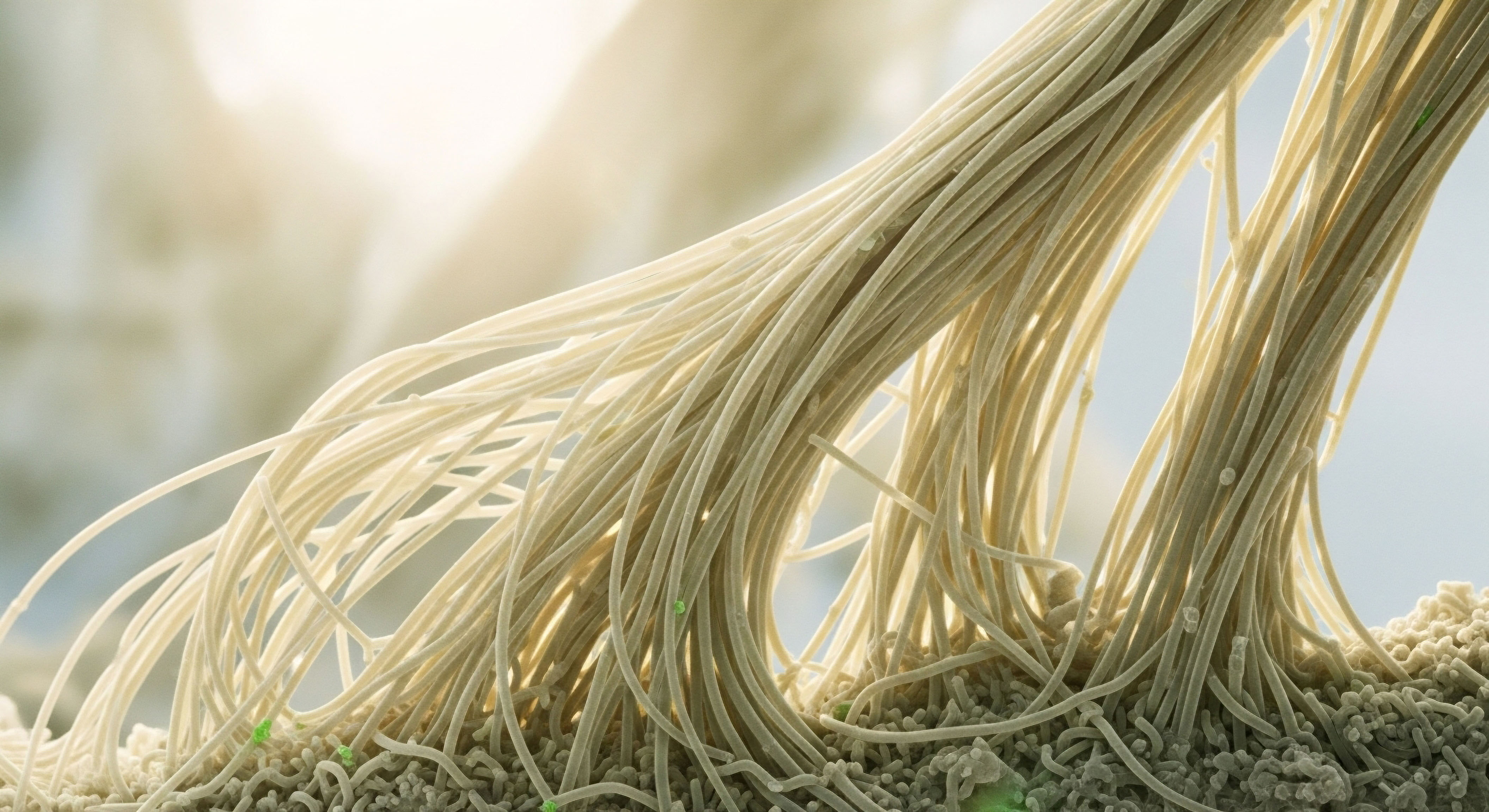
The satellite cell is the fulcrum of muscle adaptation. Its journey from quiescence to activation, proliferation, differentiation, and eventual fusion is tightly regulated by a complex web of intrinsic and extrinsic factors. Hormones are primary extrinsic regulators that dictate the fate of this cellular pool.

How Do Hormones Modulate Satellite Cell Behavior?
Testosterone exerts its pro-myogenic effects through both genomic and non-genomic signaling. Genomically, testosterone binds to the androgen receptor (AR) in the cytoplasm of satellite cells. This hormone-receptor complex translocates to the nucleus, where it acts as a transcription factor, binding to androgen response elements (AREs) on DNA.
This directly upregulates the expression of key myogenic regulatory factors (MRFs) like MyoD, which commits the satellite cell to the myogenic lineage. It also increases the expression of IGF-1 within the muscle tissue itself, creating a localized autocrine/paracrine anabolic loop that amplifies the growth signal.
The GH/IGF-1 axis provides a complementary and synergistic signal. Systemic IGF-1, produced by the liver in response to pituitary GH, and local IGF-1, produced by muscle fibers, both bind to the IGF-1 receptor on the satellite cell membrane. This binding triggers the phosphorylation and activation of the Phosphatidylinositol 3-kinase (PI3K)/Akt signaling pathway.
Akt is a central node in cellular regulation, and its activation promotes cell survival, proliferation, and growth. One of its key downstream targets is the mammalian target of rapamycin (mTOR), a protein kinase that orchestrates protein synthesis. By activating the PI3K/Akt/mTOR pathway, IGF-1 provides a powerful stimulus for satellite cell proliferation and subsequent differentiation into mature myocytes capable of fusing with existing fibers.
The fate of muscle tissue is decided by the molecular balance between the PI3K/Akt/mTOR growth pathway and the cortisol-driven ubiquitin-proteasome degradation pathway.

The Biochemical Machinery of Catabolism
Cortisol exerts its catabolic effects by tipping the molecular scales in favor of degradation. It binds to the glucocorticoid receptor (GR), which, upon activation, translocates to the nucleus and upregulates the transcription of genes central to muscle atrophy. The most critical of these are the E3 ubiquitin ligases, muscle RING-finger protein-1 (MuRF1) and Atrogin-1 (also known as MAFbx).
These enzymes are responsible for tagging contractile proteins, such as myosin and actin, with ubiquitin molecules. This “tag” marks the proteins for destruction by the 26S proteasome, a cellular complex that degrades proteins into their constituent amino acids.
Simultaneously, cortisol actively suppresses anabolic signaling. Glucocorticoid signaling has been shown to directly interfere with the PI3K/Akt pathway, creating a state of anabolic resistance. It can increase the expression of REDD1, an inhibitor of mTOR, and KLF15, a transcription factor that promotes the expression of branched-chain amino acid (BCAA) catabolizing enzymes.
This coordinated molecular assault both dismantles existing muscle protein and prevents the synthesis of new protein, creating a powerful net catabolic state that completely undermines the potential for recovery and growth.
| Hormone/Peptide | Primary Receptor | Key Signaling Pathway | Primary Molecular Outcome in Muscle Recovery |
|---|---|---|---|
| Testosterone | Androgen Receptor (AR) | AR Nuclear Translocation; Akt/mTOR | Upregulation of Myogenic Regulatory Factors (MyoD); Suppression of Myostatin. |
| IGF-1 | IGF-1 Receptor (IGF-1R) | PI3K/Akt/mTOR | Stimulation of protein synthesis; satellite cell proliferation and differentiation. |
| Cortisol | Glucocorticoid Receptor (GR) | GR Nuclear Translocation | Upregulation of MuRF1 & Atrogin-1; Inhibition of PI3K/Akt pathway. |
| Sermorelin/CJC-1295 | GHRH Receptor (GHRH-R) | cAMP/PKA in pituitary somatotrophs | Increased pulsatile release of endogenous Growth Hormone. |
| Ipamorelin | Ghrelin Receptor (GHS-R1a) | Gq/PLC/IP3/DAG in pituitary somatotrophs | Amplified release of endogenous Growth Hormone. |

Why Are Post-TRT Protocols Necessary for Fertility?
The administration of exogenous testosterone, while effective for restoring systemic levels, creates a negative feedback loop within the Hypothalamic-Pituitary-Gonadal (HPG) axis. The hypothalamus detects high levels of testosterone and estrogen (from aromatization) and ceases production of Gonadotropin-Releasing Hormone (GnRH).
This lack of GnRH signal causes the pituitary to stop producing Luteinizing Hormone (LH) and Follicle-Stimulating Hormone (FSH). The absence of LH signaling to the Leydig cells in the testes leads to a shutdown of endogenous testosterone production and spermatogenesis, causing infertility. A post-TRT or fertility-stimulating protocol is designed to restart this axis.
It uses agents like Gonadorelin to mimic GnRH, Clomid (Clomiphene Citrate) and Tamoxifen to block estrogen’s negative feedback at the hypothalamus, and sometimes Anastrozole to further reduce estrogen, all in a concerted effort to stimulate the pituitary to resume LH and FSH production.

How Can Peptide Therapies Be More Precise than HGH?
Direct administration of recombinant Human Growth Hormone (rHGH) introduces a constant, supraphysiological level of the hormone into the body, bypassing the body’s natural regulatory mechanisms. This can lead to side effects and desensitization of receptors. Peptide therapies like Sermorelin, CJC-1295, and Ipamorelin represent a more nuanced, biomimetic approach.
They work upstream by stimulating the pituitary gland itself. Sermorelin and CJC-1295 are GHRH analogs, providing the “release” signal. Ipamorelin is a ghrelin mimetic that acts on a separate receptor to amplify the release and inhibit somatostatin, the hormone that shuts off GH production.
This dual-pathway stimulation results in a release of the body’s own GH in a pulsatile manner that mimics natural physiology. This preserves the sensitivity of the HPG axis and reduces the risk of side effects, making it a more precise tool for optimizing the GH/IGF-1 axis for recovery.

References
- Bhasin, S. et al. “Testosterone Therapy in Men with Hypogonadism ∞ An Endocrine Society Clinical Practice Guideline.” The Journal of Clinical Endocrinology & Metabolism, vol. 103, no. 5, 2018, pp. 1715 ∞ 1744.
- Vierck, J. et al. “Satellite cell regulation following myotrauma caused by resistance exercise.” Cell Biology International, vol. 24, no. 5, 2000, pp. 263-72.
- Kraemer, W. J. & Ratamess, N. A. “Hormonal responses and adaptations to resistance exercise and training.” Sports Medicine, vol. 35, no. 4, 2005, pp. 339-61.
- Schakman, O. Kalista, S. Barbé, C. Loumaye, A. & Thissen, J. P. “Glucocorticoid-induced skeletal muscle atrophy.” The International Journal of Biochemistry & Cell Biology, vol. 45, no. 10, 2013, pp. 2163-72.
- Sinha-Hikim, I. et al. “Testosterone-induced increase in muscle size in healthy young men is associated with muscle fiber hypertrophy and an increase in myonuclear number.” Journal of Clinical Endocrinology & Metabolism, vol. 87, no. 8, 2002, pp. 3555-63.
- Chikani, V. & Ho, K. K. Y. “Action of GH on skeletal muscle function ∞ molecular and metabolic mechanisms.” Journal of Molecular Endocrinology, vol. 52, no. 1, 2014, pp. T107-23.
- Raue, U. et al. “Effects of Ipamorelin and CJC-1295 on growth hormone and insulin-like growth factor 1 in human subjects.” Journal of Clinical Endocrinology & Metabolism, vol. 93, no. 1, 2008, pp. 129-35.
- Walker, R. F. “Sermorelin ∞ a better approach to management of adult-onset growth hormone insufficiency?” Clinical Interventions in Aging, vol. 1, no. 4, 2006, pp. 307-8.

Reflection
The information presented here provides a map of the intricate biological territory that governs your physical adaptation and recovery. It details the molecular conversations, the cellular machinery, and the systemic signals that translate your physical efforts into tangible results. This knowledge transforms the abstract feelings of fatigue or frustration into understandable physiological processes. It offers a new lens through which to view your body, one that sees it as a complex, interconnected system striving for equilibrium.
This map is a powerful tool for understanding. It allows you to connect the dots between your lifestyle, your symptoms, and your underlying biology. The next step in this personal exploration is to ask what your unique biological landscape looks like. What signals is your body sending?
Is the conversation between your anabolic and catabolic systems clear and balanced, or is there static on the line? The science provides the framework, but your lived experience provides the context. This synthesis of knowledge and self-awareness is the true starting point for building a personalized protocol that allows you to reclaim your vitality and function at your full biological potential.