
Fundamentals
The sensation of vitality, of feeling supple and responsive, originates deep within your circulatory system. Your arteries are active, flexible conduits, designed to expand and recoil with every beat of your heart. This inherent elasticity is a cornerstone of cardiovascular health, ensuring that oxygenated blood reaches every cell in your body with steady, controlled pressure.
This process is governed by an intricate signaling network, with hormones acting as the primary chemical messengers. When this internal communication system functions optimally, your vascular network remains pliable and efficient. The lived experience of this efficiency is energy, mental clarity, and physical resilience.
Hormonal fluctuations, however, introduce a profound shift in this biological terrain. Consider estrogen, a key regulator of vascular health in both women and men. It directly supports the production of nitric oxide, a molecule that signals the smooth muscles in artery walls to relax, promoting flexibility and healthy blood flow.
A decline in estrogen, most notably during the menopausal transition, corresponds with a measurable increase in arterial stiffness. This is a physical manifestation of a communication breakdown. The clear signal for vasodilation becomes muted, and the arterial walls begin to lose their responsive, elastic quality. The body experiences this shift as a gradual loss of its youthful buffer against cardiovascular strain.
The body’s hormonal messaging system is the primary architect of arterial flexibility and resilience.
Similarly, testosterone contributes to maintaining a healthy vascular tone. It influences the structural integrity of the arterial wall and supports endothelial function, the health of the delicate inner lining of your blood vessels. When testosterone levels decline, as seen in andropause or hypogonadism, the structural support system for the arteries can weaken.
This contributes to a state where the vessels are less able to manage the pulsatile force of blood flow, leading to a progressive stiffening. This is a structural problem born from a chemical deficit, a tangible change in tissue integrity resulting from altered hormonal signals.
Conversely, an excess of certain hormones, particularly stress hormones like cortisol, can actively degrade vascular health. Chronic stress places the body in a persistent state of alert, flooding the system with cortisol. This hormone promotes processes that directly lead to arterial stiffening, including inflammation and the retention of sodium, which can elevate blood pressure.
The architecture of the artery wall itself begins to change, with a decrease in pliable elastin fibers and an increase in rigid collagen. This transforms a once-flexible tube into a more brittle pipe, a change that has profound implications for long-term health. Understanding this connection is the first step in recognizing that symptoms of fatigue or a decline in physical performance may be rooted in the silent stiffening of your vascular system, orchestrated by a shifting hormonal landscape.

Intermediate
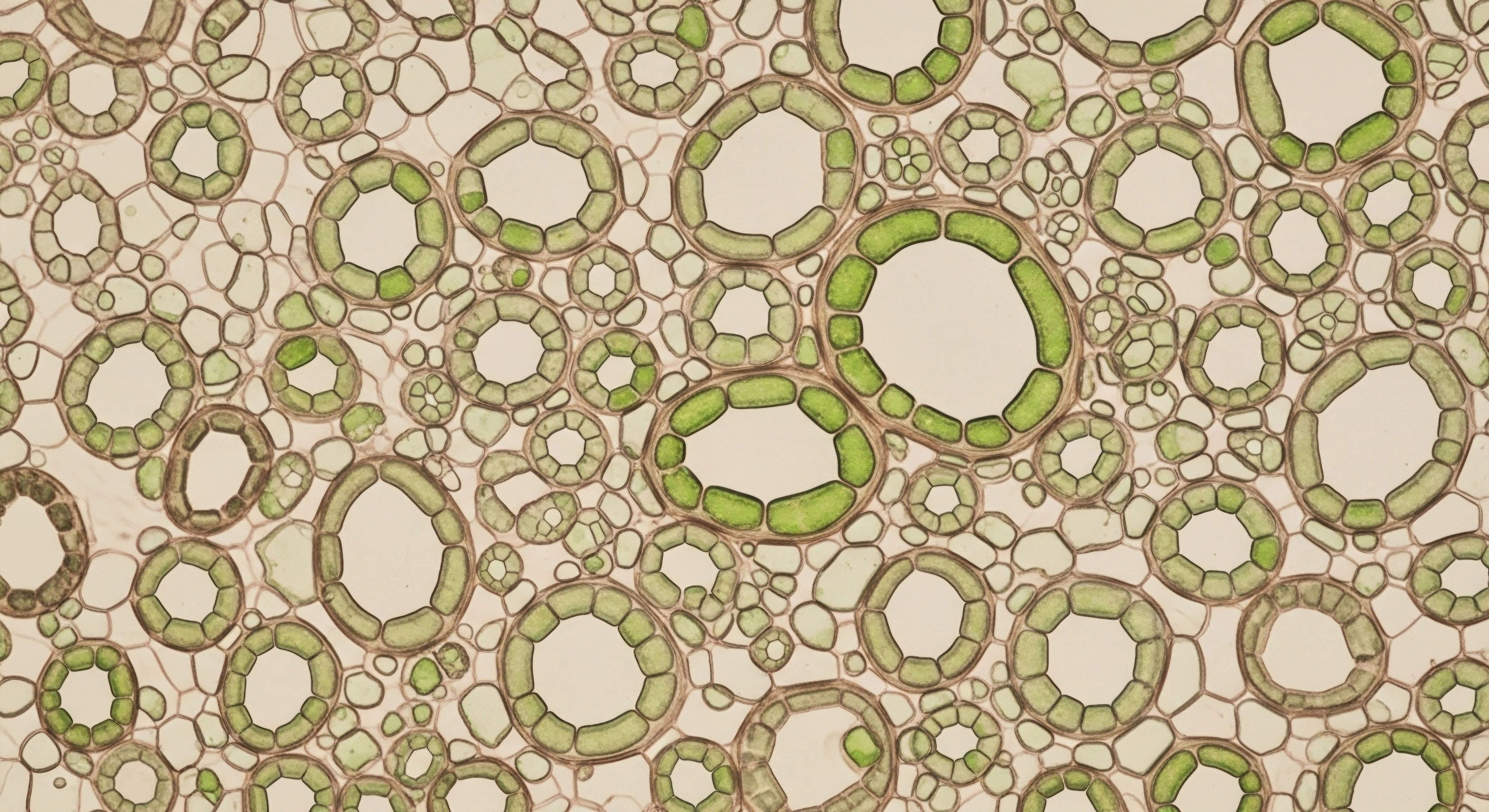
To comprehend how hormonal shifts translate into the physical reality of arterial stiffening, we must examine the cellular and molecular environment of the blood vessel wall. The central player in this arena is the endothelium, the single layer of cells lining every artery.
A healthy endothelium is a dynamic biochemical factory, producing substances that regulate vascular tone, prevent blood clots, and manage inflammation. Hormones are the master regulators of this factory. Estrogen, for instance, directly stimulates endothelial nitric oxide synthase (eNOS), the enzyme responsible for producing nitric oxide (NO).
NO is a potent vasodilator; its presence signals the vascular smooth muscle cells (VSMCs) within the artery wall to relax, resulting in a compliant, flexible vessel. A decline in estrogen starves the endothelium of this critical stimulus, leading to reduced NO bioavailability and a default state of vascular constriction and rigidity.

The Cellular Mechanisms of Hormonal Influence
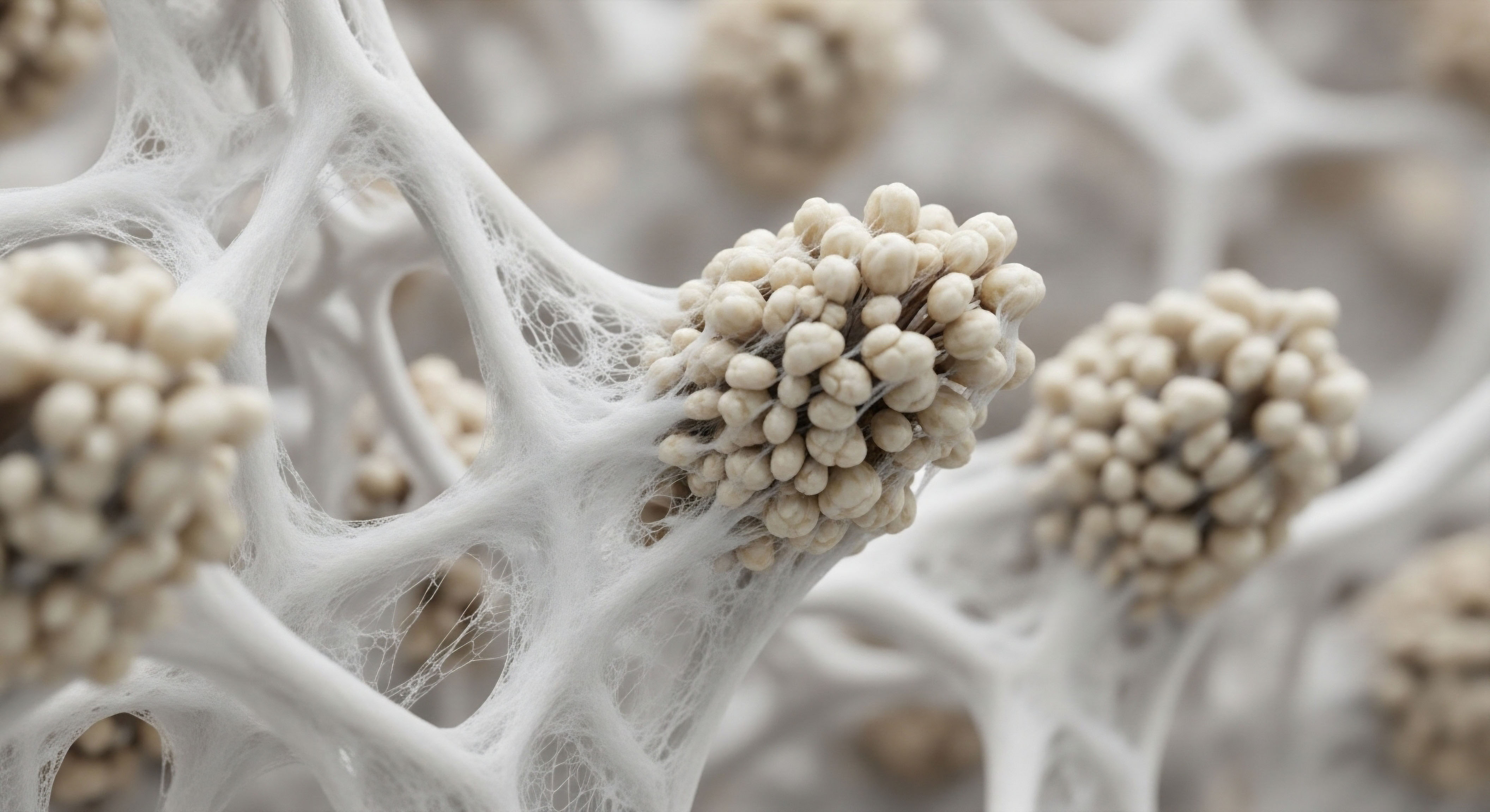
The interplay between hormones and vascular cells extends beyond nitric oxide. Hormonal balance dictates the structural composition of the artery wall itself, influencing the ratio of elastin to collagen. Elastin provides flexibility and the ability to recoil, while collagen provides tensile strength. An optimal hormonal environment maintains a healthy equilibrium. However, hormonal imbalances can trigger a cascade of events that favor collagen deposition and elastin degradation.
Key Hormonal Pathways Affecting Arterial Walls
- Inflammatory Signaling ∞ A decline in sex hormones, particularly estrogen, is associated with a rise in pro-inflammatory cytokines. These inflammatory messengers circulate in the blood and can damage the endothelium, further impairing its function and promoting a fibrotic state where rigid collagen replaces elastic tissue.
- The Renin-Angiotensin-Aldosterone System (RAAS) ∞ This hormonal system is a primary regulator of blood pressure and fluid balance. Aldosterone, its final product, can promote fibrosis and hypertrophy in the vascular wall when levels are chronically elevated. Sex hormones like estrogen help to modulate the RAAS, and their decline can lead to system overactivity, contributing directly to vascular stiffening.
- Oxidative Stress ∞ Hormonal imbalances can increase the production of reactive oxygen species (ROS), unstable molecules that cause cellular damage. ROS can degrade nitric oxide, reducing its availability, and directly damage endothelial cells and VSMCs, accelerating the aging process of the arteries.
How Do Clinical Protocols Address Vascular Health?
Understanding these mechanisms provides the rationale for hormonal optimization protocols. The goal of these interventions is to restore the biochemical signaling that preserves vascular compliance. For instance, Testosterone Replacement Therapy (TRT) in men with diagnosed hypogonadism aims to restore testosterone to physiological levels.
This supports endothelial function, modulates inflammation, and helps maintain the structural integrity of the artery wall. For perimenopausal and postmenopausal women, hormone therapy can reintroduce the protective effects of estrogen, supporting nitric oxide production and mitigating the inflammatory processes that drive arterial stiffening.
Hormone optimization protocols are designed to restore the precise biochemical signals that command arteries to remain flexible.
These are not one-size-fits-all solutions. A carefully calibrated protocol for a woman might involve low-dose Testosterone Cypionate to support vascular integrity alongside progesterone, while a man’s protocol might include Testosterone Cypionate with an aromatase inhibitor like Anastrozole to manage its conversion to estrogen, ensuring a balanced hormonal profile. The objective is always to re-establish the body’s endogenous signaling environment, thereby interrupting the progression from hormonal decline to vascular rigidity.
| Hormone | Primary Positive Vascular Effect | Effect of Deficiency or Excess |
|---|---|---|
| Estrogen | Stimulates Nitric Oxide (NO) Production | Deficiency leads to endothelial dysfunction and increased stiffness. |
| Testosterone | Supports Vascular Smooth Muscle Health | Deficiency is linked to reduced vasodilation and structural changes. |
| Progesterone | Modulates Vascular Tone | Imbalance can affect endothelial response and vessel reactivity. |
| Cortisol | Regulates Acute Stress Response | Chronic excess promotes inflammation, fibrosis, and hypertension. |


Academic
The acceleration of arterial stiffness due to hormonal dysregulation is a process rooted in the intricate molecular biology of the vascular wall. The interaction between steroid hormones and their cognate receptors within endothelial cells and vascular smooth muscle cells (VSMCs) initiates complex signaling cascades with both rapid, non-genomic effects and long-term, genomic consequences.
A deep analysis reveals that the loss of hormonal homeostasis dismantles the protective mechanisms that preserve vascular compliance, initiating a pathological shift toward a fibrotic and dysfunctional phenotype.

Genomic and Non-Genomic Actions on the Vasculature
Estrogen’s vasoprotective effects are mediated through its binding to estrogen receptors (ERα and ERβ), which are expressed in both endothelial cells and VSMCs. The classical, or genomic, pathway involves the hormone-receptor complex translocating to the nucleus, where it acts as a transcription factor to regulate the expression of genes involved in vascular health.
This includes upregulating the expression of endothelial nitric oxide synthase (eNOS) and prostacyclin synthase, both of which produce vasodilating molecules. Simultaneously, this pathway downregulates the expression of pro-inflammatory molecules and endothelin-1, a potent vasoconstrictor.
Beyond this, estrogen exerts rapid, non-genomic effects by activating membrane-associated ERs. This leads to the swift activation of intracellular signaling pathways, such as the PI3K/Akt pathway, which can phosphorylate and activate eNOS within seconds to minutes. This dual-action mechanism provides both immediate regulation of vascular tone and long-term maintenance of a healthy vascular environment.
The decline of estrogen during menopause effectively silences both of these protective pathways, leaving the vasculature vulnerable to vasoconstrictive and pro-inflammatory stimuli.

What Is the Role of Matrix Metalloproteinases?
The structural remodeling of the arterial wall is a key feature of hormonal-driven stiffening. This process is heavily influenced by the activity of matrix metalloproteinases (MMPs), a family of enzymes responsible for degrading extracellular matrix components like elastin and collagen. Hormonal balance maintains a check on MMP activity.
Estrogen, for example, has been shown to downregulate the expression of MMP-12 in macrophages, an enzyme that degrades elastin. When estrogen levels fall, MMP-12 activity can increase, leading to a net degradation of the elastic fibers that give arteries their compliance. This creates a permissive environment for VSMCs to proliferate and deposit disorganized, rigid collagen, fundamentally altering the mechanical properties of the artery from elastic to stiff.
The loss of hormonal signaling integrity at the cellular level directly precipitates the biomechanical failure known as arterial stiffness.
This pathological remodeling is exacerbated by the interplay with other systemic factors. For instance, in a state of insulin resistance, another common consequence of hormonal imbalance, the accumulation of advanced glycation end products (AGEs) becomes a significant factor. AGEs can form cross-links between collagen fibers, further increasing the rigidity of the arterial wall.
They also bind to their receptor (RAGE) on endothelial cells and macrophages, promoting oxidative stress and inflammation. This creates a vicious cycle ∞ hormonal decline promotes insulin resistance, which leads to AGE formation, which in turn amplifies the vascular damage initiated by the loss of direct hormonal protection.
| Mediator | Function in Health (Hormonally Balanced) | Dysfunction in Hormonal Imbalance |
|---|---|---|
| eNOS (Endothelial Nitric Oxide Synthase) | Produces nitric oxide for vasodilation. | Expression and activation are downregulated, leading to vasoconstriction. |
| Endothelin-1 (ET-1) | Expression is suppressed. | Upregulated expression promotes potent vasoconstriction. |
| MMP-12 (Matrix Metalloproteinase-12) | Activity is inhibited, preserving elastin. | Overactivity leads to degradation of elastin fibers. |
| NF-κB (Nuclear Factor kappa B) | Inflammatory pathway is quiescent. | Activated, leading to chronic vascular inflammation and fibrosis. |
| AGEs (Advanced Glycation End Products) | Cleared effectively from the system. | Accumulate, causing collagen cross-linking and inducing inflammation. |
Ultimately, the acceleration of arterial stiffness is a manifestation of systemic signaling failure. The loss of precise hormonal regulation removes the brakes on inflammation, oxidative stress, and pathological matrix remodeling. Therapeutic interventions such as hormone replacement therapy or peptide therapies like Sermorelin, which can influence downstream metabolic health, are attempts to restore this regulatory control at a fundamental level.
By re-engaging the appropriate cellular receptors and signaling pathways, these protocols aim to shift the vascular environment away from a pro-fibrotic, pro-inflammatory state and back toward one that favors compliance, endothelial health, and dynamic responsiveness.
- Receptor Activation ∞ Hormones like testosterone and estrogen bind to specific receptors on vascular cells, initiating protective signaling.
- Gene Expression ∞ The hormone-receptor complex influences DNA transcription, altering the production of proteins that affect vascular structure and function.
- Enzymatic Activity ∞ Key enzymes like eNOS are activated, while degradative enzymes like certain MMPs are suppressed, maintaining a healthy balance.
- Systemic Crosstalk ∞ Hormonal balance influences other systems, like insulin sensitivity and RAAS, which in turn impact vascular health, demonstrating a deeply interconnected biological network.

References
- Laurent, Stéphane, and Pierre Boutouyrie. “Mechanisms of Arterial Stiffening.” Arteriosclerosis, Thrombosis, and Vascular Biology, vol. 40, no. 5, 2020, pp. 1047-1049.
- El Khoudary, Samar R. et al. “Arterial Stiffness Accelerates within One Year of the Final Menstrual Period ∞ The SWAN Heart Study.” Arteriosclerosis, Thrombosis, and Vascular Biology, vol. 41, no. 1, 2021, pp. 481-490.
- Laitinen, Tomi T. et al. “Associations of Sex Hormones and Hormonal Status With Arterial Stiffness in a Female Sample From Reproductive Years to Menopause.” Frontiers in Cardiovascular Medicine, vol. 8, 2021.
- Lacolley, Patrick, et al. “Mechanisms of Arterial Stiffening ∞ Updates and Perspectives.” Arteriosclerosis, Thrombosis, and Vascular Biology, vol. 40, no. 5, 2020, pp. 1050-1054.
- Moreau, Kerrie L. et al. “Association between Arterial Stiffness and Variations in Estrogen-Related Genes.” Advances in Vascular Medicine, vol. 2013, 2013, pp. 1-8.

Reflection
The information presented here provides a map of the biological processes connecting your internal chemistry to your physical vitality. It translates the silent, cellular events into the tangible feelings of energy, resilience, and well-being. This knowledge serves as a powerful tool, moving the conversation about health from one of passive observation to one of active participation.
Your body is constantly communicating its needs and its state of balance. The journey now involves learning to listen to these signals with a new level of understanding, recognizing that the path to sustained health is paved with informed, personalized choices guided by the unique language of your own physiology.
