


Fundamentals
The feeling is distinct. One moment, you are moving through your day with clarity, and the next, a fog descends. The name you were about to say vanishes, the reason you entered a room dissolves, and a pervasive sense of mental slowness settles in.
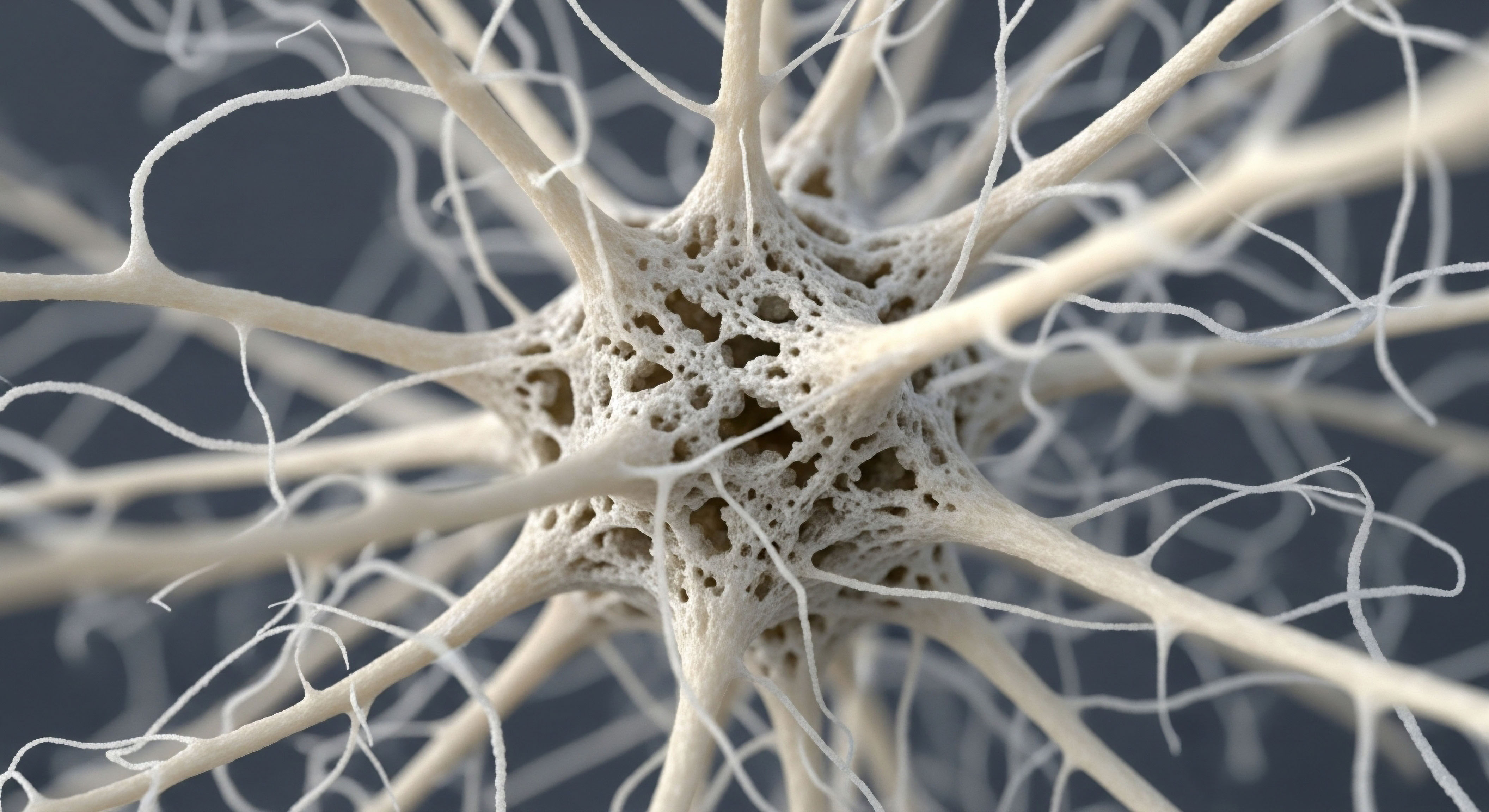
This experience, often labeled as “brain fog,” is a deeply personal and disorienting reality for many women navigating perimenopause. It is a tangible shift in cognitive function that originates from profound biological changes within the brain itself. Your brain is an organ rich with receptors for hormones, particularly estrogen.
It relies on a steady hormonal environment to regulate its energy supply, manage communication between neurons, and maintain its intricate architecture. Perimenopause introduces a period of dramatic fluctuation in these essential chemical messengers, directly disrupting the brain’s established operational rhythm.
This transitional phase is a neurological event. The cognitive symptoms that arise are direct biological signals of a system in adaptation. The primary hormones of the female reproductive cycle, estradiol and progesterone, perform extensive roles within the central nervous system. Estradiol, the most potent form of estrogen, is a master regulator of brain energy.
It facilitates the brain’s ability to uptake and use glucose, its primary fuel. When estradiol levels become erratic and begin to decline, the brain’s capacity to power itself efficiently is compromised. This creates a state of energetic stress, which manifests as difficulty with concentration, memory retrieval, and mental speed. You are feeling the direct consequence of a change in your brain’s metabolic vitality.
The cognitive and mood-related symptoms of perimenopause are neurological in origin, stemming directly from the brain’s adaptation to a shifting hormonal landscape.
Concurrently, progesterone and its powerful neuroactive metabolite, allopregnanolone, modulate the brain’s primary calming neurotransmitter system, known as GABA (gamma-aminobutyric acid). This system acts like a dimmer switch, tempering anxiety and promoting emotional stability and restful sleep. The unpredictable rise and fall of progesterone during perimenopause disrupts this calming influence.
This can lead to heightened feelings of anxiety, irritability, and disturbed sleep patterns. These sleep disturbances further compound cognitive challenges, creating a cycle where hormonal imbalance affects sleep, and poor sleep worsens brain fog and mood. Understanding these mechanisms is the first step in recognizing that these symptoms are not a personal failing but a physiological process of adaptation that can be understood and supported.

What Are the Primary Hormonal Drivers of Change?
The perimenopausal transition is defined by the changing behavior of the ovaries, which leads to significant shifts in two key hormones that have profound effects on brain function. These are not just reproductive hormones; they are integral neuro-regulators.
- Estradiol This is the most active form of estrogen in the female body. In the brain, it acts as a powerful growth factor, supporting the health and connectivity of neurons. It is fundamental for memory formation, particularly verbal memory, and plays a significant part in regulating mood-influencing neurotransmitters like serotonin and dopamine. Its role in facilitating glucose metabolism means it is directly linked to the brain’s energy levels.
- Progesterone This hormone is best known for its role in the menstrual cycle and pregnancy. In the brain, its influence is largely mediated through its conversion to allopregnanolone, a potent neurosteroid. Allopregnanolone enhances the activity of GABA, the brain’s main inhibitory neurotransmitter, which produces a calming, anti-anxiety effect. Fluctuations in progesterone can therefore lead to increased anxiety and sleep disruption.
The erratic nature of these hormonal fluctuations during perimenopause, with their unpredictable peaks and valleys, creates a state of instability within the brain’s internal environment. This biochemical disruption is the root cause of the cognitive and emotional symptoms that define the perimenopausal experience for many women.


Intermediate
To comprehend the brain’s response to perimenopause, we must examine the specific biological systems that are recalibrated by fluctuating hormone levels. The experience of brain fog and mood instability is the macroscopic symptom of microscopic changes in cellular energy processing and neurotransmitter signaling.
The transition forces the brain to adapt its long-standing metabolic and chemical wiring, a process that is often disruptive before a new equilibrium is found post-menopause. This section details the precise mechanisms behind these changes and introduces the clinical logic for interventions designed to support the brain through this demanding transition.

The Brain’s Energy Crisis the Shift from Glucose Hypometabolism
For its entire adult life, the female brain is optimized to run on glucose, facilitated by the constant presence of estradiol. Estradiol enhances the transport of glucose into neurons and promotes its efficient use in generating ATP, the cell’s energy currency.
This process is so fundamental that regions of the brain crucial for higher cognitive functions, like the hippocampus and prefrontal cortex, are densely populated with estrogen receptors. During perimenopause, as estradiol levels fluctuate and decline, this efficient system of glucose utilization is disrupted.
Positron Emission Tomography (PET) scans of perimenopausal women reveal a distinct pattern of cerebral glucose hypometabolism, meaning a measurable reduction in the brain’s ability to use glucose for energy. This reduction can be significant, creating an energy deficit in key cognitive circuits. The brain, in response, must seek alternative fuel.
It begins to shift toward using ketone bodies, derived from fats, as a backup energy source. This metabolic switch is an elegant adaptation, yet the transition period itself is metabolically stressful and is experienced subjectively as brain fog, mental fatigue, and slower processing speed.
Perimenopause initiates a bioenergetic shift in the brain, marked by reduced glucose metabolism that necessitates an adaptation to using alternative fuel sources like ketones.

How Do Hormones Regulate Brain Neurotransmitters?
The emotional and cognitive symptoms of perimenopause are also tightly linked to the disruption of key neurotransmitter systems. Hormones act as powerful modulators of these chemical messengers, and their decline unbalances a finely tuned system.
Estradiol directly influences the synthesis and receptor availability for several critical neurotransmitters ∞
- Serotonin Estradiol supports serotonin production and function, which is integral to mood regulation, sleep, and appetite. Declining levels can contribute to the onset of depressive symptoms and mood lability.
- Dopamine This neurotransmitter is associated with motivation, focus, and executive function. Estradiol helps modulate dopamine activity in the prefrontal cortex, and its decline can impair concentration and drive.
- Acetylcholine Essential for learning and memory, acetylcholine systems are also supported by estrogen. A reduction in estrogenic support can impact memory encoding and retrieval.
Progesterone’s primary neurological influence comes from its metabolite, allopregnanolone, a potent positive allosteric modulator of GABA-A receptors. This means it enhances the calming effect of GABA. When progesterone levels become erratic and fall, the brain experiences a loss of this powerful endogenous anti-anxiety signal. This can result in a lower threshold for stress, increased anxiety, irritability, and significant sleep disruption, as the brain’s primary “braking” system is less effective.
| Hormone | Primary Brain Function | Effect of Decline in Perimenopause |
|---|---|---|
| Estradiol | Promotes glucose uptake and energy metabolism; supports neuron health and connectivity; modulates serotonin, dopamine, and acetylcholine. | Reduced brain energy (hypometabolism); impaired memory, focus, and mood regulation; contributes to “brain fog”. |
| Progesterone (via Allopregnanolone) | Enhances the calming effect of the GABA neurotransmitter system; promotes sleep and emotional stability. | Increased anxiety, irritability, and restlessness; significant disruption of sleep architecture. |

An Introduction to Hormonal Support Protocols
Given that the symptoms of perimenopause are a direct result of hormonal fluctuation and decline, a primary therapeutic strategy involves restoring a more stable hormonal environment. This is the principle behind menopausal hormone therapy (MHT). The “critical window” hypothesis suggests that initiating MHT during perimenopause or early post-menopause may have the most beneficial effects, particularly for brain health. By providing the brain with the hormones it is losing, MHT can help mitigate the bioenergetic crisis and stabilize neurotransmitter systems.
Protocols are tailored to the individual’s specific needs and symptoms:
- Estradiol Therapy Administered via transdermal patches or gels, this replaces the declining estrogen, directly addressing the root cause of glucose hypometabolism and supporting cognitive function and mood.
- Progesterone Therapy For women with a uterus, progesterone is necessary to protect the uterine lining. Beyond this, micronized progesterone can help restore the calming influence of the GABA system, improving sleep and reducing anxiety. Its use is timed to mimic natural cycles or taken continuously.
- Testosterone Therapy Though often considered a male hormone, testosterone is also vital for women’s health, contributing to mental clarity, energy, and libido. Low-dose testosterone supplementation is sometimes included in a comprehensive protocol to address persistent cognitive fog and low motivation that may not fully resolve with estrogen and progesterone alone.
These biochemical recalibration strategies are designed to ease the brain’s transition, reducing the severity of neurological symptoms and supporting long-term cognitive vitality.


Academic
The perimenopausal transition represents a critical period of neurological remodeling driven by the withdrawal of foundational gonadal steroid support. An academic analysis moves beyond symptom description to a systems-biology perspective, investigating the molecular mechanisms and structural brain changes that underlie the cognitive and affective symptomatology.
This exploration focuses on the interplay between endocrine aging, cerebral bioenergetics, and neuroinflammation, framing perimenopause as a potential window of vulnerability for age-related neurodegenerative processes. The evidence from neuroimaging and clinical studies provides a detailed map of this transition, highlighting specific pathways and structures that are most susceptible to hormonal change.

Neuroanatomical and Bioenergetic Correlates of Perimenopausal Brain Aging
The brain is a profoundly estrogen-receptive organ. The decline in circulating 17β-estradiol during perimenopause uncouples a distributed network of estrogen receptors (ERα and ERβ) from the brain’s bioenergetic systems. This uncoupling has measurable consequences. Multimodal neuroimaging studies reveal significant alterations in brain structure and function across the menopausal transition, independent of chronological aging.
Key findings point to reduced gray matter volume and altered white matter connectivity in regions integral to the default mode network and executive control networks, including the precuneus, posterior cingulate, and prefrontal cortex.
The most consistent functional finding is a decline in cerebral glucose metabolic rate, as measured by fluorodeoxyglucose positron emission tomography (FDG-PET). This hypometabolism is not uniform; it preferentially affects the same temporoparietal regions that are vulnerable in the early stages of Alzheimer’s disease (AD).
This observation has led to the hypothesis that the bioenergetic deficit induced by menopause may represent an early event that, in susceptible individuals, increases the risk for late-life dementia. The brain attempts to compensate for this glucose deficit by upregulating the transport and metabolism of ketone bodies, a process that may be neuroprotective but indicates an underlying state of metabolic stress. Furthermore, this metabolic shift is associated with mitochondrial dysfunction, a core feature of brain aging and neurodegeneration.
Neuroimaging confirms that perimenopause induces a state of cerebral hypometabolism and structural changes in brain networks critical for cognition, mirroring early markers of neurodegenerative risk.

What Is the Role of Progesterone Metabolites in Neurological Stability?
The neurological impact of progesterone withdrawal is mediated primarily by its 5α-reduced metabolite, allopregnanolone. Allopregnanolone is a potent endogenous positive allosteric modulator of the GABA-A receptor, the primary mediator of fast synaptic inhibition in the brain. During the luteal phase of a normal menstrual cycle, rising allopregnanolone levels exert anxiolytic, sedative, and mood-stabilizing effects.
The erratic progesterone production during perimenopause leads to unpredictable fluctuations and an overall decline in allopregnanolone levels. This results in a functional withdrawal from a key neurosteroid, which can precipitate symptoms of anxiety, emotional dysregulation, and insomnia. The brain adapts to sustained high levels of allopregnanolone (e.g.
during pregnancy) by altering GABA-A receptor subunit expression to reduce sensitivity. Conversely, the fluctuating withdrawal during perimenopause can leave the brain in a state of GABAergic hypofunction, contributing to a state of neuronal hyperexcitability that manifests as anxiety and poor sleep.
| Finding | Imaging Modality | Affected Brain Regions | Underlying Mechanism |
|---|---|---|---|
| Glucose Hypometabolism | FDG-PET | Temporal Lobe, Precuneus, Frontal Lobe, Posterior Cingulate | Estradiol withdrawal impairs glucose transport and mitochondrial function, forcing a shift to ketone metabolism. |
| Gray Matter Volume Reduction | MRI (VBM) | Hippocampus, Precuneus, Higher-order cognitive regions | Loss of estrogen’s neurotrophic support, potentially reflecting synaptic pruning or neuronal atrophy. Some recovery may occur post-menopause. |
| Altered Functional Connectivity | fMRI | Default Mode Network, Executive Control Network | Disruption of synchronized activity between brain regions due to changes in hormonal modulation and metabolic stress. |
| Increased Amyloid-β Deposition | PET (Amyloid Imaging) | Global, more pronounced in AD-vulnerable regions | Observed particularly in APOE4 carriers, suggesting estrogen loss may accelerate AD pathology in genetically susceptible women. |

Evaluating Clinical Intervention Data on Hormone Therapy and Cognition
The clinical evidence regarding menopausal hormone therapy (MHT) and cognition is complex, with outcomes heavily influenced by the timing of initiation, formulation of therapy, and the cognitive domains assessed. The “critical window” hypothesis posits that MHT initiated during the perimenopausal or early postmenopausal period may confer neuroprotective benefits, while later initiation may be neutral or even detrimental.
Observational studies generally support this, suggesting that women who begin MHT around the time of menopause have a reduced risk of developing AD. Randomized controlled trials (RCTs) have yielded more mixed results. Studies like the Kronos Early Estrogen Prevention Study (KEEPS) found no significant long-term cognitive benefit or harm from short-term MHT initiated in early postmenopause.
However, it is important to note that these trials were often not specifically designed to detect subtle cognitive changes in healthy women. There is a lack of large-scale RCTs focused specifically on cognitive outcomes in symptomatic perimenopausal women.
The type of progestogen used also matters; synthetic progestins may have different, and potentially less favorable, effects on the brain compared to bioidentical micronized progesterone, which can be metabolized to the beneficial neurosteroid allopregnanolone. Therefore, while MHT is effective for vasomotor and other systemic symptoms, its prescription solely for cognitive preservation is not currently standard practice, pending more definitive data.
The existing evidence does, however, strongly suggest that for symptomatic women, timely initiation of MHT is unlikely to be harmful to cognition and may support the brain’s transition to a low-hormone state.

References
- Brinton, Roberta D. and Lisa Mosconi. “Perimenopause as a neurological transition state.” Nature Reviews Endocrinology, vol. 18, no. 10, 2022, pp. 631-649.
- Mosconi, Lisa, et al. “Menopause impacts the human brain structure, connectivity, energy metabolism, and amyloid-beta deposition.” Scientific Reports, vol. 11, no. 1, 2021, p. 10867.
- Gleason, Carey E. et al. “Short-term menopausal hormone therapy has no long-term cognitive impact ∞ findings from the KEEPS-Continuation study.” PLOS Medicine, vol. 20, no. 11, 2023, e1004313.
- Maki, Pauline M. and Susan M. Resnick. “The Critical Window Hypothesis of Hormone Therapy and Cognition ∞ A Scientific Update on Clinical Studies.” Journal of Gerontology ∞ Series A, vol. 73, no. 6, 2018, pp. 745-758.
- Scheyer, T. K. et al. “The Allopregnanolone to Progesterone Ratio Across the Menstrual Cycle and in Menopause.” Neurobiology of Stress, vol. 11, 2019, 100182.
- Wharton, Whitney, et al. “Hormone therapy and cognition ∞ A scientific update on clinical studies.” The Journal of Steroid Biochemistry and Molecular Biology, vol. 167, 2017, pp. 91-104.
- Catenaccio, E. et al. “Brain functional changes in perimenopausal women ∞ an amplitude of low-frequency fluctuation study.” Menopause, vol. 26, no. 10, 2019, pp. 1135-1142.
- Genazzani, A. R. and F. Bernardi. “Estradiol and the Brain ∞ a new perspective on an old story.” Climacteric, vol. 21, no. 3, 2018, pp. 223-228.
- Schaffir, J. et al. “Hormone therapy and cognitive function in the menopausal transition.” Obstetrics & Gynecology, vol. 127, no. 5, 2016, pp. 889-896.
- Wang, M. et al. “Transitions in metabolic and immune systems from pre-menopause to post-menopause ∞ implications for age-associated neurodegenerative diseases.” Cell & Bioscience, vol. 10, no. 1, 2020, p. 12.

Reflection

Charting Your Own Neurological Path
The information presented here provides a biological blueprint for the changes occurring within your brain. It validates the lived experience of cognitive and emotional shifts with concrete, evidence-based mechanisms. This knowledge is the foundational tool for transforming your relationship with this life transition.
It moves the conversation from one of passive endurance to one of active, informed self-advocacy. Understanding that your brain is undergoing a significant metabolic and neurochemical adaptation allows you to reframe your symptoms. They are signals from a system in flux, a system that can be supported.
Your personal health journey is unique, and the path forward involves a partnership between your subjective experience and objective clinical data. This understanding empowers you to ask precise questions, seek personalized evaluations, and build a strategy that honors the intricate connection between your hormones and your neurological well-being, allowing you to reclaim vitality and function on your own terms.

Glossary

perimenopause

brain fog

cognitive symptoms

allopregnanolone

estrogen receptors

prefrontal cortex

glucose hypometabolism

menopausal hormone therapy