


Fundamentals
Your body possesses an innate, deeply intelligent communication network. Every signal, every sensation, every shift in your energy and mood is a piece of data. When you experience symptoms like fatigue, brain fog, or a persistent feeling of being unwell, it is your biology speaking to you, reporting on the status of its internal environment.
The sense that something is misaligned is often the first and most important indicator that the conversation between your hormones and your cells has been disrupted. This conversation is profoundly influenced by the very materials you provide your body, particularly the dietary fats that form the physical structures where these critical dialogues happen.
At its most elemental level, every cell in your body is encased in a dynamic, fluid barrier known as the plasma membrane. This membrane is composed primarily of lipids, or fats. It functions as the cell’s intelligent gatekeeper, controlling what enters and exits, and housing the vast array of receptors that receive messages from the outside world.
Hormone receptors are specialized proteins embedded within this lipid sea. For a hormone to deliver its message ∞ whether it’s testosterone signaling for muscle repair, estrogen influencing mood, or thyroid hormone setting the pace of your metabolism ∞ it must first bind to its specific receptor. The integrity and composition of the surrounding cell membrane directly dictate how efficiently this connection occurs.
The quality of dietary fats consumed directly shapes the cellular environment where hormonal communication takes place.
The Cell Membrane a Dynamic Lipid Environment
Imagine the cell membrane not as a static wall, but as a flowing, flexible mosaic. Its primary components are phospholipids, which are fat molecules with a water-loving head and a water-fearing tail. These molecules arrange themselves into a double layer, creating a fluid environment.
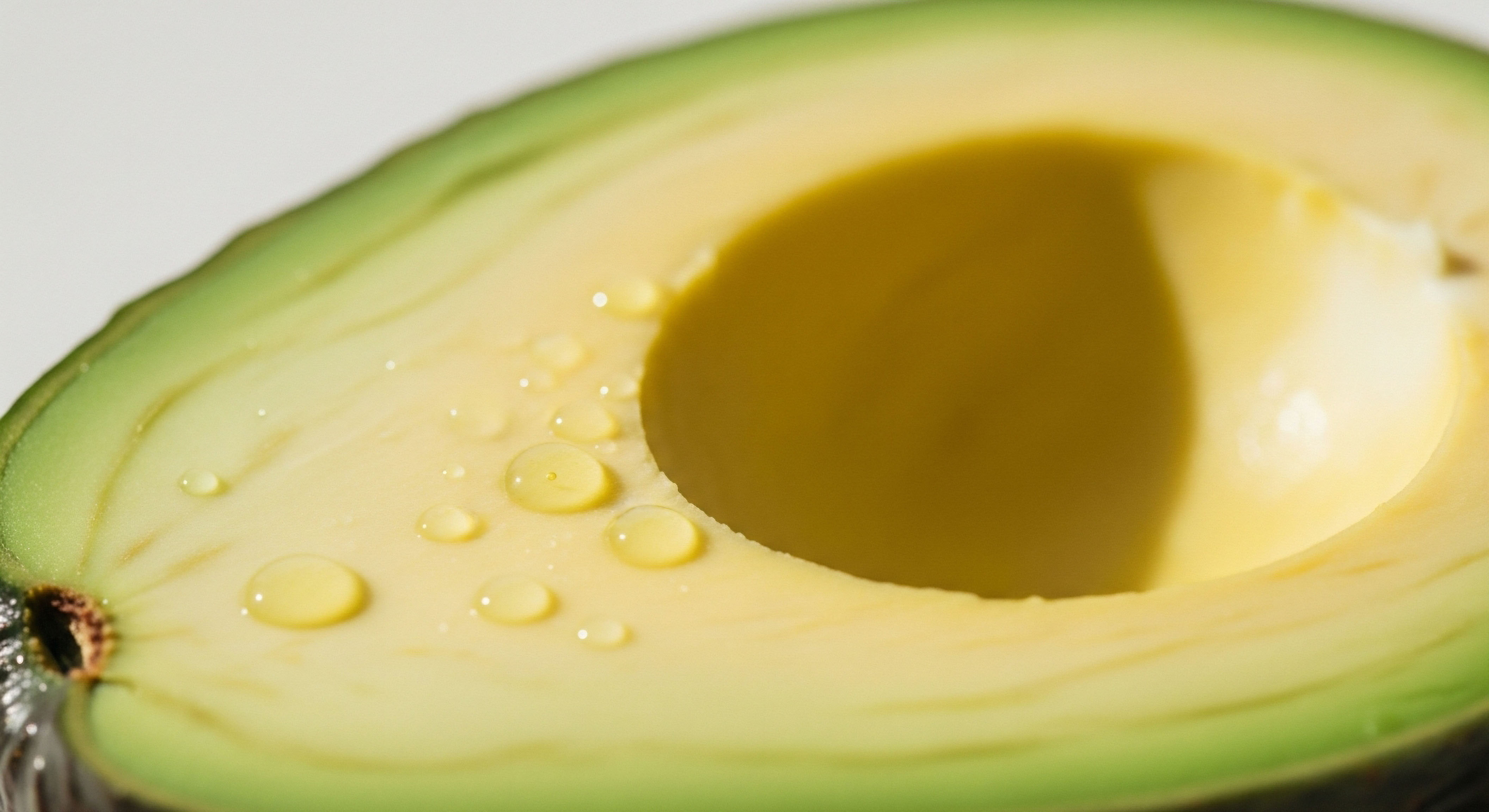
The type of fatty acid tails in these phospholipids ∞ whether they are straight and rigid (saturated) or bent and flexible (unsaturated) ∞ determines the membrane’s physical state. A membrane rich in saturated fats tends to be more viscous and tightly packed. A membrane incorporating more unsaturated fats, like those found in olive oil and avocados, is more fluid and flexible. This property, known as membrane fluidity, is paramount for receptor function.

Hormone Receptors as Mobile Messengers
Hormone receptors are not fixed in one position. They must be able to move laterally through the fluid membrane to find their hormonal partners, cluster together to amplify signals, and initiate cascades of communication within the cell. When the membrane becomes too rigid due to a high intake of certain types of fats, the movement of these receptors is impeded.
Their ability to conform their shape to bind effectively with a hormone is reduced. This physical hindrance can lead to a state of diminished hormonal sensitivity, where even if hormone levels are adequate, the cells cannot properly receive their instructions. Your body might be producing the right chemical messengers, but the receiving stations are effectively offline, leading to the very symptoms that signal a system in distress.


Intermediate
Understanding that dietary fats build your cellular architecture is the first step. The next level of comprehension involves recognizing how specific classes of lipids directly modulate the biophysical properties of the cell membrane and, consequently, the functional efficacy of hormone receptors.
The distinction between saturated, monounsaturated, and polyunsaturated fatty acids extends beyond simple nutritional labels; it represents a set of instructions for building cellular machinery that is either rigid and slow or fluid and responsive. This has direct implications for anyone on a hormonal optimization protocol, as the sensitivity of the target receptors is as important as the dose of the hormone itself.

How Does Lipid Composition Alter Receptor Mechanics?
The functionality of a hormone receptor is a physical process. The receptor, a complex protein, must be able to change its three-dimensional shape ∞ a process called conformational change ∞ when a hormone binds to it. This change is what activates the signaling cascade inside the cell.
The fluidity of the surrounding lipid membrane is a key permissive factor for this mechanical action. A diet high in saturated fatty acids can lead to their incorporation into the cell membrane, increasing its viscosity and restricting the receptor’s ability to move and change shape. Conversely, a diet rich in omega-3 polyunsaturated fatty acids (PUFAs), like EPA and DHA, increases membrane fluidity, allowing for more dynamic receptor movement and more efficient signal transduction.
Optimizing the lipid composition of cell membranes can enhance the effectiveness of hormonal therapies by improving receptor sensitivity.

The Role of Lipid Rafts in Signal Amplification
Within the general fluidity of the cell membrane exist specialized, highly organized microdomains known as lipid rafts. These platforms are enriched in cholesterol and specific types of lipids like sphingolipids. They function as signaling hubs, concentrating hormone receptors and their associated downstream signaling molecules in one location.
This proximity dramatically increases the speed and efficiency of the hormonal signal. The formation and stability of these crucial rafts are directly dependent on the availability of cholesterol and the balance of fatty acids in the membrane. An improper lipid balance can disrupt these platforms, scattering the signaling components and weakening the hormonal response. This explains why protocols focused on hormonal health must also address the foundational importance of dietary lipid quality.

Practical Implications for Hormonal Health Protocols
For an individual undergoing Testosterone Replacement Therapy (TRT) or utilizing peptide therapies, the cellular response is the ultimate determinant of success. The prescribed molecule is only one half of the equation; the receptivity of the target tissue is the other.
A diet that creates rigid, unresponsive cell membranes can effectively blunt the impact of the therapy, requiring higher dosages and increasing the potential for side effects. A structured nutritional approach that emphasizes specific types of lipids can prepare the cellular environment to receive these signals optimally.
The following table outlines the functional impact of different dietary lipid families on the cellular environment relevant to hormone receptor function.
| Lipid Family | Primary Dietary Sources | Effect on Membrane Fluidity | Impact on Receptor Function |
|---|---|---|---|
| Saturated Fatty Acids (SFAs) | Red meat, butter, coconut oil | Decreases fluidity (more rigid) | Can impede receptor movement and conformational changes. |
| Monounsaturated Fatty Acids (MUFAs) | Olive oil, avocados, nuts | Increases fluidity (more flexible) | Promotes efficient receptor mobility and signaling. |
| Omega-6 Polyunsaturated Fatty Acids (PUFAs) | Vegetable oils, seeds | Increases fluidity; can be pro-inflammatory in excess | Supports receptor function but requires balance with Omega-3s. |
| Omega-3 Polyunsaturated Fatty Acids (PUFAs) | Fatty fish, flaxseed, walnuts | Significantly increases fluidity; anti-inflammatory | Enhances receptor sensitivity and signaling efficiency. |
| Cholesterol | Eggs, meat, dairy | Acts as a fluidity buffer | Essential for the formation of lipid raft signaling platforms. |
A nutritional strategy for someone on a hormonal optimization protocol should therefore be designed to create a favorable membrane lipid profile. This involves a conscious effort to balance the intake of these lipid families.
- Prioritize Omega-3s ∞ Conscious inclusion of sources like wild-caught salmon, sardines, and high-quality fish oil supplements provides the foundational lipids for highly fluid and responsive membranes.
- Moderate Omega-6s ∞ While necessary, the typical modern diet often contains an excess of omega-6 fatty acids from processed seed oils. Shifting towards whole food sources helps maintain a healthy inflammatory balance.
- Utilize MUFAs ∞ Extra virgin olive oil and avocados should be staple fats, contributing to membrane fluidity and providing antioxidant benefits.
- Manage SFAs ∞ Saturated fats are necessary for hormone production and membrane structure, but their intake should be balanced with unsaturated fats to prevent excessive membrane rigidity.


Academic
The dialogue between dietary lipids and the endocrine system transcends general effects on membrane fluidity, extending into the precise molecular choreography of signal transduction and gene expression. The plasma membrane is a complex, heterogenous environment where the lateral organization of lipids into distinct domains dictates the spatiotemporal regulation of hormone receptor signaling.
A sophisticated understanding requires an appreciation of the membrane as an active participant in hormonal communication, with lipid molecules themselves acting as allosteric regulators and precursors to potent signaling molecules that influence the genomic and non-genomic actions of hormones.

Lipid Rafts as Steroid Receptor Signaling Hubs
Steroid hormones, such as testosterone and estrogen, traditionally are understood to act on nuclear receptors, directly modulating gene transcription over hours or days. There is a growing body of evidence for rapid, non-genomic actions that are initiated by a subpopulation of these receptors located at the plasma membrane.
These membrane-associated steroid receptors are preferentially localized within lipid rafts. The unique biochemical environment of the raft ∞ a liquid-ordered phase rich in cholesterol and sphingomyelin ∞ is indispensable for the functional integrity of these signaling pathways. Within this domain, the receptor is brought into close proximity with G-proteins, kinases like Src, and effector enzymes such as adenylyl cyclase, allowing for the rapid initiation of intracellular signaling cascades upon hormone binding.
The composition of dietary fatty acids directly influences the stability and composition of these rafts. Incorporation of docosahexaenoic acid (DHA), an omega-3 PUFA, into membrane phospholipids can alter the phase behavior of the membrane, potentially displacing receptors from rafts or modifying the raft’s protein and lipid composition.
This perturbation can selectively attenuate rapid, non-genomic signaling while leaving the slower, genomic pathways intact. This phenomenon illustrates a mechanism by which dietary choices can fine-tune the character of a hormonal response, shifting the balance between rapid cellular adjustments and long-term genomic programming.
The specific fatty acid profile of the cell membrane can selectively filter and modify the signals initiated by hormone-receptor binding events.

What Is the Role of Phosphoinositides in Hormone Signaling?
Beyond structural roles, specific membrane lipids function as critical second messengers. Phosphoinositides, a minor class of phospholipids, are central to this process. The enzyme Phospholipase C (PLC), which is often activated by G-protein coupled receptors linked to hormone signaling, cleaves phosphatidylinositol 4,5-bisphosphate (PIP2) into two potent signaling molecules ∞ inositol trisphosphate (IP3) and diacylglycerol (DAG).
DAG remains in the membrane and activates Protein Kinase C (PKC), while IP3 diffuses into the cytoplasm to trigger calcium release. The fatty acid chains of the parent PIP2 molecule, determined by dietary intake, affect the potency and downstream metabolism of the resulting DAG.
A DAG molecule containing an omega-3 fatty acid, for instance, may have different activation kinetics for PKC isoforms compared to one containing a saturated or omega-6 fatty acid. This provides another layer of regulatory control where diet composition directly informs the nature of intracellular signaling.

Lipids as Precursors to Endocrine Modulators
The influence of dietary lipids extends to their role as precursors for eicosanoids ∞ a family of signaling molecules including prostaglandins, thromboxanes, and leukotrienes. These molecules are generated from 20-carbon PUFAs, primarily arachidonic acid (an omega-6) and eicosapentaenoic acid (an omega-3). The balance of dietary omega-6 to omega-3 intake determines the predominant class of eicosanoids produced.
Those derived from arachidonic acid are generally pro-inflammatory, while those from EPA are anti-inflammatory. This is critically relevant to endocrinology, as the local inflammatory state of a tissue profoundly impacts its sensitivity to hormones. Chronic low-grade inflammation, driven by an excess of omega-6-derived eicosanoids, can induce a state of hormone resistance in tissues like adipose and muscle, contributing to metabolic dysfunction.
The following table details the distinct signaling pathways originating from key dietary polyunsaturated fatty acids.
| Precursor Fatty Acid | Primary Eicosanoid Series | General Biological Action | Relevance to Hormonal Health |
|---|---|---|---|
| Arachidonic Acid (Omega-6) | Prostaglandin E2, Leukotriene B4 | Pro-inflammatory, Vasoconstrictive | Can promote hormone resistance and disrupt tissue sensitivity. |
| Eicosapentaenoic Acid (Omega-3) | Prostaglandin E3, Leukotriene B5 | Anti-inflammatory, Vasodilatory | Reduces cellular stress and improves hormonal signaling environment. |
Therefore, the dietary lipid profile exerts a multi-layered influence on hormone receptor function. It sets the physical stage through membrane fluidity, organizes the actors through lipid raft integrity, directs the downstream plot through second messenger generation, and modulates the entire production through the inflammatory milieu. A clinical approach to hormonal optimization that neglects the foundational role of lipid biochemistry is addressing only a fraction of the system.

References
- Berlin, E. S. J. Bhathena, and M. T. Clandinin. “Dietary fat and hormonal effects on erythrocyte membrane fluidity and lipid composition in adult women.” Journal of the American College of Nutrition, vol. 8, no. 6, 1989, pp. 547-55.
- Corsetto, P. A. A. Montorfano, A. Kopecka, et al. “Changes in the plasma membrane in metabolic disease ∞ impact of the membrane environment on G protein-coupled receptor structure and function.” The FASEB Journal, vol. 31, no. 8, 2017, pp. 3258-71.
- Levental, K. R. M. Malmberg, and I. Levental. “Dietary lipids in membrane organization and function.” Nature Reviews Molecular Cell Biology, vol. 21, 2020, pp. 361-76.
- Hulbert, A. J. and P. L. Else. “Membranes as metabolic pacemakers.” Journal of Experimental Biology, vol. 199, Pt 11, 1996, pp. 2585-9.
- Stillwell, W. and S. R. Wassall. “Docosahexaenoic acid ∞ membrane properties of a unique fatty acid.” Chemistry and Physics of Lipids, vol. 126, no. 1, 2003, pp. 1-27.

Reflection
The information presented here offers a map of the intricate connections between your plate and your physiology. It translates the abstract language of biochemistry into a tangible understanding of how your daily choices construct the very foundation of your hormonal health. This knowledge is the starting point.
The path toward optimal function is one of personal inquiry, observing how your own body responds to these inputs. Consider the architecture of your cells not as a fixed state, but as a dynamic system you consciously rebuild with every meal. Your personal journey to vitality is a process of learning your body’s unique language and providing it with the precise materials it needs to communicate with clarity and strength.