
Fundamentals
You may have arrived here carrying a constellation of symptoms that feel both personal and frustratingly vague. Perhaps it is a persistent layer of body fat that resists your best efforts, a subtle but draining sense of fatigue, or shifts in mood and libido that seem to have no clear origin.
Your lived experience of these changes is the most important data point we have. It is the starting point of a logical inquiry into the body’s intricate internal communication network, the endocrine system. The connection between what you eat, specifically the fats in your diet, and how you feel is profoundly intimate. This connection is arbitrated by a multitude of biological processes, one of the most significant being the activity of an enzyme called aromatase.
Aromatase is a key player in the body’s hormonal architecture. Its primary function is to convert a class of hormones called androgens into another class called estrogens. Think of testosterone as a primary androgen and estradiol as a primary estrogen. Aromatase is the biochemical mechanism that facilitates this transformation.
This process is a fundamental aspect of human physiology, occurring in both men and women in various tissues, including the gonads, brain, bone, and, most critically for this discussion, adipose tissue, which is body fat. The amount of aromatase activity in your body directly influences your hormonal balance, particularly the ratio of testosterone to estrogen.
When this system is functioning optimally, it maintains a delicate equilibrium essential for everything from bone density and cardiovascular health to cognitive function and body composition.

The Central Role of Adipose Tissue
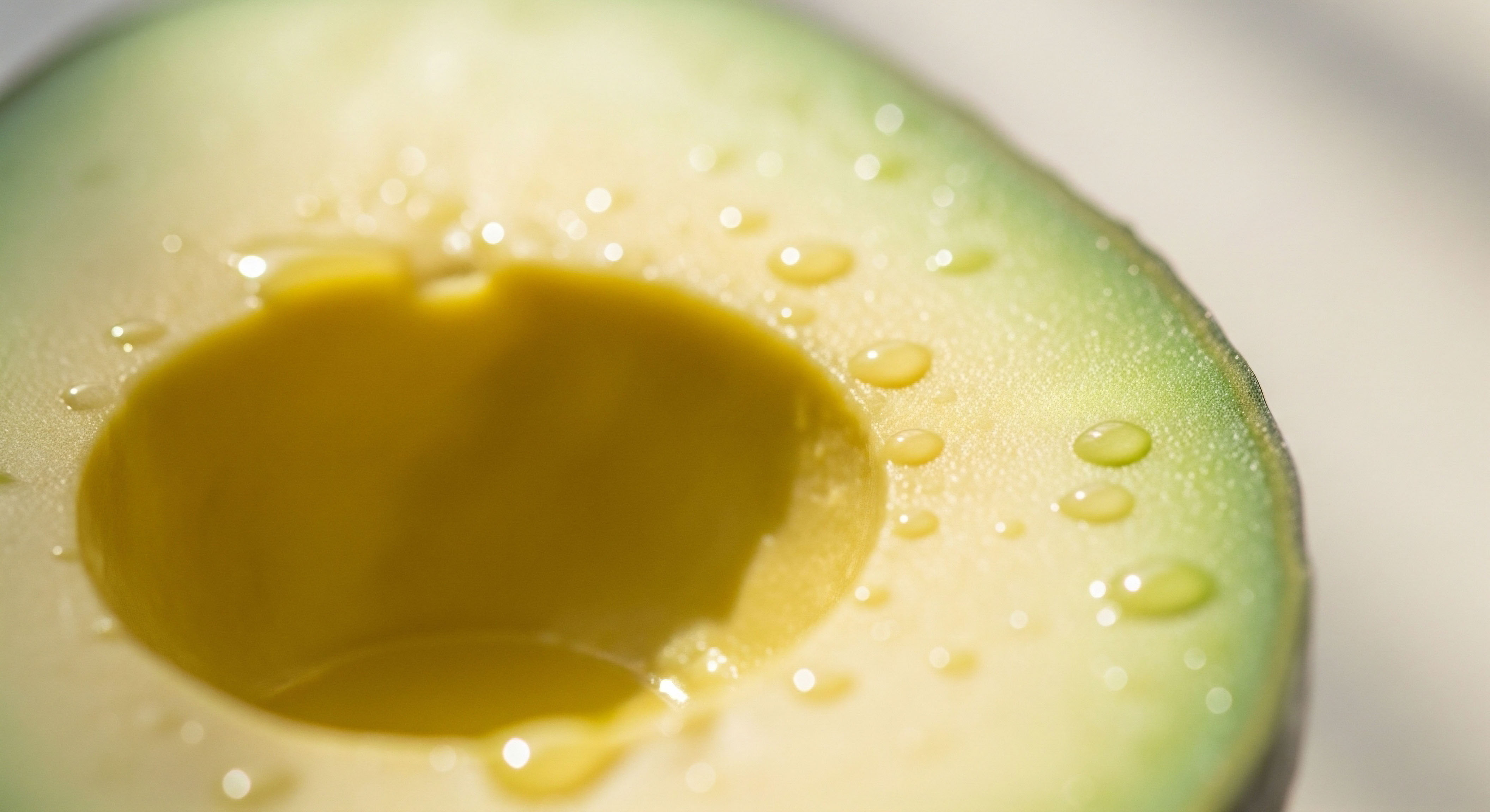
Understanding where dietary fats enter this picture requires us to look closely at adipose tissue. Your body fat is an active endocrine organ. It produces and responds to a wide array of signaling molecules.
When you consume more calories than your body expends, particularly from energy-dense dietary fats and refined carbohydrates, this excess energy is stored in adipocytes, or fat cells, causing them to expand in size and number. An increase in total body fat, especially visceral fat that surrounds the internal organs, creates a larger reservoir for aromatase production.
Consequently, a higher body fat percentage often correlates directly with increased systemic aromatase activity. This means more of your body’s androgens are being converted into estrogens. In men, this can lead to a reduction in free testosterone and an elevation in estrogen, contributing to symptoms like reduced muscle mass, increased fat deposition in the chest and hips, and diminished vitality.
In postmenopausal women, whose ovaries have ceased being the primary source of estrogen, adipose tissue becomes the main site of estrogen production through this very process. The type and quantity of dietary fats you consume directly influence the amount of adipose tissue you carry, thereby setting the stage for your baseline level of aromatase activity.
The body’s fat tissue functions as an active endocrine organ, and its expansion through diet directly increases the production of the aromatase enzyme.
This biochemical reality places dietary strategy at the center of hormonal health management. The choices you make at every meal have a cascading effect on your body’s hormonal milieu. A diet high in processed foods and certain types of fats can promote the expansion of adipose tissue, which in turn elevates aromatase activity.
This creates a self-perpetuating cycle ∞ higher estrogen levels can promote further fat storage, which then generates even more aromatase. This is a physiological loop that can feel like a frustrating battle against your own body. However, viewing it through this mechanistic lens transforms the narrative. It provides a clear biological target.
By understanding that dietary fats influence aromatase primarily through their impact on body composition and inflammation, you gain a powerful lever for intervention. The goal becomes one of creating a metabolic environment that discourages the storage of excess fat and quiets the inflammatory signals that drive aromatase expression. This perspective shifts the focus from a generic concept of “weight loss” to a more precise objective of recalibrating your body’s hormonal machinery through targeted nutritional protocols.

Inflammation the Bridge between Diet and Aromatase
What is the specific trigger that tells fat cells to produce more aromatase? The answer is inflammation. Diet-induced obesity is closely linked to a state of chronic, low-grade inflammation. Certain dietary fats, particularly an excess of saturated fats and omega-6 polyunsaturated fats found in many processed foods, can promote an inflammatory response within adipose tissue itself.
This is not the acute inflammation you experience with an injury; it is a persistent, systemic state of alert. Immune cells called macrophages are recruited to expanding adipose tissue, where they release a cascade of pro-inflammatory signaling molecules, known as cytokines. Key among these are tumor necrosis factor-alpha (TNF-α) and interleukin-1β (IL-1β).
These cytokines act as potent stimulators of the gene that produces aromatase. Essentially, an inflammatory environment created by certain dietary patterns sends a loud and clear signal to your fat cells, instructing them to ramp up the conversion of testosterone to estrogen. This provides a direct molecular link between your plate and your hormonal profile, a pathway that we can strategically influence.


Intermediate
Moving beyond the foundational understanding that excess body fat increases aromatase, we can now dissect the specific roles that different types of dietary fats play in this complex process. The biochemical structure of a fatty acid determines how it is metabolized and what signals it sends within the body.
This granular perspective is essential for developing a sophisticated dietary strategy aimed at managing aromatase activity. The primary categories of dietary fats ∞ saturated, monounsaturated, and polyunsaturated ∞ each have distinct relationships with the inflammatory pathways that govern aromatase expression. Recognizing these differences allows for a more precise and effective nutritional intervention, one that supports hormonal optimization protocols like Testosterone Replacement Therapy (TRT) by addressing the underlying biochemical environment.

A Comparative Look at Dietary Fat Families
The impact of dietary fats on aromatase is mediated less by the fats themselves and more by the inflammatory response they can provoke. Let us examine the major types of fatty acids and their characteristic effects on the body’s inflammatory tone, which is a key regulator of aromatase.
- Saturated Fatty Acids (SFAs) ∞ Found predominantly in animal products like red meat and full-fat dairy, as well as in some tropical oils like coconut and palm oil, SFAs have been directly implicated in stimulating pro-inflammatory responses. Research shows that an excess of certain SFAs, such as palmitic acid, can activate immune receptors on macrophages, leading to the production of TNF-α and other cytokines that directly upregulate the aromatase gene in surrounding fat cells. This makes a diet rich in processed meats and high-fat dairy a potential driver of increased estrogen conversion.
-
Polyunsaturated Fatty Acids (PUFAs) ∞ This family is divided into two main classes, omega-6 and omega-3, which have opposing effects on inflammation.
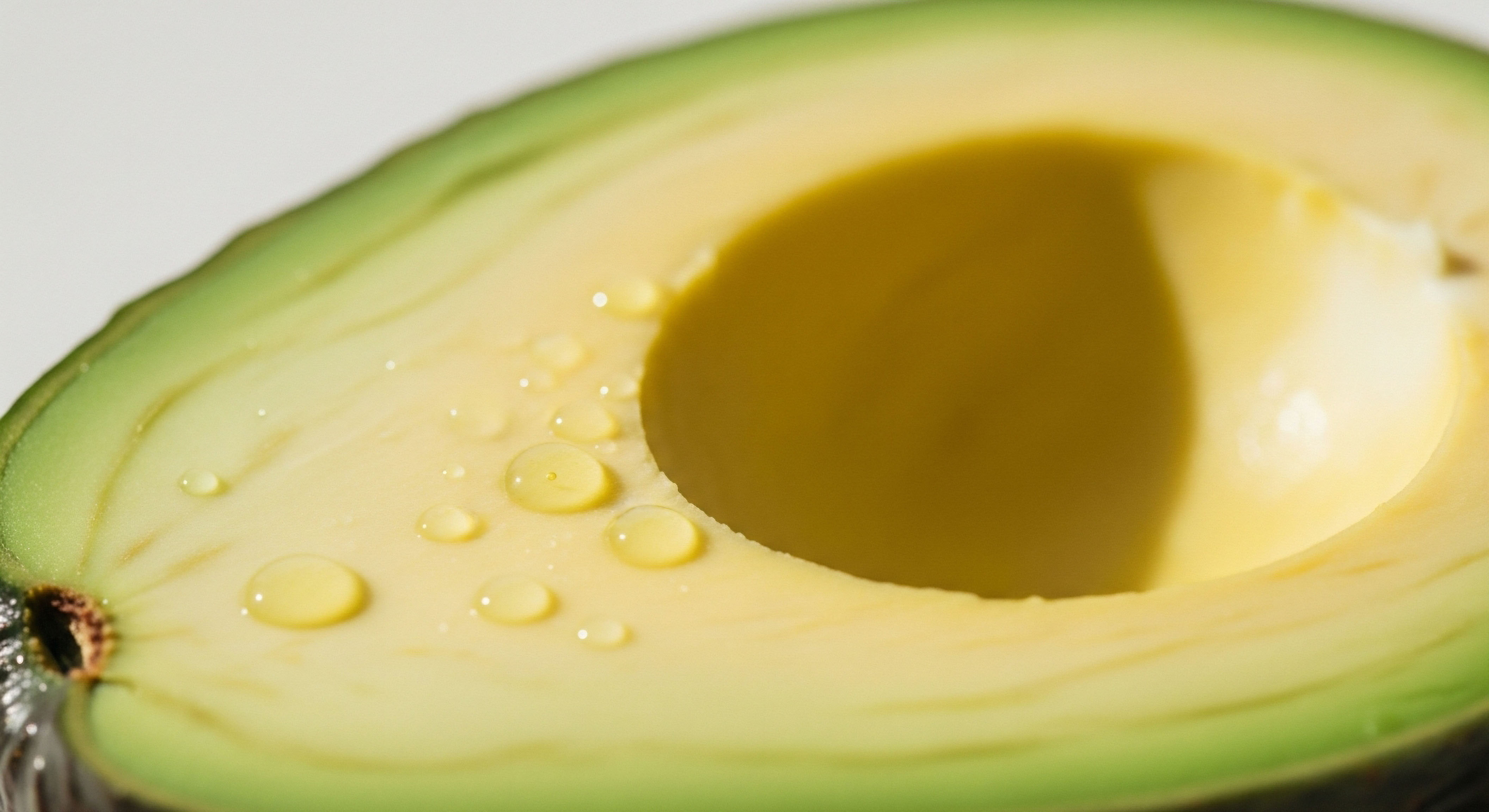
Omega-6 PUFAs, abundant in many industrial seed oils (corn, soybean, safflower), are precursors to arachidonic acid, a molecule that can be converted into pro-inflammatory eicosanoids. A high intake of omega-6s relative to omega-3s can create a net inflammatory state, thereby promoting aromatase activity. Omega-3 PUFAs, found in fatty fish (salmon, arctic char, herring), flaxseed, and walnuts, are precursors to anti-inflammatory signaling molecules. They actively compete with omega-6s and help resolve inflammation. A diet with a balanced or higher ratio of omega-3 to omega-6 fats can help suppress the inflammatory signals that drive aromatase expression. - Monounsaturated Fatty Acids (MUFAs) ∞ The hallmark of the Mediterranean diet, MUFAs are found in olive oil, avocados, and nuts. Generally considered to be neutral or mildly anti-inflammatory, MUFAs can support a healthy inflammatory response when they replace excess SFAs and omega-6s in the diet. Their role is one of supportive balance, helping to create an internal environment less conducive to aromatase upregulation.

How Does Diet Complement Clinical Protocols?
For individuals undergoing hormonal optimization, such as men on TRT or women using hormone therapies during perimenopause, dietary management of aromatase is a critical component of a successful protocol. Clinical interventions like the use of anastrozole, an aromatase inhibitor, are designed to block the conversion of testosterone to estrogen directly.
While effective, a supportive dietary strategy can enhance the protocol’s efficacy and potentially reduce the required dosage of medication. By minimizing the inflammatory triggers that stimulate aromatase production in the first place, a targeted diet works synergistically with the medication.
For example, a man on TRT who also adopts a diet lower in processed foods and saturated fats, while increasing his intake of omega-3s, is tackling the issue from two angles. The anastrozole provides a direct biochemical block, while the diet reduces the fundamental stimulus for producing the enzyme.
This integrated approach leads to a more stable and predictable hormonal environment, optimizing the benefits of the therapy while minimizing potential side effects associated with estrogen excess, such as water retention or gynecomastia.
A diet rich in omega-3 fatty acids and low in processed saturated fats can significantly reduce the inflammatory signals that command fat cells to produce more aromatase.
This principle is equally relevant for protocols involving peptides that stimulate growth hormone release, like Sermorelin or Ipamorelin. These therapies are often aimed at improving body composition by reducing fat mass and increasing lean muscle. Since excess adipose tissue is the primary site of inflammation-driven aromatase, any protocol that successfully reduces body fat will inherently help in managing aromatase.
A diet that supports this goal ∞ one rich in lean protein, fiber, and anti-inflammatory fats ∞ accelerates progress and reinforces the therapy’s benefits. The body’s systems are deeply interconnected; a therapeutic intervention in one area is always amplified by supportive changes in another.
The table below provides a simplified comparison of how different dietary fat sources can influence the inflammatory environment and, by extension, aromatase activity. This is a practical guide for making conscious food choices that align with the goal of hormonal balance.
| Dietary Fat Type | Common Food Sources | Primary Effect on Inflammation | Potential Impact on Aromatase Activity |
|---|---|---|---|
| Saturated Fat (SFA) | Processed meats, full-fat dairy, palm oil | Can be pro-inflammatory in excess | Stimulatory (via inflammatory pathways) |
| Omega-6 PUFA | Corn oil, soybean oil, processed snacks | Pro-inflammatory when ratio to omega-3 is high | Stimulatory (via inflammatory pathways) |
| Omega-3 PUFA | Fatty fish, flaxseed, walnuts, chia seeds | Anti-inflammatory | Inhibitory (by reducing inflammation) |
| Monounsaturated Fat (MUFA) | Olive oil, avocados, almonds, macadamia nuts | Neutral to mildly anti-inflammatory | Supportive of a balanced state |

Practical Dietary Adjustments for Aromatase Management
Translating this biochemical understanding into daily practice involves a strategic selection of foods that either inhibit aromatase directly or quell the inflammation that drives it. Certain foods contain potent phytochemicals that have been shown to have a natural aromatase-inhibiting effect. Incorporating these into a diet that is already structured to be anti-inflammatory can provide a powerful, multi-pronged approach.
- Prioritize Omega-3 Sources ∞ Actively include sources of omega-3 fatty acids in your diet. This could mean consuming fatty fish like salmon or arctic char two to three times per week, or adding ground flaxseed and walnuts to your meals. This directly counters the pro-inflammatory cascade from omega-6 fats.
- Increase Cruciferous Vegetable Intake ∞ Vegetables like broccoli, cauliflower, cabbage, and Brussels sprouts contain compounds such as indole-3-carbinol and diindolylmethane (DIM), which help support healthy estrogen metabolism. While not direct aromatase inhibitors, they assist the body in processing estrogen efficiently.
- Utilize Natural Aromatase Inhibitors ∞ Certain foods have been found to contain compounds that can modestly inhibit the aromatase enzyme. These include quercetin (found in onions and apples), grape seed extract, and celery. White button mushrooms have also been studied for their aromatase-inhibiting properties. Incorporating a wide variety of these plant foods can contribute to a favorable hormonal balance.
- Reduce Pro-Inflammatory Inputs ∞ A critical step is to consciously reduce the intake of foods that promote inflammation. This includes limiting processed foods, sugary beverages, and industrial seed oils high in omega-6 fatty acids. Reducing red meat and high-fat dairy, sources of saturated fat, can also help lower the inflammatory burden on the body.
By implementing these strategies, you are actively curating your body’s biochemical environment. You are providing the raw materials for anti-inflammatory pathways while simultaneously removing the inputs that drive inflammation and aromatase production. This is a proactive stance on health, one that recognizes the profound ability of nutrition to shape our physiology from the cellular level upwards.


Academic
An academic exploration of the relationship between dietary fats and aromatase activity requires a descent into the molecular mechanisms that govern gene expression within the adipose tissue microenvironment. The conversion of androgens to estrogens is catalyzed by the aromatase enzyme, which is the protein product of the CYP19A1 gene.
The regulation of this gene is extraordinarily complex, involving multiple tissue-specific promoters that are controlled by a sophisticated network of signaling pathways. In adipose tissue, the primary driver of CYP19A1 expression is a promoter known as I.4, which is exquisitely sensitive to inflammatory signals. The type and quantity of dietary fatty acids consumed are primary determinants of the inflammatory tone within adipose tissue, thereby creating a direct link between lipid metabolism and sex steroid biosynthesis.

The NF-κB Pathway the Master Regulator of Inflammatory Gene Expression
At the heart of obesity-induced inflammation and subsequent aromatase upregulation lies the transcription factor Nuclear Factor-kappa B (NF-κB). In a quiescent state, NF-κB is held inactive in the cytoplasm of cells, bound to an inhibitory protein called IκB.
A variety of stimuli, including exposure to certain saturated fatty acids like palmitate, can trigger a signaling cascade that leads to the phosphorylation and subsequent degradation of IκB. This frees NF-κB to translocate into the nucleus, where it binds to specific DNA sequences in the promoter regions of target genes. These target genes include a host of pro-inflammatory cytokines, chief among them being TNF-α and IL-1β.
Saturated fatty acids can initiate this cascade by engaging with Toll-like receptor 4 (TLR4), a pattern recognition receptor on the surface of macrophages within adipose tissue. The binding of SFAs to TLR4 initiates an intracellular signaling cascade that activates IκB kinase (IKK). IKK then phosphorylates IκB, marking it for destruction and liberating NF-κB.
This mechanism positions NF-κB as the central node translating a dietary signal (excess SFA) into a transcriptional response (cytokine production). The cytokines produced and secreted by these activated macrophages then act in a paracrine fashion on the surrounding preadipocytes and adipocytes, which are the primary sites of aromatase expression in adipose tissue.

Cytokine-Mediated Upregulation of the CYP19A1 Gene
The cytokines TNF-α and IL-1β, once secreted by adipose tissue macrophages, bind to their respective receptors on the surface of adipocytes. This binding event triggers another set of intracellular signaling pathways that converge on the I.4 promoter of the CYP19A1 gene. One of the key pathways involves the cyclooxygenase-2 (COX-2) enzyme.
TNF-α and IL-1β stimulate the expression and activity of COX-2, which in turn leads to the synthesis of prostaglandin E2 (PGE2). PGE2 is a potent signaling molecule that, upon binding to its receptor on the adipocyte, increases intracellular levels of cyclic AMP (cAMP).
This rise in cAMP activates Protein Kinase A (PKA), which then phosphorylates and activates transcription factors like CREB (cAMP response element-binding protein). These activated transcription factors can then bind to the I.4 promoter region of the CYP19A1 gene, dramatically increasing its rate of transcription. The result is a marked increase in the synthesis of aromatase mRNA and, subsequently, the aromatase enzyme itself. This entire sequence represents a detailed molecular pathway connecting a high-fat diet to elevated estrogen production.
The molecular conversation begins with saturated fats activating immune cells, which then release signals that command nearby fat cells to transcribe the aromatase gene.
The table below outlines this intricate signaling cascade, providing a step-by-step view of the molecular events that link dietary fat intake to aromatase expression in adipose tissue. This is the biological blueprint of how diet directly modulates hormonal balance at the cellular level.
| Step | Cell Type Involved | Molecular Event | Key Molecules | Outcome |
|---|---|---|---|---|
| 1. Initial Stimulus | Macrophage | Excess saturated fatty acids bind to cell surface receptors. | Palmitic Acid, Toll-like Receptor 4 (TLR4) | Activation of intracellular signaling. |
| 2. NF-κB Activation | Macrophage | The IKK complex is activated, leading to the degradation of the IκB inhibitor. | IKK, IκB, NF-κB | NF-κB translocates to the nucleus. |
| 3. Cytokine Transcription | Macrophage | NF-κB binds to the promoter regions of inflammatory genes. | TNF-α gene, IL-1β gene | Increased production and secretion of pro-inflammatory cytokines. |
| 4. Paracrine Signaling | Adipocyte | Cytokines bind to receptors on adjacent adipocytes. | TNF-α, IL-1β, their respective receptors | Activation of downstream signaling within the adipocyte. |
| 5. PGE2 Synthesis | Adipocyte | Cytokine signaling upregulates the COX-2 enzyme, leading to prostaglandin synthesis. | COX-2, Prostaglandin E2 (PGE2) | PGE2 is released and acts on the adipocyte. |
| 6. Aromatase Gene Transcription | Adipocyte | PGE2 signaling increases cAMP levels, activating transcription factors that bind to the aromatase gene promoter. | cAMP, PKA, CREB, CYP19A1 Promoter I.4 | Markedly increased synthesis of aromatase enzyme. |

What Is the Role of Lipid Species in This Process?
A more sophisticated analysis moves beyond broad categories of fats to consider the specific effects of individual fatty acid species. The biological activity of a fatty acid is determined by its chain length and degree of saturation. For instance, while the saturated fatty acid palmitate (C16:0) is strongly pro-inflammatory, stearate (C18:0) is considered less so.
Within the polyunsaturated family, the balance between omega-6 arachidonic acid (AA) and omega-3 eicosapentaenoic acid (EPA) is paramount. AA is the substrate for the synthesis of pro-inflammatory eicosanoids like prostaglandin E2 and leukotriene B4. Conversely, EPA competes with AA for the same enzymes (COX and LOX) and produces less inflammatory eicosanoids and specialized pro-resolving mediators (SPMs) like resolvins and protectins.
Therefore, a high dietary intake of omega-6 fats relative to omega-3s creates a biochemical environment that is primed for inflammation. An increase in the EPA/AA ratio, achieved through higher consumption of omega-3s, can directly reduce the substrate pool for PGE2 synthesis, thereby dampening the final and most potent stimulus for aromatase expression in adipocytes.

Tissue Specificity and the Prostate Gland
While adipose tissue is a major site of diet-influenced aromatase activity, other tissues are also affected. Research has demonstrated that a high-fat diet can induce inflammation and increase aromatase expression within the prostate gland itself. This process appears to involve a distinct but related molecular pathway.
Studies suggest that a high-fat diet leads to increased expression of estrogen receptor alpha (ER-α) and the signal transducer and activator of transcription 3 (Stat3) protein in the prostate. The activated, phosphorylated form of Stat3 (p-Stat3) and ER-α can form a complex that binds directly to the aromatase promoter, driving its expression.
This creates a local feed-forward loop within the prostate, where diet-induced inflammation increases local estrogen production, which can then contribute to proliferative signaling pathways implicated in prostate health issues. This highlights the systemic nature of dietary influences, showing how nutritional inputs can modulate hormone signaling in a tissue-specific manner, with significant clinical implications for men’s health.

References
- Subbaramaiah, K. et al. “Obesity is associated with inflammation and elevated aromatase expression in the mouse mammary gland.” Cancer Prevention Research, vol. 4, no. 11, 2011, pp. 1834-44.
- “Improving Low Testosterone Naturally.” Whole Health Library, U.S. Department of Veterans Affairs, 2020.
- Bhaskaran, Natarajan, et al. “Abstract 5452 ∞ High-fat diet induces inflammation by increasing estrogen levels through Stat3, estrogen receptor alpha and aromatase in the mouse prostate.” Cancer Research, vol. 73, no. 8 Supplement, 2013, p. 5452.
- “Foods To Eat & Avoid During Aromatase Inhibitor Treatment.” Foodforbreastcancer.com, 20 Jul. 2025.
- DeLauer, Thomas. “Eat These Foods to Lower Estrogen, Lose Fat & Increase Testosterone (aromatase inhibitors).” YouTube, 10 Nov. 2023.

Reflection
You have now traveled from the lived experience of symptoms to the intricate molecular ballet occurring within your cells. This knowledge provides a detailed map of the biological territory. It shows precisely how the fats on your plate are translated into the hormonal signals that shape your health, your body composition, and your sense of vitality.
The purpose of this detailed exploration is to move beyond generalized advice and toward a place of specific, actionable understanding. You can now see the logic behind prioritizing certain foods and minimizing others, viewing these choices not as restrictions, but as precise instructions you are giving to your own physiology.
This information forms a new foundation for the conversations you have with yourself and with your clinical team. It allows you to ask more targeted questions and to understand the ‘why’ behind the protocols you may undertake. Your personal health path is unique, shaped by your genetics, your history, and your goals.
The biological principles are universal, but their application is deeply personal. What does this understanding mean for your next meal? For your next workout? For your next consultation? The journey forward is one of continued learning and self-aware application, using this knowledge as a compass to navigate your own path toward sustained wellness.

Glossary

aromatase

aromatase activity

hormonal balance

body composition

adipose tissue

dietary fats

estrogen production

inflammatory signals that drive aromatase expression

inflammation

within adipose tissue

saturated fats

cytokines

tnf-α
testosterone replacement therapy

aromatase expression

fatty acids

saturated fatty acids

inflammatory signals that drive aromatase

anastrozole
dietary fat

omega-3 fatty acids

estrogen metabolism

aromatase enzyme

cyp19a1 gene

cyp19a1