


Fundamentals
You may feel a profound sense of frustration, a feeling that your body is working against you. The persistent weight that clings to your midsection, the relentless cravings that sabotage your best intentions, and the pervasive fatigue that clouds your days are real, tangible experiences. These are not failures of discipline.
These sensations are the downstream consequences of a silent, microscopic process occurring within the most sophisticated control tower of your body ∞ the hypothalamus. This small, diamond-shaped structure, located deep within your brain, acts as the master regulator of your internal universe. It is the central command that dictates hunger, thirst, body temperature, sleep cycles, and the symphony of hormones that govern your metabolism and vitality.
The hypothalamus functions by listening. It constantly monitors a stream of incoming data from your body ∞ hormones like leptin from your fat cells that signal fullness, and insulin from your pancreas that reports on blood sugar. When this communication network is clear, the hypothalamus responds with precision, adjusting your appetite and energy expenditure to maintain a state of equilibrium.
Your dietary choices, specifically the types of fats you consume, act as a primary modulator of this communication system. Certain fats can introduce static and interference, while others can enhance the clarity of the signal. This is the biological reality behind why some foods leave you feeling satiated and energetic, while others seem to trigger a cascade of cravings and lethargy.

The Nature of Dietary Fats
Dietary fats are a diverse group of molecules, each with a unique chemical structure that determines its function within your body. They are essential for absorbing vitamins, producing hormones, and building healthy cells. For the purposes of understanding their impact on the hypothalamus, we can categorize them into broad groups based on their molecular architecture. This structure is what determines how they interact with the cellular machinery in your brain.
Saturated fatty acids, commonly found in animal products and some tropical oils, possess a straight, rigid structure. This physical characteristic allows them to pack together tightly, influencing cell membrane fluidity. When they arrive in the hypothalamus in high concentrations, they can interact with specialized sensor proteins on the surface of the brain’s resident immune cells, the microglia.
This interaction is interpreted as a danger signal, initiating a protective, yet ultimately damaging, inflammatory response. It is a case of mistaken identity, where a nutrient is perceived as a threat.
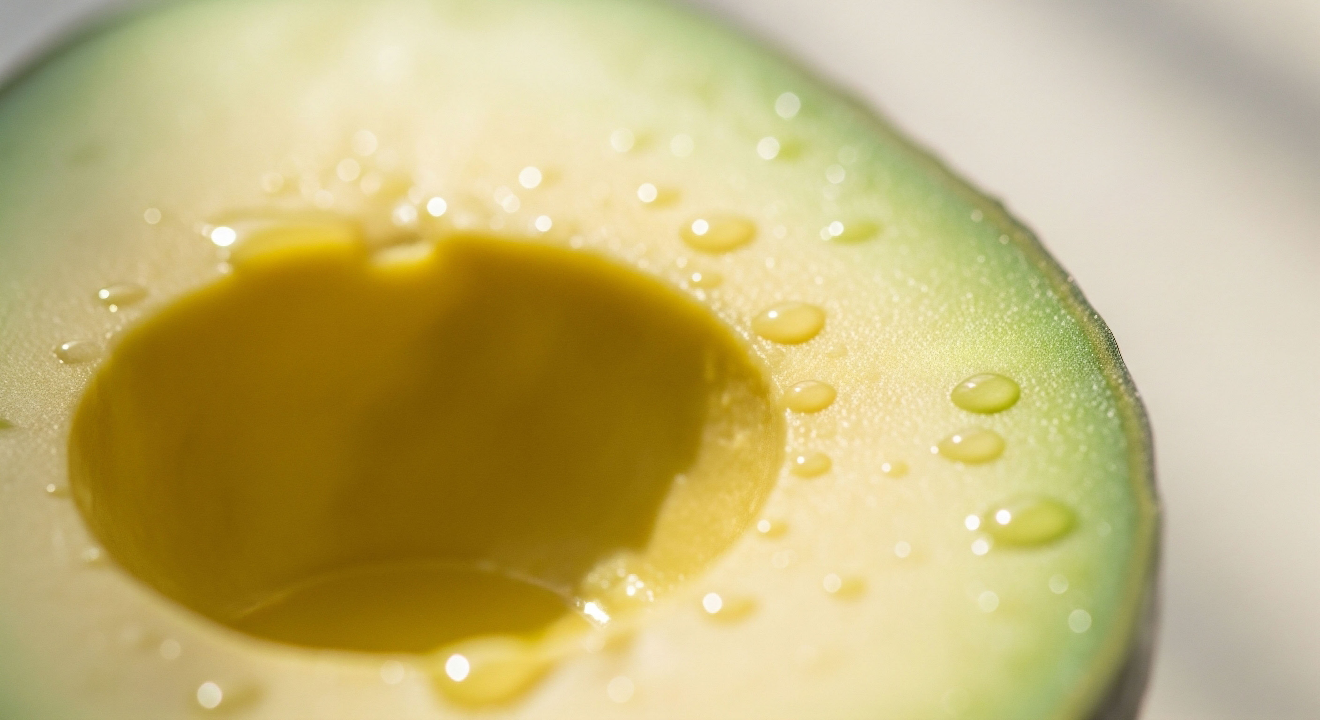
Conversely, unsaturated fatty acids, which include both monounsaturated (found in olive oil and avocados) and polyunsaturated fats (found in fatty fish, nuts, and seeds), have a different shape. Their molecular structure includes one or more double bonds, which create “kinks” or bends in the fatty acid chain.
This prevents them from packing together tightly, contributing to more fluid and flexible cell membranes. Within the hypothalamus, these fats interact with a different set of receptors, often triggering pathways that actively resolve inflammation and enhance neuronal function. They can be thought of as agents of communication clarity, helping to ensure the signals of satiety and energy balance are received and understood.
The types of fats consumed in your diet directly influence the inflammatory status of the brain’s primary metabolic control center.

Inflammation a Disrupted Dialogue
Hypothalamic inflammation is a state of chronic, low-grade activation of the brain’s immune system within this specific region. Think of it as a persistent, low-level hum of static that disrupts the clear communication between your body and your brain.
When inflammatory processes are active, the neurons in the hypothalamus become less sensitive to the hormonal signals they are meant to receive. This is the very definition of receptor insensitivity. The messages of “I am full” or “energy stores are sufficient,” carried by hormones like leptin and insulin, are sent but they are not properly received. The hypothalamus, hearing only silence or a muffled message, continues to send out its own powerful signals ∞ “seek food” and “conserve energy.”
This creates a vicious cycle. The brain, believing the body is in a state of starvation, drives behaviors that lead to increased consumption of energy-dense foods, which often contain the very saturated fats that initiated the inflammation in the first place. Simultaneously, it slows down metabolic rate to conserve resources.
The result is weight gain, particularly visceral fat, which in turn produces more inflammatory signals, further amplifying the static in the hypothalamus. Your personal experience of this is the frustrating battle against cravings and a sluggish metabolism. Understanding this biological loop is the first step toward breaking it, shifting the focus from a battle against your own body to a process of restoring clear communication within it.
The journey to reclaiming your vitality begins with recognizing that your symptoms are a logical response to a specific biological environment. By consciously selecting dietary fats that promote clarity and quiet the inflammatory hum, you are directly participating in a conversation with your own physiology. You are providing the raw materials needed to rebuild sensitive receptors, restore fluid cell membranes, and re-establish the precise, elegant feedback loops that govern your well-being.


Intermediate
Moving beyond the foundational understanding of hypothalamic inflammation, we can begin to examine the precise molecular mechanisms through which dietary fats exert their influence. The process is an elegant, if sometimes detrimental, example of how our bodies interpret chemical signals from our environment.
The hypothalamus does not simply react to the quantity of fat, but to its quality and chemical structure, initiating distinct intracellular signaling cascades in response to different fatty acid profiles. This cellular dialogue determines whether the hypothalamus maintains its sensitivity to metabolic hormones or descends into a state of chronic inflammation and resistance.
The central players in this drama are the specialized immune cells of the brain, known as microglia and astrocytes, and the neurons themselves. These cells are equipped with a variety of receptors that act as gatekeepers, sensing the composition of nutrients that cross the blood-brain barrier. It is the activation of these receptors that translates the chemistry of a meal into a biological response, setting in motion a chain of events that can either support or sabotage metabolic health.

Toll-Like Receptor 4 the Inflammatory Trigger
One of the most well-characterized pathways in diet-induced hypothalamic inflammation involves a receptor called Toll-Like Receptor 4 (TLR4). TLR4 is a key component of the innate immune system, found on the surface of cells like microglia. Its primary evolutionary purpose is to recognize specific molecular patterns associated with pathogens, such as the lipopolysaccharide (LPS) found on the outer membrane of gram-negative bacteria. When TLR4 detects LPS, it triggers a powerful inflammatory response to neutralize the threat.
Scientific investigations have revealed that long-chain saturated fatty acids, such as palmitic acid (abundant in palm oil and animal fats) and stearic acid, can also directly bind to and activate the TLR4 receptor. From the receptor’s perspective, the molecular signature of these fats is similar enough to that of a bacterial threat to sound the alarm.
This activation initiates a downstream signaling cascade inside the microglial cell, leading to the production and release of pro-inflammatory cytokines like Tumor Necrosis Factor-alpha (TNF-α), Interleukin-1beta (IL-1β), and Interleukin-6 (IL-6). These cytokines are the chemical messengers of inflammation. They circulate within the local environment of the hypothalamus, binding to receptors on nearby neurons and disrupting their function.
This process has profound implications for metabolic regulation. Both leptin and insulin rely on specific signaling pathways within hypothalamic neurons to exert their effects on appetite and energy balance. The inflammatory cytokines released via TLR4 activation directly interfere with these pathways.
For instance, TNF-α can phosphorylate a specific serine residue on the Insulin Receptor Substrate-1 (IRS-1) molecule, which effectively blocks the insulin signal from being transmitted. A similar interference occurs with the leptin signaling pathway (JAK-STAT). The result is cellular resistance; the hormones are present, but the neurons are functionally deaf to their messages.
Saturated fatty acids can directly activate immune receptors in the brain, triggering an inflammatory cascade that blunts the neurons’ ability to sense satiety hormones.

GPR120 the Anti-Inflammatory Counterpart
The body’s systems are rarely one-sided. In contrast to the inflammatory role of TLR4, the hypothalamus is also equipped with receptors that mediate protective, anti-inflammatory responses. One such key receptor is G-protein coupled receptor 120 (GPR120), also known as Free Fatty Acid Receptor 4 (FFAR4). This receptor is highly responsive to polyunsaturated fatty acids (PUFAs), particularly the omega-3 fatty acids eicosapentaenoic acid (EPA) and docosahexaenoic acid (DHA), found abundantly in fatty fish.
When EPA or DHA bind to GPR120 on the surface of a microglial cell, it initiates a signaling cascade that actively suppresses inflammation. It effectively intercepts and deactivates the inflammatory pathway triggered by TLR4. This provides a clear biochemical basis for the anti-inflammatory properties of omega-3 fatty acids.
By activating GPR120, these PUFAs can prevent the release of inflammatory cytokines like TNF-α and IL-1β, even in the presence of saturated fats. This protects the hypothalamic neurons from the inflammatory damage and preserves their sensitivity to leptin and insulin.
This creates a dynamic push-and-pull system within the hypothalamus, where the balance between saturated and polyunsaturated fat intake can directly influence the net inflammatory tone. A diet rich in saturated fats continuously activates the TLR4 pathway, promoting a pro-inflammatory state and driving receptor insensitivity.
A diet rich in omega-3 PUFAs, conversely, activates the GPR120 pathway, promoting an anti-inflammatory state and protecting neuronal function. This explains how dietary shifts can have such a potent impact on appetite regulation and metabolic health.

Comparing Fatty Acid Effects on Hypothalamic Signaling
The divergent roles of different fat types can be summarized by their interaction with key cellular components. The following table outlines these opposing effects.
| Feature | Saturated Fatty Acids (e.g. Palmitic Acid) | Polyunsaturated Fatty Acids (e.g. DHA/EPA) |
|---|---|---|
| Primary Receptor Interaction | Activates Toll-Like Receptor 4 (TLR4) on microglia. | Activates G-protein coupled receptor 120 (GPR120). |
| Immune Cell Response | Promotes microglial activation and a pro-inflammatory state. | Inhibits microglial activation and promotes an anti-inflammatory state. |
| Cytokine Production | Increases production of TNF-α, IL-1β, and IL-6. | Inhibits the production of pro-inflammatory cytokines. |
| Effect on Neuronal Receptors | Induces resistance to insulin and leptin signaling. | Protects and preserves insulin and leptin sensitivity. |
| Overall Metabolic Outcome | Drives appetite, promotes energy storage, and contributes to weight gain. | Supports satiety, normalizes energy expenditure, and aids in metabolic balance. |

The Progression to Neuronal Dysfunction
If the inflammatory state persists, the consequences can escalate beyond simple receptor insensitivity. The chronic presence of inflammatory cytokines and other damaging molecules, like ceramides, which are also produced in response to excess saturated fats, creates a toxic environment for the neurons. This can lead to a more severe form of cellular dysfunction known as endoplasmic reticulum (ER) stress, and can ultimately result in the death of the neurons themselves.
This process is particularly damaging to a specific population of neurons in the arcuate nucleus of the hypothalamus called POMC neurons. These are the primary satiety neurons; when activated, they release signals that produce the sensation of fullness and increase energy expenditure. Chronic inflammation selectively targets and damages these POMC neurons.
As the population of these crucial satiety-signaling cells declines, the balance is tipped in favor of the opposing set of neurons, the NPY/AgRP neurons, which drive hunger and energy conservation. This results in a permanent or semi-permanent resetting of the body’s “adipostat” to a higher set point, making sustained weight loss extraordinarily difficult. The brain is now hard-wired to defend a higher body weight.
- Initial Exposure ∞ High intake of saturated fats leads to their accumulation in the hypothalamus.
- Receptor Activation ∞ Saturated fatty acids bind to TLR4 on microglia, initiating an immune response.
- Inflammatory Signaling ∞ Microglia release pro-inflammatory cytokines (TNF-α, IL-1β).
- Receptor Insensitivity ∞ Cytokines interfere with insulin and leptin signaling pathways in POMC and AgRP neurons.
- Chronic Activation ∞ Persistent inflammation leads to cellular stress, including ER stress and ceramide accumulation.
- Neuronal Damage ∞ The toxic environment causes damage and apoptosis (cell death) of sensitive POMC neurons.
- Systemic Dysregulation ∞ Loss of satiety signals and dominance of hunger signals leads to a higher defended body weight set point.
Understanding this progression from a dietary choice to a molecular event to a change in brain structure is profoundly empowering. It reframes the struggle with weight and appetite from a personal failing to a physiological process that can be influenced and, in many cases, reversed through targeted biochemical interventions, starting with the fats on your plate.


Academic
A sophisticated analysis of hypothalamic lipotoxicity reveals a multi-faceted pathophysiology that extends beyond the simplified model of receptor-mediated inflammation. The academic inquiry focuses on the intricate interplay between glial cell activation, intracellular stress pathways, and the progressive structural remodeling of hypothalamic circuitry.
The process by which dietary fats induce hypothalamic dysfunction is a cascade, where initial inflammatory signaling creates an environment conducive to more profound and lasting cellular damage, ultimately altering the homeostatic regulation of energy balance at a systems level.

The Central Role of Glial Cells in Lipotoxicity
While neurons are the ultimate victims of hypothalamic inflammation, glial cells ∞ specifically microglia and astrocytes ∞ are the primary instigators and amplifiers of the lipotoxic response. A high-fat diet (HFD) rich in saturated fatty acids (SFAs) leads to a rapid activation of these glial populations, a phenomenon termed reactive gliosis. This is not merely a passive response. Activated microglia and astrocytes undergo significant morphological and functional changes, transforming into secretory cells that release a potent cocktail of inflammatory mediators.
Microglial activation via the TLR4 pathway is the initial and most studied event. However, the process is more complex, involving the synergistic action of other pattern recognition receptors and intracellular lipid-sensing mechanisms. Astrocytes, once thought to be simple support cells, are now understood to be critical participants.
They respond to both the SFAs themselves and to the initial wave of cytokines released by microglia. Activated astrocytes contribute to the inflammatory milieu by releasing their own cytokines and chemokines, further amplifying the signal. Moreover, they can release gliotransmitters like glutamate, which can lead to excitotoxicity in adjacent neurons, adding another layer of damage.

What Are the Long-Term Consequences of Chronic Hypothalamic Inflammation?
Chronic reactive gliosis has severe long-term consequences. The persistent release of inflammatory mediators creates a neurotoxic environment that directly contributes to neuronal apoptosis, particularly of the vulnerable POMC neurons. Furthermore, activated glial cells begin to physically remodel the neuronal circuits. In a healthy state, astrocytes form precise sheaths around synapses, regulating neurotransmitter uptake and synaptic plasticity.
In a state of reactive gliosis, these astrocytic processes retract, leading to aberrant synaptic connections and impaired communication between key neuronal populations, such as the anorexigenic POMC neurons and the orexigenic AgRP neurons. This structural disruption provides a physical basis for the permanent dysregulation of energy homeostasis seen in chronic obesity.

Intracellular Stress Pathways Endoplasmic Reticulum and Ceramide Synthesis
The damaging effects of SFAs are not limited to receptor-mediated inflammation. Once inside the cell, high levels of SFAs, particularly palmitate, exert direct cytotoxic effects by inducing endoplasmic reticulum (ER) stress. The ER is a critical organelle responsible for protein folding and lipid synthesis. An overwhelming influx of SFAs can disrupt its function, leading to an accumulation of misfolded proteins. This triggers a cellular defense mechanism known as the Unfolded Protein Response (UPR).
While initially protective, chronic activation of the UPR becomes maladaptive. It can inhibit global protein synthesis, including the synthesis of crucial receptors and signaling molecules, and can ultimately trigger apoptosis through the activation of pathways involving CHOP and JNK.
JNK (c-Jun N-terminal kinase) is a particularly important node in this process, as it is activated by both ER stress and the TLR4 inflammatory pathway. Activated JNK can phosphorylate IRS-1 at an inhibitory site, directly contributing to insulin and leptin resistance, thus linking intracellular stress directly to hormonal insensitivity.
In parallel, excess SFAs are shunted into non-oxidative metabolic pathways, leading to the de novo synthesis of bioactive lipids like ceramides. Ceramides are potent signaling molecules that can induce lipoapoptosis (cell death induced by lipids). They can activate protein phosphatase 2A (PP2A), which dephosphorylates and inactivates Akt/PKB, a key pro-survival and insulin-signaling protein.
The accumulation of ceramides within hypothalamic neurons is now recognized as a significant contributor to both metabolic resistance and neuronal cell death in the context of an HFD.

Key Molecular Pathways in Hypothalamic Lipotoxicity
The convergence of these pathways creates a robust and self-amplifying cycle of dysfunction. The following table details the primary mechanisms involved.
| Pathway | Primary Trigger | Key Cellular Players | Molecular Mediators | Functional Consequence |
|---|---|---|---|---|
| Innate Immune Signaling | Saturated Fatty Acids (SFAs) | Microglia, Astrocytes | TLR4, MyD88, NF-κB | Production of TNF-α, IL-1β; Initiation of inflammation. |
| Endoplasmic Reticulum Stress | Intracellular SFA accumulation | Neurons, Glia | PERK, IRE1, ATF6, JNK, CHOP | Inhibition of protein synthesis, induction of apoptosis, promotion of insulin/leptin resistance. |
| Ceramide Synthesis | Excess intracellular SFAs | Neurons | Serine palmitoyltransferase (SPT) | Activation of PP2A, inhibition of Akt signaling, induction of lipoapoptosis. |
| Reactive Gliosis | Chronic exposure to SFAs and cytokines | Astrocytes, Microglia | STAT3, IKKβ/NF-κB | Synaptic remodeling, impaired neurotransmitter clearance, sustained neuroinflammation. |
Systemic Integration the Blood-Brain Barrier and Gut-Brain Axis
The hypothalamus is uniquely positioned to be vulnerable to circulating metabolites due to its location adjacent to the median eminence, a region with a more permeable blood-brain barrier (BBB). An HFD itself can further increase the permeability of the BBB.
This allows for greater influx of not only fatty acids but also other inflammatory molecules from the periphery, such as LPS from the gut microbiome. An HFD is known to alter the composition of the gut microbiota, favoring gram-negative bacteria that produce LPS.
This “leaky gut” allows for increased translocation of LPS into the bloodstream, which can then cross the compromised BBB and act as a powerful additional activator of the TLR4 pathway in the hypothalamus, synergizing with the effects of SFAs.
This highlights that hypothalamic inflammation is not an isolated central nervous system event. It is part of a systemic, diet-driven inflammatory state that involves a complex interplay between the gut, the peripheral immune system, and the brain.
The sensitivity of hypothalamic receptors is thus influenced by a network of factors that begins with dietary intake and extends through gut microbial composition and BBB integrity, culminating in the molecular events within the hypothalamic circuitry itself. This systems-biology perspective is essential for developing effective therapeutic strategies that target multiple nodes within this pathological network.
Ultimately, the influence of dietary fats on hypothalamic inflammation and receptor sensitivity is a story of molecular mimicry, cellular stress, and network-level disruption. Saturated fats, through their unique chemical structure, initiate a multi-pronged assault on the homeostatic control centers of the brain, leveraging both immune and metabolic stress pathways to create a self-perpetuating cycle of inflammation, resistance, and neuronal damage.
Understanding this intricate cascade at an academic level provides the necessary framework for designing targeted nutritional and pharmacological interventions aimed at restoring central metabolic control.

References
- Valdearcos, M. Robblee, M. M. & Koliwad, S. K. (2014). MECHANISMS IN ENDOCRINOLOGY ∞ Hypothalamic inflammation and nutrition. European Journal of Endocrinology, 171(5), R13-R28.
- Fido, M. Le Foll, C. & Kavaliers, M. (2021). Behavioral Feeding Circuit ∞ Dietary Fat-Induced Effects of Inflammatory Mediators in the Hypothalamus. Frontiers in Neuroscience, 15, 769537.
- Cai, D. & Liu, T. (2011). Hypothalamic inflammation ∞ a unifying theory for obesity and related diseases. Trends in Endocrinology & Metabolism, 22(2), 47-54.
- Thaler, J. P. Yi, C. X. Schur, E. A. Guyenet, S. J. Hwang, B. H. Dietrich, M. O. & Schwartz, M. W. (2012). Obesity is associated with hypothalamic injury in rodents and humans. Journal of Clinical Investigation, 122(1), 153-162.
- Posey, K. A. Clegg, D. J. Printz, R. L. Byun, J. Morton, G. J. Vivekanandan-Giri, A. & Niswender, K. D. (2009). Hypothalamic proinflammatory lipid accumulation, inflammation, and insulin resistance in rats fed a high-fat diet. American Journal of Physiology-Endocrinology and Metabolism, 296(5), E1003-E1012.

Reflection
You have now traveled through the intricate biological landscape that connects the food you eat to the fundamental command center of your brain. This knowledge is more than a collection of scientific facts; it is a new lens through which to view your own body and its responses.
The sensations of hunger, cravings, and fatigue are not abstract enemies to be fought with sheer will. They are signals in a complex system, a conversation that may have been disrupted. Understanding the language of this conversation ∞ the molecular dialogue between dietary fats and hypothalamic neurons ∞ is the foundational step toward changing its tone and outcome.

What Is the First Step in Applying This Knowledge?
The path forward begins with a shift in perspective. The goal becomes one of fostering clear communication within your body. This is achieved by consciously choosing the building blocks that support sensitive receptors and calm inflammatory pathways. It is a deliberate act of providing your internal systems with the resources they need to function with precision.
Consider your next meal not in terms of restriction, but in terms of the information you are sending to your hypothalamus. Are you sending signals of static and alarm, or signals of clarity and peace?
This journey of recalibration is deeply personal. While the biological principles are universal, your individual genetic makeup, lifestyle, and history create a unique context. The information presented here is a map, illuminating the territory.
Navigating that territory to find your optimal path requires careful observation of your own body’s responses, and often, partnership with a guide who can help interpret the signals and tailor the approach. You possess the remarkable capacity to influence your own physiology. The potential for renewed vitality and function lies within the choices you make, a process that begins now, with this understanding.