


Fundamentals
You have likely felt it. That sensation of a mental fog lifting, when thoughts suddenly arrange themselves with crisp precision. Or perhaps you know its opposite all too well a persistent static that dulls focus and makes clarity feel just out of reach. These states of mind are not abstract experiences.
They are the direct output of intricate biological conversations happening at a microscopic level, and the quality of those conversations is profoundly shaped by the fats you consume. Your brain is the most fat-rich organ in your body, a complex electrical grid where every wire, every connection, and every signal receiver is built from and managed by lipids. Understanding this principle is the first step toward reclaiming your cognitive and hormonal vitality.
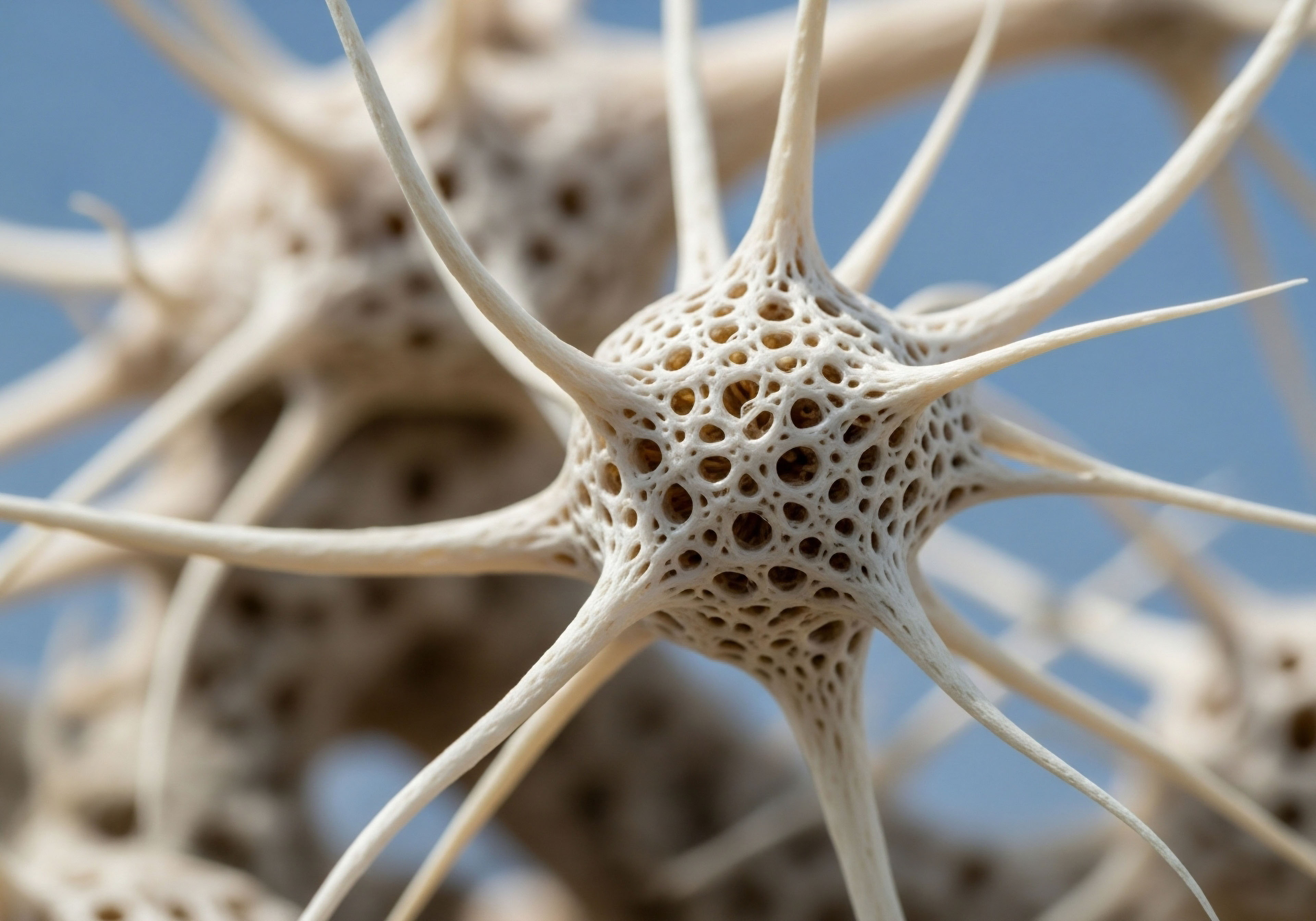
At the heart of this entire system is the cell membrane of your neurons. This is a dynamic, fluid barrier constructed primarily from a double layer of fats, or phospholipids. Embedded within this fatty sea are receptors, the specialized proteins that act as docking stations for hormones and neurotransmitters.
These are the gatekeepers of cellular communication. When a hormone like testosterone or a neurotransmitter like serotonin arrives, it must bind to its specific receptor to deliver its message. The integrity and function of this entire signaling process depend on the physical structure of the membrane itself.

The Building Blocks of Your Brain’s Receptors
The fats from your diet are not merely fuel; they are the literal architectural components of these neural membranes. The type of fatty acid incorporated into the phospholipid layer dictates the membrane’s physical characteristics, most importantly its fluidity. A fluid membrane allows receptors to move freely, change shape, and couple with other proteins efficiently, ensuring a clear and strong signal.
A rigid, inflexible membrane can hinder this process, effectively muffling the message before it is even fully received. This is a foundational concept in understanding how diet directly translates to brain function and hormonal response.
The primary categories of dietary fats each contribute a unique structural property to your neural architecture. Recognizing their distinct roles is essential for building a biological environment conducive to optimal signaling.
- Saturated Fats These fats, found in animal products and tropical oils, have straight, uniform structures. This uniformity allows them to pack together tightly, which can increase the rigidity of cell membranes when consumed in excess.
- Monounsaturated Fats Found in foods like olive oil and avocados, these fats have a single bend in their structure. This kink prevents them from packing as tightly as saturated fats, contributing a moderate level of fluidity to cell membranes.
- Polyunsaturated Fats (PUFAs) This category includes two critical sub-types, Omega-3 and Omega-6 fatty acids. Their multiple bends, or “kinks,” are what make them so powerful in shaping membrane function. They create significant space within the membrane, promoting a high degree of fluidity that is essential for rapid and efficient receptor signaling.
The fats you consume become the physical environment where every hormonal and neural signal is either clearly received or fundamentally impaired.
This direct, physical relationship between dietary lipids and cell structure provides a powerful framework for understanding your own body. The feelings of mental sharpness, mood stability, and hormonal balance are deeply connected to the structural integrity of your brain’s cellular membranes.
By supplying your body with the correct building materials, you are directly investing in the quality of your neurological and endocrine function. This is the groundwork upon which more targeted wellness protocols are built, as the efficacy of any therapeutic intervention relies on the receptivity of the cells it aims to influence.


Intermediate
Building upon the foundational knowledge that dietary fats are the primary architects of neural membranes, we can examine the specific mechanisms through which they modulate receptor function. The interaction is a sophisticated dance of biophysics and biochemistry.
The right lipid composition creates an environment where hormonal signals are received with high fidelity, a critical factor for anyone on a hormonal optimization protocol, such as Testosterone Replacement Therapy (TRT) or peptide therapy. The hormone itself is only one part of the equation; the cell’s ability to receive its message is the other.

How Does Fat Composition Alter Membrane Dynamics?
The biophysical properties of the neuronal membrane govern how receptors behave. These properties are directly regulated by the fatty acid tails of the phospholipids that comprise the bulk of the membrane. Each type of fat imparts a distinct quality, influencing everything from receptor movement to the assembly of complex signaling platforms.

Omega-3 Fatty Acids the Conductors of Fluidity
Docosahexaenoic acid (DHA), a primary omega-3 fatty acid, is the undisputed star of neuronal membrane health. Its unique molecular structure, which contains six double bonds, creates a highly kinked and flexible tail. When DHA is incorporated into the phospholipid bilayer, its bulky, curved shape pushes adjacent fatty acids apart, creating space and increasing the overall fluidity of the membrane.
This fluidity is essential for several aspects of receptor function. It allows receptor proteins to move laterally across the membrane, find their signaling partners, and change their three-dimensional shape, a process required for activation.
For a man on TRT, a fluid membrane ensures that testosterone receptors can efficiently bind to testosterone and initiate the downstream signaling cascade that leads to improved energy, libido, and muscle mass. A deficiency in DHA can lead to a more rigid membrane, impairing this process and potentially reducing the effectiveness of the therapy.

Saturated and Trans Fats the Architects of Rigidity
Saturated fatty acids, lacking any double bonds, are straight and rigid molecules. This structure allows them to pack together very tightly, much like bricks in a wall. While some level of saturated fat is necessary for structural integrity, an overabundance in the diet leads to its excessive incorporation into cell membranes.
This results in a stiff, less dynamic membrane environment. Trans fats, which are artificially hydrogenated, have a similar straight structure and produce an even more pronounced rigidifying effect. This rigidity can physically constrain receptors, locking them into a conformation that makes it difficult to bind their target ligand or to move and interact with other necessary proteins. This can dampen cellular communication and contribute to symptoms like brain fog and fatigue, as neuronal signaling becomes less efficient.

Cholesterol the Master Modulator
Cholesterol is another critical lipid component, acting as a buffer for membrane fluidity. It inserts itself between phospholipids, and its function changes based on the surrounding environment. In a membrane rich with saturated fats, cholesterol prevents the fatty acids from packing too tightly, thus increasing fluidity.
Conversely, in a membrane rich with fluid omega-3s, cholesterol provides structural integrity, preventing the membrane from becoming too flimsy. The brain maintains its own cholesterol supply, largely independent of dietary cholesterol, but overall metabolic health, which is influenced by diet, affects the brain’s ability to synthesize and manage this essential molecule. Proper cholesterol homeostasis is vital for creating stable platforms for receptor activity.
Optimal receptor function depends on a membrane that is fluid enough to permit signaling yet stable enough to maintain structural integrity.
This balance is directly managed by the interplay of dietary fats and endogenous cholesterol. The following table illustrates the contrasting effects of different dietary fat profiles on the key characteristics of a neuronal membrane.
| Membrane Characteristic | High Omega-3 (DHA/EPA) Profile | High Saturated Fat Profile |
|---|---|---|
| Membrane Fluidity |
High. The kinked structure of PUFAs creates space, allowing for free movement of embedded proteins. |
Low. The straight structure of saturated fats allows for tight packing, creating a rigid environment. |
| Receptor Mobility |
Enhanced. Receptors can move laterally to form signaling complexes efficiently. |
Impaired. Receptor movement is restricted, slowing down or preventing signal transduction. |
| Signal Transduction |
Efficient and clear. Conformational changes in receptors occur easily, leading to robust downstream signaling. |
Muffled or blunted. The physical constraint on receptors can weaken the signal’s intensity. |
| Inflammatory Potential |
Low. Omega-3s are precursors to anti-inflammatory signaling molecules. |
High. Certain saturated fats can activate pro-inflammatory pathways directly within the brain. |

Lipid Rafts the Brain’s Signaling Hotspots
Within the fluid mosaic of the cell membrane exist specialized microdomains known as lipid rafts. These are small, highly organized areas that are enriched in cholesterol and specific lipids like sphingolipids. They function as floating platforms or command centers where receptors and signaling molecules are concentrated to make communication more rapid and efficient.
The integrity of these rafts is paramount for complex processes like synaptic plasticity, which underlies learning and memory. Dietary fat composition directly influences the formation and stability of these rafts. A healthy balance of fats ensures these crucial signaling hubs can assemble correctly, supporting robust neurotransmission and hormonal signaling. An imbalanced lipid profile can disrupt raft integrity, scattering the components and weakening the cell’s communication network.


Academic
A sophisticated examination of how dietary lipids influence brain receptor function requires moving beyond general principles of membrane fluidity and into the precise molecular interactions between specific fatty acids and receptor superfamilies. The dialogue between dietary intake and cellular signaling is not merely structural; it is an active, ligand-receptor-mediated process.
Two areas of intense research provide a clear window into this mechanism ∞ the direct activation of G-protein coupled receptors (GPCRs) by omega-3 fatty acids and the indispensable role of cholesterol in the mechanics of synaptic vesicle fusion and neurotransmitter release.

Omega-3 Fatty Acids as Direct Signaling Ligands for GPR120
The conventional view holds that the benefits of omega-3s like DHA and EPA are primarily derived from their structural incorporation into membranes and their role as precursors to anti-inflammatory molecules. A more advanced understanding reveals that these fatty acids also function as direct signaling molecules, or ligands, for a specific class of receptors.
The G-protein coupled receptor 120 (GPR120), also known as Free Fatty Acid Receptor 4 (FFA4), has been identified as a high-affinity receptor for long-chain unsaturated fatty acids, particularly DHA. This discovery fundamentally reframes the role of DHA from a passive structural element to an active participant in signal transduction.
When DHA binds to GPR120 on the surface of brain cells, particularly immune cells like microglia, it initiates a powerful anti-inflammatory signaling cascade. The binding event causes a conformational change in the GPR120 receptor, which in turn activates its associated intracellular G-proteins.
This activation triggers a series of downstream events, most notably the inhibition of the Toll-like receptor 4 (TLR4) signaling pathway. TLR4 is a key initiator of the innate immune response and is potently activated by molecules like lipopolysaccharide (LPS) from bacteria and certain saturated fatty acids.
Activation of TLR4 leads to a pro-inflammatory cascade mediated by transcription factors like NF-κB. The GPR120 pathway directly counteracts this by preventing the recruitment of key adapter proteins necessary for TLR4 signaling. This mechanism demonstrates that DHA actively suppresses neuroinflammation at the receptor level, protecting neurons from inflammatory damage and preserving the integrity of other receptor systems.
| Step | Molecular Action in the GPR120 Pathway | Physiological Outcome |
|---|---|---|
| 1. Ligand Binding |
DHA, circulating from dietary sources, binds to the extracellular domain of the GPR120 receptor on a microglial cell. |
Initiation of an anti-inflammatory signal. |
| 2. Receptor Activation |
The receptor undergoes a conformational change, activating its coupled intracellular Gq/11 protein. |
The signal is transduced across the cell membrane. |
| 3. Downstream Inhibition |
The activated pathway leads to the phosphorylation and binding of β-arrestin2, which interacts with and blocks the TAK1 binding protein (TAB1). |
The central pro-inflammatory pathway (NF-κB) is inhibited. |
| 4. Gene Transcription |
The suppression of NF-κB prevents the transcription of pro-inflammatory cytokines like TNF-α and IL-6. |
Reduction of the local inflammatory state in the brain tissue. |

Why Is Cholesterol Essential for Synaptic Function?
The brain’s relationship with cholesterol is unique and profound. While the blood-brain barrier isolates the central nervous system from peripheral cholesterol, the brain itself is the most cholesterol-rich organ, synthesizing its own supply primarily in astrocytes. This cholesterol is absolutely vital for the fundamental mechanics of neurotransmission.
Its role extends far beyond simply modulating membrane fluidity. Cholesterol is a key structural determinant in the formation and function of synaptic vesicles, the tiny lipid bubbles that store and release neurotransmitters like acetylcholine and dopamine.
Cholesterol’s rigid, planar structure is essential for inducing the high degree of membrane curvature required to form these vesicles. Without sufficient cholesterol, the membrane cannot bend properly to bud off and create these critical storage units. Furthermore, cholesterol is directly involved in the process of exocytosis, where the vesicle fuses with the presynaptic membrane to release its contents into the synapse.
It helps organize the specific proteins (SNARE complexes) that mediate this fusion event. Therefore, a disruption in the brain’s delicate cholesterol homeostasis can directly impair the release of neurotransmitters, leading to significant deficits in cognition, mood, and neuromuscular control. This highlights how overall metabolic health, which impacts the brain’s synthetic machinery, is inextricably linked to the very foundation of neuronal communication.
The brain’s independent cholesterol metabolism is a cornerstone of synaptic transmission, governing the physical release of neurotransmitters at the presynaptic terminal.

Neuroinflammation a Lipid-Mediated Process
The brain’s resident immune cells, the microglia, are exquisitely sensitive to the surrounding lipid environment. High levels of certain saturated fatty acids, such as palmitic acid, can act as danger signals, directly activating microglia through the same TLR4 receptors involved in bacterial infections.
This triggers a shift in microglia from their resting, housekeeping state to an activated, pro-inflammatory phenotype. Activated microglia release a barrage of inflammatory cytokines and reactive oxygen species. This inflammatory environment can cause widespread damage. It can directly impair the function of neuronal receptors, reduce synaptic density, and even trigger neuronal cell death.
This process of diet-induced neuroinflammation is a key mechanism linking metabolic dysfunction with cognitive decline and mood disorders. Conversely, the presence of DHA, acting through GPR120, actively suppresses this microglial activation, creating a neuroprotective shield. This illustrates a direct antagonism at the cellular level between different classes of dietary fats, with the balance determining the inflammatory tone of the brain and the subsequent health of its receptor systems.

References
- Oh, D. Y. et al. “GPR120 is an Omega-3 Fatty Acid Receptor Mediating Potent Anti-Inflammatory and Insulin-Sensitizing Effects.” Cell, vol. 142, no. 5, 2010, pp. 687-698.
- Zhang, J. and L. Liu. “Cholesterol Metabolism and Homeostasis in the Brain.” Protein & Cell, vol. 6, no. 4, 2015, pp. 254-264.
- Di Paolo, G. and T. W. Kim. “Linking Lipids to Alzheimer’s Disease ∞ Cholesterol and Beyond.” Nature Reviews Neuroscience, vol. 12, no. 5, 2011, pp. 284-296.
- Valdearcos, M. et al. “Microglia Dictate the Impact of Saturated Fat Consumption on Hypothalamic Inflammation and Neuronal Function.” Cell Reports, vol. 9, no. 6, 2014, pp. 2124-2138.
- Inamori, K. et al. “Omega-3 Fatty Acids and Inflammatory Processes.” Progress in Lipid Research, vol. 51, no. 3, 2012, pp. 232-237.
- Chianese, R. et al. “Impact of Dietary Fats on Brain Functions.” Current Neuropharmacology, vol. 16, no. 7, 2018, pp. 1059-1085.
- Suzuki, T. et al. “Administration of Phosphatidylcholine Increases Brain Acetylcholine Concentration and Improves Memory in Mice with Dementia.” The Journal of Nutrition, vol. 125, no. 6, 1995, pp. 1484-1489.
- Thayer, D. A. et al. “High-fat diet is a risk factor for cognitive decline in aging C57BL/6J mice.” Physiology & Behavior, vol. 227, 2020, 113175.

Reflection

From Knowledge to Personal Protocol
You now possess a detailed map illustrating the profound connection between the fats on your plate and the function of your brain’s most intricate signaling systems. You can see how the architecture of a single fatty acid molecule can scale up to influence your mood, your clarity of thought, and your body’s response to hormonal guidance.
This knowledge is the critical first step. It moves the locus of control from a place of uncertainty to a position of informed action. The question now becomes a personal one. How does this biological reality manifest in your own life?
Consider the patterns of your own cognitive and emotional landscape. Are there times of sharp focus and effortless mental flow? Are there periods of static, fatigue, or emotional dysregulation? The information presented here invites you to look at these experiences through a new lens, one that connects your internal state to your external choices.
This is not about assigning blame or demanding perfection. It is about recognizing the power you have to influence your own biology. Your body is in constant dialogue with its environment, and your diet is one of the most intimate and powerful forms of that conversation.
By understanding the language of lipids, you can begin to guide that conversation toward a state of balance and vitality. This journey of biological self-awareness is unique to each individual, and the path forward is one of personalized, deliberate calibration.

Glossary

dietary fats

saturated fats

fatty acids

structural integrity

receptor function

testosterone replacement therapy

peptide therapy

saturated fatty acids

saturated fat
membrane fluidity

cholesterol homeostasis

signal transduction

lipid rafts
omega-3 fatty acids

synaptic vesicle

certain saturated fatty acids

neuroinflammation