
Fundamentals
The feeling of being “off” ∞ the persistent fatigue, the mental fog, the subtle loss of drive ∞ is a deeply personal and often isolating experience. When you suspect your testosterone levels might be low, the immediate question that surfaces is a critical one ∞ Is this something I was born with, or is it a product of my life?
This question is the starting point of a personal investigation into your own biology. It is the first step toward understanding the intricate communication network within your body, the endocrine system, and reclaiming your vitality. The answer is rarely a simple “yes” or “no.” Instead, it is a complex interplay between your genetic blueprint and the life you have lived.
Your body’s ability to produce testosterone is governed by a sophisticated command chain known as the Hypothalamic-Pituitary-Gonadal (HPG) axis. Think of it as a finely tuned internal messaging service. The hypothalamus in your brain sends a signal, Gonadotropin-Releasing Hormone (GnRH), to the pituitary gland.
The pituitary, in turn, releases two messenger hormones ∞ Luteinizing Hormone (LH) and Follicle-Stimulating Hormone (FSH). LH is the primary signal that travels to the testes, instructing them to produce testosterone. When this system is working optimally, your body produces what it needs. When you feel the symptoms of low testosterone, it means there is a disruption somewhere along this chain of command.
Distinguishing between a genetic predisposition and a lifestyle-driven hormonal imbalance begins with a comprehensive evaluation of your body’s endocrine signaling pathways.
A genetic cause for low testosterone often implies a fundamental issue with the hardware of this system. These are typically congenital conditions, meaning they have been present since birth. For example, conditions like Klinefelter syndrome, where a male is born with an extra X chromosome (47,XXY), can lead to abnormal testicular development and an impaired ability to produce testosterone.
Another genetic condition, Kallmann syndrome, affects the development of the GnRH neurons in the brain, meaning the very first signal in the testosterone production chain is never properly sent. These conditions are intrinsic to your biological makeup. They are part of your personal genetic code.
Conversely, lifestyle-related low testosterone suggests that the hardware is intact, but the operating software has been corrupted by external factors. Chronic stress, poor sleep, a diet high in processed foods, and a sedentary lifestyle can all disrupt the delicate hormonal symphony.
For instance, excess body fat, particularly visceral fat around the organs, is a potent endocrine disruptor. It increases the activity of an enzyme called aromatase, which converts testosterone into estrogen, thereby lowering active testosterone levels. Chronic sleep deprivation directly impacts the brain’s ability to send those crucial LH signals during the night, when a significant portion of testosterone production occurs.
These are not issues with the fundamental machinery but rather with the conditions under which that machinery is forced to operate.
How, then, do you begin to untangle these threads? The initial step is a conversation with a clinician who understands the language of the endocrine system. This will involve a detailed personal and family medical history, a physical examination, and, most importantly, a specific set of blood tests.
These initial tests will measure your total and free testosterone levels, but just as critically, they will also measure your LH and FSH levels. The relationship between these hormones provides the first major clue in determining the origin of the problem. This initial diagnostic process is the gateway to a deeper understanding of your unique physiology and the first concrete step toward developing a personalized protocol to restore your body’s intended function.


Intermediate
To clinically differentiate between a genetic and a lifestyle-induced origin for low testosterone, we must move beyond the symptoms and examine the biochemical dialogue of the Hypothalamic-Pituitary-Gonadal (HPG) axis. The diagnostic process is a form of biological investigation, and the key evidence lies within your bloodwork.
Specifically, the relationship between testosterone, Luteinizing Hormone (LH), and Follicle-Stimulating Hormone (FSH) allows us to pinpoint the location of the dysfunction. This distinction is the foundation for classifying hypogonadism into two primary categories ∞ primary and secondary.

Understanding Primary and Secondary Hypogonadism
Primary hypogonadism suggests a problem originating in the testes themselves. The testes are failing to produce sufficient testosterone despite receiving the correct signals from the brain. In this scenario, the brain recognizes the low testosterone levels and attempts to compensate by increasing the output of LH and FSH.
It is akin to turning up the volume on a faulty speaker; the signal is loud and clear, but the output remains low. Therefore, the hallmark laboratory finding for primary hypogonadism is low testosterone in the presence of high LH and FSH levels. This pattern strongly points toward an intrinsic, and often genetic, issue with testicular function.
Secondary hypogonadism, on the other hand, indicates that the problem lies within the brain, specifically the hypothalamus or the pituitary gland. In this case, the testes are perfectly capable of producing testosterone, but they are not receiving the necessary hormonal instructions (LH) to do so.
The initial signal from the brain is weak or absent. The laboratory signature for secondary hypogonadism is low testosterone accompanied by low or inappropriately normal LH and FSH levels. The brain is not recognizing the need to send a stronger signal. This type of hypogonadism is frequently associated with lifestyle factors, although some genetic conditions like Kallmann syndrome also fall into this category.
The interplay between testosterone, LH, and FSH levels is the critical diagnostic tool for pinpointing the source of hormonal disruption.

Genetic Determinants of Primary Hypogonadism
Many cases of primary hypogonadism have a genetic basis. The most common is Klinefelter syndrome (47,XXY), a chromosomal abnormality that occurs in approximately 1 in 500 to 1,000 male births. The presence of an extra X chromosome leads to abnormal development of the seminiferous tubules and Leydig cells within the testes, directly impairing their ability to produce testosterone and sperm.
Other causes can include physical injury to the testes, certain infections like mumps orchitis, or treatments like chemotherapy or radiation that damage testicular tissue.

Lifestyle Factors and Secondary Hypogonadism
Lifestyle-induced low testosterone most often manifests as secondary hypogonadism. Obesity is a primary driver of this condition, creating what is sometimes termed Male Obesity-Associated Secondary Hypogonadism (MOSH). The mechanisms are multifaceted:
- Aromatization ∞ Adipose (fat) tissue contains the enzyme aromatase, which converts testosterone to estradiol (a form of estrogen). Increased body fat leads to increased aromatase activity, effectively reducing the pool of available testosterone.
- Leptin and Insulin Resistance ∞ Obesity often leads to resistance to the hormones leptin and insulin. These metabolic derangements can interfere with the signaling of GnRH in the hypothalamus, suppressing the entire HPG axis.
- Inflammation ∞ Chronic, low-grade inflammation associated with obesity can also disrupt hypothalamic function and testosterone production.
Other lifestyle factors contribute significantly as well. Chronic stress elevates cortisol levels, which can suppress GnRH and LH secretion. Poor sleep quality, particularly a lack of deep sleep, directly blunts the nocturnal LH pulse necessary for robust testosterone synthesis.
The following table outlines the key diagnostic differences between these two conditions:
| Diagnostic Marker | Primary Hypogonadism | Secondary Hypogonadism |
|---|---|---|
| Origin of Dysfunction | Testes | Hypothalamus or Pituitary Gland (Brain) |
| Total Testosterone | Low | Low |
| LH & FSH Levels | High | Low or Inappropriately Normal |
| Common Genetic Causes | Klinefelter Syndrome (47,XXY) | Kallmann Syndrome |
| Common Lifestyle Causes | Less common, usually due to direct testicular injury or toxins. | Obesity, Chronic Stress, Poor Sleep, Poor Diet |
A thorough clinical evaluation, including a detailed history and these critical lab tests, provides a clear roadmap. It allows a clinician to move from a general diagnosis of low testosterone to a specific understanding of the underlying mechanism. This clarity is essential because the therapeutic approach for restoring testicular function in a man with a genetic condition is different from recalibrating the HPG axis in a man whose system is suppressed by lifestyle factors.


Academic
A sophisticated analysis of hypogonadism requires a deep dive into the molecular and genetic underpinnings that differentiate congenital from acquired functional deficiencies. The distinction rests upon the integrity of the Hypothalamic-Pituitary-Gonadal (HPG) axis at a cellular and genetic level. While a patient’s symptoms may be identical, the pathophysiological pathways are fundamentally distinct.
We will explore two illustrative examples ∞ the genetic architecture of Klinefelter syndrome as a model of primary hypogonadism, and the complex neuroendocrine disruptions of obesity-induced secondary hypogonadism.

What Is the Genetic Basis of Klinefelter Syndrome?
Klinefelter syndrome (KS), characterized by a 47,XXY karyotype, represents a clear-cut example of primary hypergonadotropic hypogonadism. The presence of a supernumerary X chromosome is not a benign addition; it profoundly alters testicular development and function from an early stage.
The core pathology involves a progressive hyalinization and fibrosis of the seminiferous tubules, which house the Sertoli cells responsible for sperm production and various endocrine support functions. Concurrently, there is a progressive depletion and dysfunction of the Leydig cells, the primary producers of testosterone.
The molecular mechanisms are complex. The extra X chromosome leads to the overexpression of certain genes that escape X-inactivation, creating a state of genetic imbalance. This disrupts the delicate process of testicular cell differentiation and maturation.
While testosterone levels may be normal during early to mid-puberty, the progressive testicular failure leads to declining testosterone and rising gonadotropin (LH and FSH) levels in adulthood. The elevated LH is a direct physiological response by the pituitary to the failing testicular output, a classic hallmark of a primary defect. The infertility seen in nearly all men with 47,XXY KS is a direct consequence of the severe disruption to spermatogenesis.

How Does Obesity Induce Secondary Hypogonadism?
In contrast, male obesity-associated secondary hypogonadism (MOSH) is a functional, and often reversible, state of hypogonadotropic hypogonadism. The testicular machinery is intact, but its function is suppressed by systemic metabolic dysregulation originating from excess adiposity. Several interconnected molecular pathways contribute to this suppression:
- Hypothalamic Kisspeptin Suppression ∞ The neuropeptide kisspeptin, produced by Kiss1 neurons in the hypothalamus, is the master regulator of GnRH secretion. In states of obesity, elevated levels of leptin and insulin lead to central leptin and insulin resistance. This resistance has been shown to decrease the expression of Kiss1 in the arcuate nucleus of the hypothalamus. The reduction in kisspeptin signaling leads to inadequate GnRH pulsatility, which in turn results in diminished LH and FSH secretion from the pituitary and, consequently, lower testosterone production.
- Aromatase-Mediated Hyperestrogenism ∞ Visceral adipose tissue is a primary site of extragonadal aromatase expression. This enzyme catalyzes the conversion of androgens (like testosterone) into estrogens (like estradiol). In obesity, the sheer mass of adipose tissue leads to a significant increase in this conversion, resulting in elevated circulating estrogen levels. This hyperestrogenism exerts a potent negative feedback on the HPG axis at both the hypothalamic and pituitary levels, further suppressing LH secretion.
- Inflammatory Cytokine Interference ∞ Obesity is a state of chronic, low-grade systemic inflammation, characterized by elevated levels of pro-inflammatory cytokines such as Tumor Necrosis Factor-alpha (TNF-α) and Interleukin-1 (IL-1). These cytokines can directly inhibit GnRH neuron function and interfere with pituitary gonadotropin secretion, adding another layer of suppression to the HPG axis.
The following table provides a comparative summary of the pathophysiological features:
| Feature | Klinefelter Syndrome (Primary) | Obesity-Induced Hypogonadism (Secondary) |
|---|---|---|
| Karyotype | 47,XXY | 46,XY |
| Primary Site of Defect | Testes (Leydig and Sertoli cell failure) | Hypothalamus (GnRH/Kisspeptin neuron dysfunction) |
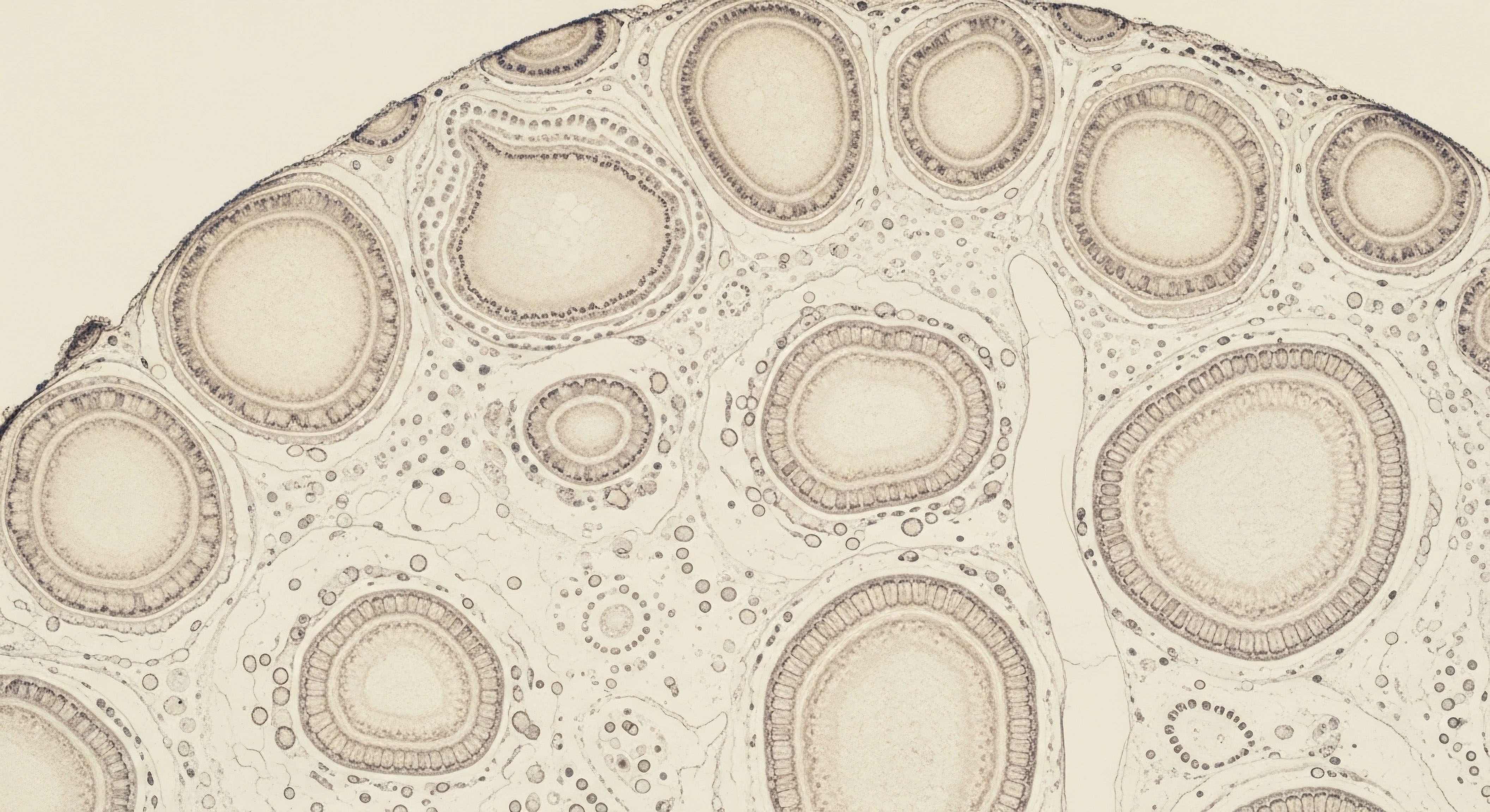
| Testicular Histology | Seminiferous tubule hyalinization, fibrosis, Leydig cell depletion | Generally normal histology, capable of function |
| Key Molecular Driver | Overexpression of genes on extra X chromosome | Leptin/insulin resistance, inflammation, aromatase activity |
| Kisspeptin Signaling | Not the primary defect | Suppressed due to metabolic dysregulation |
| Serum Estradiol | Relatively elevated due to altered T/E ratio | Absolutely elevated due to peripheral aromatization |
| Reversibility | Irreversible testicular failure | Potentially reversible with significant weight loss |
The molecular diagnosis distinguishes an irreversible genetic hardware failure from a reversible, system-wide software suppression.
This academic distinction is paramount for clinical management. For a man with Klinefelter syndrome, the therapeutic goal is hormonal optimization through exogenous testosterone administration to mitigate the symptoms of androgen deficiency, as endogenous production cannot be restored. For the man with MOSH, the primary intervention is addressing the root cause ∞ the metabolic dysfunction driven by obesity.
Lifestyle modification, including diet and exercise, can restore HPG axis function and normalize testosterone levels without the need for lifelong hormonal support. In some cases, therapies like Gonadorelin may be used to directly stimulate the HPG axis, but this is only effective if the downstream components (pituitary and testes) are functional, as they are in secondary hypogonadism.

References
- Bhasin, S. et al. “Testosterone Therapy in Men With Hypogonadism ∞ An Endocrine Society Clinical Practice Guideline.” The Journal of Clinical Endocrinology & Metabolism, vol. 103, no. 5, 2018, pp. 1715 ∞ 1744.
- Millar, A. C. et al. “Genetics of Hypogonadotropic Hypogonadism.” Translational Andrology and Urology, vol. 10, no. 3, 2021, pp. 1401-1409.
- Groth, K. A. et al. “Klinefelter Syndrome ∞ A Clinical Update.” The Journal of Clinical Endocrinology & Metabolism, vol. 98, no. 1, 2013, pp. 20-30.
- Nieschlag, E. et al. “Klinefelter Syndrome ∞ The Lower End of the Spectrum of Male Infertility.” Human Reproduction Update, vol. 5, no. 5, 1999, pp. 499-504.
- Rochira, V. et al. “Male Obesity and Androgen-Related Disorders.” Journal of Endocrinological Investigation, vol. 31, no. 8, 2008, pp. 732-741.
- Cohen, J. et al. “The role of the intestinal microbiome in mediating the benefits of bariatric surgery.” Nature, vol. 497, no. 7451, 2013, pp. 581-585.
- Kumagai, H. et al. “Increased physical activity has a greater effect than reduced energy intake on lifestyle modification-induced increases in testosterone.” Journal of Clinical Biochemistry and Nutrition, vol. 58, no. 1, 2016, pp. 84-89.
- Fechner, A. Fong, S. & McGovern, P. “A review of Kallmann syndrome ∞ genetics, pathophysiology, and clinical management.” Obstetrical & Gynecological Survey, vol. 63, no. 3, 2008, pp. 189-194.
- Navarro, V. M. et al. “Obesity-induced hypogonadism in the male ∞ premature reproductive neuroendocrine senescence and contribution of Kiss1-mediated mechanisms.” Endocrinology, vol. 155, no. 4, 2014, pp. 1067-79.
- Santi, D. et al. “Molecular Mechanisms Underlying the Relationship between Obesity and Male Infertility.” International Journal of Molecular Sciences, vol. 21, no. 24, 2020, p. 9630.

Reflection
You have now explored the biological pathways that separate a genetic predisposition from a lifestyle-driven reality in the context of hormonal health. This knowledge is a powerful tool. It transforms vague feelings of unwellness into a set of specific, answerable questions about your own body.
The information presented here is the map; your personal health journey is the territory. Understanding the distinction between a hardware and a software problem is the first step. The next is to consider which path of investigation resonates most with your own experience. This process of self-discovery, guided by clinical expertise, is the foundation of reclaiming your biological potential and living a life of uncompromised function.

Glossary

testosterone levels

endocrine system

low testosterone

klinefelter syndrome

testosterone production

kallmann syndrome

aromatase

fsh levels

primary hypogonadism

secondary hypogonadism

lifestyle factors

male obesity-associated secondary hypogonadism

hpg axis

kisspeptin


