

Fundamentals
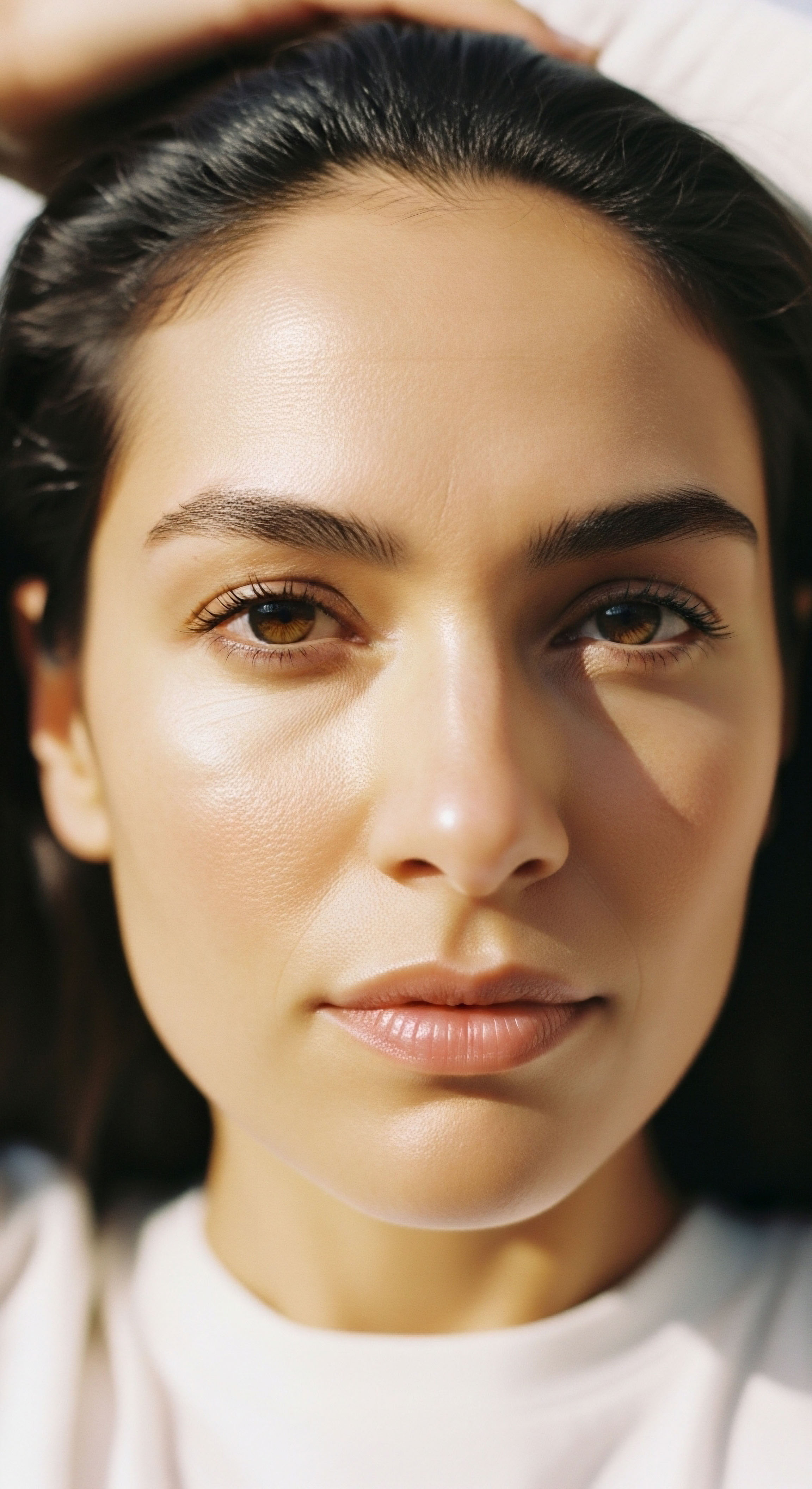
You look in the mirror and see a tiredness that sleep does not seem to resolve. The puffiness under your eyes feels like a permanent fixture, a frustrating signal that stands at odds with the vitality you feel you should have. Your experience is valid, and the question of whether hormonal optimization can help is a gateway to a much deeper understanding of your body’s intricate communication network.
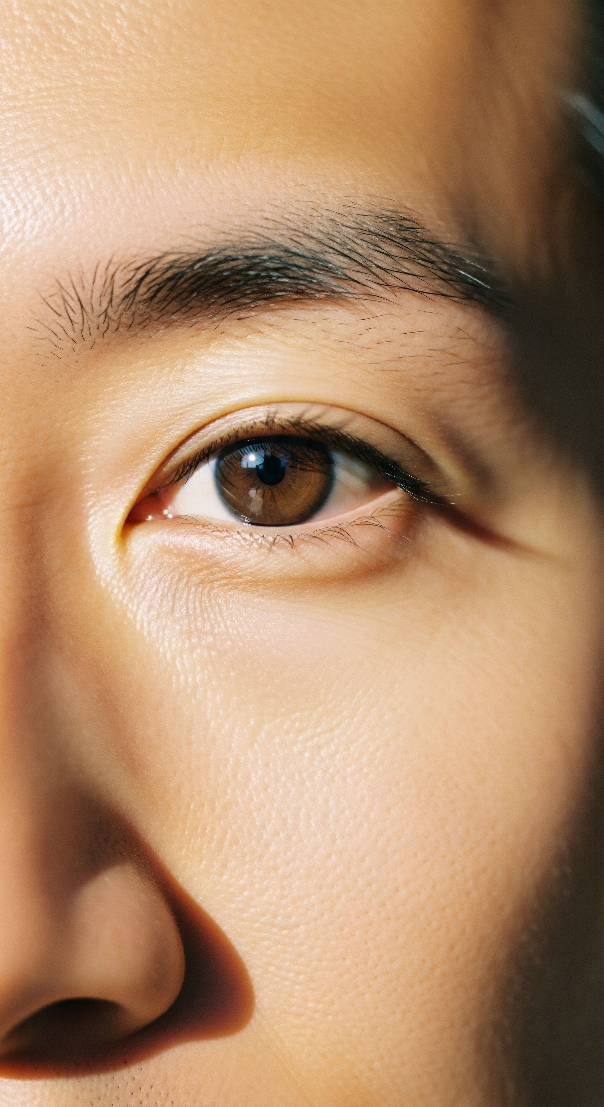
The skin around the eyes is exceptionally thin and sensitive, acting as a public-facing dashboard for your internal systemic balance. When puffiness appears, it is often telling a story about what is happening beneath the surface, a story written in the language of hormones.
To understand the connection, we can look at three primary biological narratives that unfold during hormonal shifts. The first narrative concerns the structural integrity of your skin. Think of your skin as a resilient, flexible fabric woven from protein fibers like collagen and elastin. Estrogen is a master regulator of this process, continuously signaling specialized cells called fibroblasts to produce these fibers, keeping the skin thick and elastic.
As estrogen levels decline, particularly during perimenopause and menopause, this signal weakens. The production of new fibers slows, and the existing framework begins to degrade. The skin under the eyes, being so delicate, is one of the first places to show this change, becoming thinner and less able to hold back the fatty tissue that naturally cushions the eyeball. This creates a visible bulge or “puff.”
The appearance of under-eye puffiness can be a direct reflection of changes in the skin’s foundational structure and the body’s fluid management systems, both profoundly influenced by hormones.
The second narrative is about fluid dynamics. Your body is a complex hydraulic system, and the balance between estrogen and progesterone plays a significant role in managing fluid retention. Progesterone has a natural diuretic effect, helping your body excrete excess sodium and water. When the ratio of progesterone to estrogen shifts, the body may begin to retain more fluid.
This systemic change often manifests in areas with soft, expandable tissue, such as the periorbital region. The result is edema, or swelling, which contributes to the puffy appearance, especially upon waking when fluid has had hours to accumulate horizontally.
The third story is one of local inflammation and glandular function. The eyelids are lined with tiny glands, known as meibomian glands, that produce an essential oil for the tear film. The health of these glands is regulated by multiple hormones, including androgens like testosterone. When androgen levels decline, these glands can become dysfunctional, leading to a condition called blepharitis.
This dysfunction causes inflammation, irritation, and swelling right at the eyelid margin, which presents as puffiness and redness. It is a localized inflammatory response triggered by a systemic hormonal deficit. Viewing puffy eyes through these three lenses—structural decline, fluid imbalance, and local inflammation—transforms the conversation. It becomes clear that this is a physiological signal, one that points toward a systemic recalibration as a potential solution.


Intermediate
Understanding that puffy eyes are a signal of systemic imbalance allows us to explore how specific hormonal interventions can address the root causes. A properly structured hormonal optimization protocol is designed to restore the body’s signaling architecture, targeting the precise mechanisms that lead to dermal degradation and fluid dysregulation. This involves a sophisticated understanding of how individual hormones function and interact within the body’s complex feedback loops.


The Specific Roles of Key Hormones
The decline in specific hormones during a woman’s life contributes directly to the changes seen in the periorbital area. Each hormone has a distinct yet complementary role in maintaining the skin’s youthful characteristics and the body’s homeostatic balance.


Estrogen the Architect of Dermal Health
Estrogen is the primary architect of skin structure. Its influence extends to increasing the synthesis of collagen and elastin, which provide the skin’s firmness and elasticity. Furthermore, estrogen boosts the production of hyaluronic acid, a molecule that can hold up to 1,000 times its weight in water, acting as the skin’s natural moisturizer and plumping agent. When estrogen levels fall, the skin’s ability to retain moisture and its structural scaffolding are both compromised, leading to thinning and laxity that allows under-eye fat pads to become more prominent.


Progesterone and Testosterone Supporting Players
Progesterone acts as a crucial counterpoint to estrogen, particularly in fluid management. Its mild diuretic properties help to regulate fluid retention, and maintaining adequate levels can mitigate the edema that contributes to puffiness. Testosterone, often considered a male hormone, is vital for both men and women.
In women, it contributes to skin thickness and the healthy function of sebaceous glands, including the meibomian glands in the eyelids. Optimized testosterone levels can therefore support the structural integrity of the skin and help prevent the localized inflammation associated with glandular dysfunction.
A well-designed hormonal protocol aims to restore the synergistic functions of estrogen, progesterone, and testosterone to address skin laxity, fluid retention, and localized inflammation simultaneously.

Clinical Protocols for Systemic Recalibration
Addressing these hormonal deficits requires a personalized clinical approach. For women experiencing symptoms like puffy eyes alongside other indicators of hormonal change, a protocol may involve a combination of hormones to restore systemic balance. The goal is to re-establish physiological levels that support optimal function across multiple body systems.
- Testosterone Cypionate ∞ For women, a low dose of Testosterone Cypionate (typically 0.1–0.2ml of 200mg/ml solution weekly) is often prescribed. This helps to restore androgen levels, which can improve skin thickness, support meibomian gland function to reduce eyelid inflammation, and enhance overall energy and well-being.
- Progesterone ∞ Depending on menopausal status, bioidentical progesterone is often included. It helps to balance the effects of estrogen and provides its own benefits, including supporting sleep and managing fluid retention due to its diuretic properties.
- Anastrozole ∞ In some cases, particularly with pellet therapy or higher testosterone doses, a medication like Anastrozole may be used sparingly to prevent the conversion of testosterone into estrogen, ensuring the hormonal ratios remain optimized.
This multi-faceted approach recognizes that the symptom of puffy eyes is the outcome of interconnected systemic changes. By addressing the hormonal foundation, these protocols can produce improvements that are part of a global enhancement of skin quality and physiological function.
| Hormone | Primary Effect on Skin Structure | Influence on Fluid Balance | Impact on Eyelid Glands |
|---|---|---|---|
| Estrogen | Increases collagen and hyaluronic acid synthesis, improving skin thickness and hydration. | Can contribute to fluid retention if unopposed by progesterone. | Supports overall tissue health and vascularity. |
| Progesterone | Plays a secondary role in skin elasticity. | Acts as a natural diuretic, helping to reduce systemic fluid retention and edema. | Has a calming, anti-inflammatory effect on tissues. |
| Testosterone | Contributes to dermal thickness and sebum production, maintaining skin barrier function. | Minimal direct effect on fluid retention at physiological female doses. | Essential for healthy meibomian gland function; deficiency is linked to blepharitis. |
Academic
A sophisticated analysis of periorbital puffiness requires a deep exploration of the molecular and physiological mechanisms governed by the endocrine system. The visible changes in the under-eye area are the macroscopic expression of complex cellular events involving the dermal extracellular matrix, systemic fluid regulation via the kidneys, and localized lipid metabolism in the eyelid glands. Hormone replacement therapy’s efficacy is rooted in its ability to intervene at these fundamental biological levels.


How Do Hormones Modulate the Dermal Extracellular Matrix?
The extracellular matrix Meaning ∞ The Extracellular Matrix, often abbreviated as ECM, represents the non-cellular component present within all tissues and organs, providing essential physical scaffolding for cellular constituents and initiating crucial biochemical and biomechanical signals. (ECM) of the dermis is a dynamic, bioactive scaffold primarily composed of collagen, elastin, and proteoglycans like hyaluronic acid. Its integrity is actively managed by hormones. Estrogen, acting through its receptors (ERα and ERβ) on dermal fibroblasts, directly upregulates the gene transcription for COL1A1 and COL3A1, the genes responsible for producing type I and type III collagen. This action preserves the tensile strength and structure of the skin.
Concurrently, estrogen appears to decrease the expression of matrix metalloproteinases (MMPs), enzymes that degrade collagen. The decline in estrogen during menopause reverses this protective effect, leading to a net loss of collagen estimated at 2% per year. This degradation of the ECM framework causes the dermal layer to thin and lose its supportive capacity, allowing the orbital fat pads to herniate and become visible.

The Renin-Angiotensin-Aldosterone System Connection
Periorbital edema is a direct result of fluid shifting into the interstitial space, a process heavily influenced by the Renin-Angiotensin-Aldosterone System Meaning ∞ The Renin-Angiotensin-Aldosterone System, or RAAS, is a crucial hormonal cascade regulating blood pressure, fluid volume, and electrolyte balance. (RAAS), the body’s primary regulator of blood pressure and fluid volume. Sex hormones are significant modulators of this system. Estrogen can increase the production of angiotensinogen, a precursor in the RAAS cascade, which can lead to increased sodium and water retention. Progesterone, conversely, acts as a competitive antagonist at the mineralocorticoid receptor, blocking the action of aldosterone.
This antagonism promotes sodium and water excretion, producing a natural diuretic effect. The fluctuating and ultimately declining levels of progesterone relative to estrogen during perimenopause can therefore lead to a state of net fluid retention, which manifests as puffiness in compliant tissues like the periorbital area.
The efficacy of combined hormone therapy lies in its ability to restore a favorable progesterone-to-estrogen ratio, thereby promoting diuresis and reducing the fluid component of under-eye puffiness.
Hormonal therapy, particularly protocols combining estrogen with progesterone, aims to restore this balance. By providing sufficient progesterone to counteract estrogen’s fluid-retaining tendencies, the system can be guided back toward a state of fluid homeostasis, directly reducing edema.


What Is the Role of Androgens in Meibomian Gland Dysfunction?
The inflammatory component of puffy eyes is frequently linked to Meibomian Gland Dysfunction (MGD), a leading cause of dry eye disease and blepharitis. These specialized sebaceous glands are androgen-dependent. Testosterone and its metabolites are crucial for stimulating the proliferation of meibomian gland epithelial cells Senolytics precisely target and eliminate dysfunctional senescent cells by disrupting their pro-survival pathways, reducing inflammation, and restoring cellular health. and for promoting the synthesis of the complex lipids that form the tear film’s outer layer. A decline in androgens, a hallmark of menopause and andropause, leads to gland atrophy and altered lipid production.
The meibum becomes thicker, blocking the glands and triggering a localized inflammatory response along the eyelid margin. This chronic inflammation, or blepharitis, causes the eyelids to appear red, swollen, and puffy. A therapeutic intervention with low-dose testosterone in women can directly address this pathology by restoring androgen signaling to the meibomian glands, improving their function and reducing the associated inflammation.
- Androgen Decline ∞ Systemic levels of testosterone and its precursors decrease with age.
- Glandular Atrophy ∞ Meibomian glands, being androgen-dependent, receive insufficient signaling to maintain normal cell turnover and lipid synthesis.
- Altered Meibum ∞ The lipid secretion becomes thicker and has a higher melting point, leading to obstruction of the gland orifices.
- Inflammatory Cascade ∞ The blockage and stagnant secretions trigger a chronic inflammatory response, resulting in redness and swelling of the eyelid margins.
| Hormone | Target Cell/System | Molecular Action | Physiological Outcome |
|---|---|---|---|
| Estrogen | Dermal Fibroblast | Upregulates COL1A1, COL3A1, and Hyaluronic Acid Synthase gene expression. | Increased skin thickness, firmness, and hydration. |
| Progesterone | Renal Tubule Cells | Acts as a competitive antagonist at the mineralocorticoid receptor, blocking aldosterone. | Promotes sodium and water excretion (diuresis), reducing systemic edema. |
| Testosterone | Meibomian Gland Epithelial Cells | Binds to androgen receptors, stimulating cell proliferation and lipid synthesis. | Maintains healthy tear film and prevents glandular inflammation (blepharitis). |
References
- Stevenson, S. & Thornton, J. (2007). Effect of estrogens on skin aging. American Journal of Clinical Dermatology, 8(4), 219-236.
- Khandelwal, P. & Liu, S. (2012). Androgens and dry eye in menopause. International Ophthalmology Clinics, 52(2), 57-66.
- Golebiowski, B. & Badarudin, N. (2017). The role of sex hormones in ocular physiology and pathology. Journal of the Optical Society of America A, 34(4), B15-B26.
- Thornton, M. J. (2013). The biological actions of estrogens on skin. Experimental Dermatology, 22(3), 159-163.
- Woodruff, C. M. & Kilaru, G. C. (2011). The role of hormones in the etiology of dry eye syndrome. Current Opinion in Ophthalmology, 22(3), 209-214.
- Schaumberg, D. A. Buring, J. E. & Sullivan, D. A. (2003). Prevalence of dry eye syndrome among US women. American Journal of Ophthalmology, 136(2), 318-326.
- Sator, P. G. Schmidt, J. B. & Rabe, T. (2004). Skin aging and sex hormones in women. Journal der Deutschen Dermatologischen Gesellschaft, 2(1), 36-41.
- Afonso, A. A. Sobrin, L. & Monroy, D. C. (2009). The effect of sex hormones on the ocular surface. The Ocular Surface, 7(3), 158-172.
Reflection
You arrived here with a specific question about your appearance, and in seeking an answer, you have uncovered a profound truth about the body’s interconnectedness. The skin beneath your eyes is a sensitive canvas, illustrating a much larger, more complex story of cellular communication, structural integrity, and systemic balance. The knowledge that these changes are rooted in tangible physiological processes is the first step toward reclaiming your narrative.
The path forward involves turning this objective understanding into a personal inquiry. What is your body communicating to you through these signals? The journey of hormonal optimization is one of deep listening and precise recalibration.
It asks you to look beyond the symptom and engage with the underlying systems that govern your vitality. This knowledge empowers you to ask more informed questions and to seek a partnership in health that is grounded in restoring your body’s innate potential, allowing you to function with clarity and confidence from the inside out.