


Fundamentals
The feeling can be unsettling. A word that was once on the tip of your tongue now feels miles away. You walk into a room and forget why you entered. This subtle, creeping sense of cognitive change, often dismissed as a simple consequence of aging or stress, is a deeply personal experience for many women.
It is a quiet concern that grows louder during the profound biological shifts of perimenopause and post-menopause. Your experience is valid. The perceived fog and the lapses in memory are not imagined; they are signals from a biological system in transition. Understanding this system is the first step toward reclaiming your cognitive vitality.
At the center of this transition is the endocrine system, the body’s intricate communication network. This network uses chemical messengers called hormones to regulate everything from our energy levels and mood to our bone density and brain function. For women, the primary hormonal architects are estrogen, progesterone, and, quite significantly, testosterone.
While often associated with male physiology, testosterone is a critical hormone for women, present in amounts far greater than estrogen during the reproductive years. It is a key contributor to muscle mass, bone health, energy, and libido. Its gradual decline with age is a central part of the menopausal transition.

The Brain’s Hormonal Environment
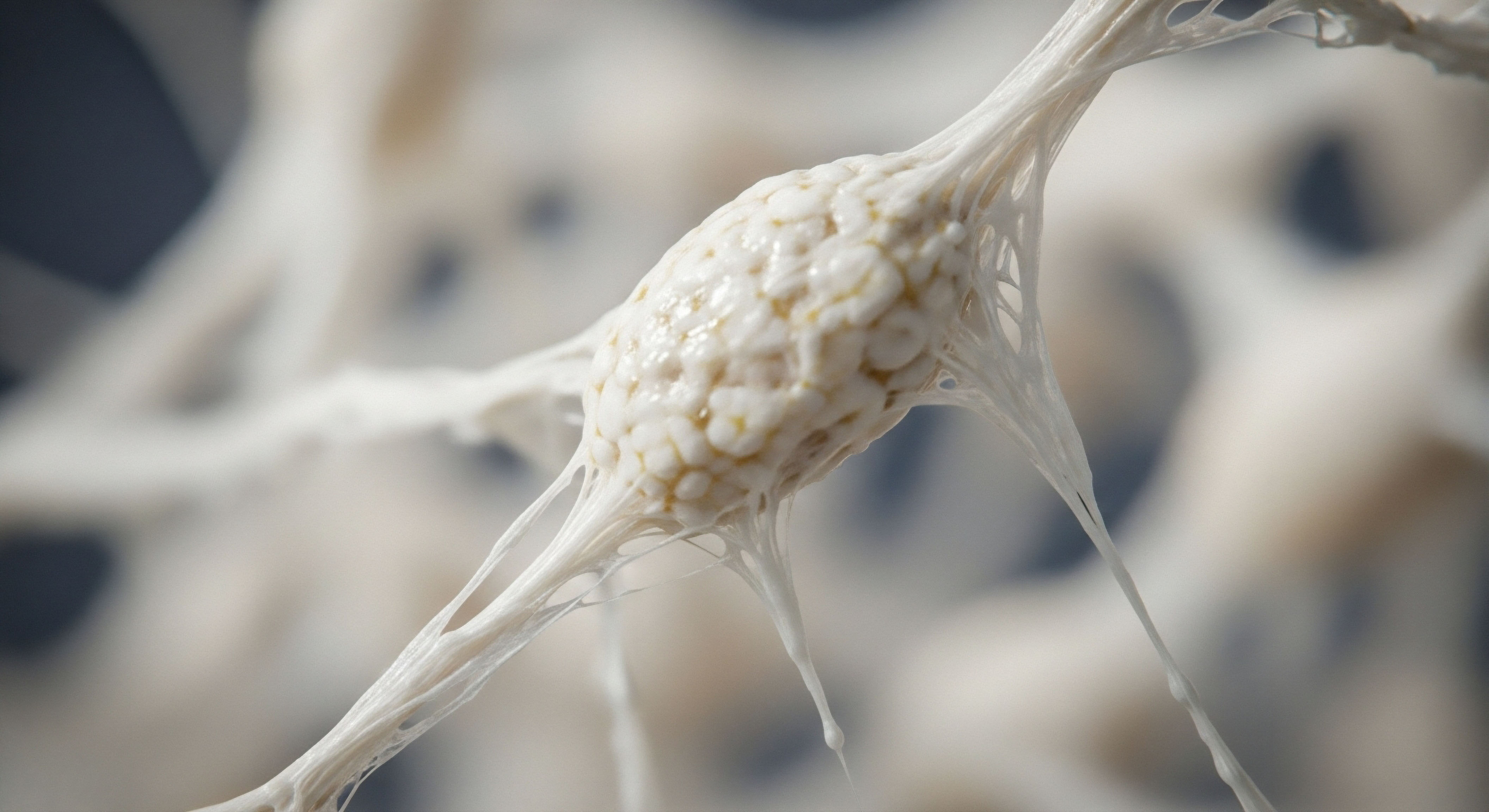
Your brain is a remarkably active hormonal environment. It does not just receive hormonal signals from elsewhere in the body; it actively produces and metabolizes them. Hormones that are active within the central nervous system are known as neurosteroids. Testosterone, along with its derivatives, is a powerful neurosteroid. It crosses the protective blood-brain barrier and directly influences the health and function of your neurons ∞ the very cells responsible for thought, memory, and learning.
Think of these hormones as dedicated maintenance workers for your brain’s complex electrical grid. They support the growth of new connections between neurons, protect existing cells from damage, and help manage inflammation. When the levels of these critical workers decline, as they do during menopause, the system’s resilience can be compromised. This hormonal shift is a key reason why women face a higher risk of neurodegenerative conditions like Alzheimer’s disease than men.
The gradual decline of testosterone in women is a physiological reality that directly impacts the brain’s ability to maintain itself and function optimally.

What Is the Connection to Neurodegeneration?
Neurodegenerative conditions are characterized by the progressive loss of structure and function of neurons. In Alzheimer’s disease, this involves the accumulation of proteins like amyloid-beta plaques and tau tangles, which disrupt communication between brain cells and lead to their eventual death.
Emerging research indicates that the age-related loss of sex steroid hormones, including both testosterone and estrogen, is a significant risk factor in the development of this pathology. The presence of these hormones appears to support the brain’s natural defense mechanisms against these degenerative processes.
The question then becomes a proactive one. If the decline of these essential hormones contributes to increased vulnerability, could restoring them to optimal levels help protect the brain? This line of inquiry moves the conversation from one of passive acceptance of age-related cognitive decline to one of active, informed biological support.
It is about understanding the machinery of your own body to provide it with the raw materials it needs to thrive, particularly within the delicate and vital ecosystem of the brain.


Intermediate
To comprehend how testosterone therapy might serve a neuroprotective role, we must examine the specific biological mechanisms at play within the female brain. The conversation moves from the general presence of hormones to their specific actions at a cellular level. The evidence points toward testosterone exerting its influence through several interconnected pathways, each contributing to a more resilient neural environment. Its potential is rooted in its ability to directly counteract some of the core pathological processes associated with neurodegenerative diseases.

Direct Mechanisms of Neural Support
Testosterone’s function in the brain is multifaceted. It acts directly through androgen receptors, which are found on neurons throughout critical brain regions, and it also serves as a precursor, or prohormone, that can be converted into other vital neurosteroids. This dual action allows it to provide comprehensive support to neural tissue.
- Amyloid-Beta Regulation ∞ A primary hallmark of Alzheimer’s disease is the accumulation of amyloid-beta protein into toxic plaques. Studies in animal models have shown that androgen depletion leads to an increase in these amyloid-beta levels. Conversely, testosterone appears to modulate the processing of the amyloid precursor protein (APP), steering it away from the pathway that produces the toxic amyloid-beta fragments.
- Anti-Inflammatory Action ∞ Chronic neuroinflammation is another key driver of neuronal damage. Testosterone has demonstrated anti-inflammatory properties within the brain, helping to reduce the activity of pro-inflammatory molecules that can damage neurons over time. This creates a less hostile environment for brain cells, allowing them to function more effectively.
- Promotion of Neuronal Survival ∞ Testosterone supports the expression of genes and proteins that promote cell survival and growth. It can protect neurons from apoptosis (programmed cell death) and encourage the growth of neurites, the projections that form synaptic connections between brain cells. This fosters a more robust and adaptable neural network, which is the physical basis of learning and memory.
By actively reducing harmful protein aggregation and inflammation while promoting cellular health, testosterone functions as a key regulator of the brain’s internal ecosystem.
Clinical Protocols for Women
When considering testosterone therapy for women, the goal is physiological restoration, not the pursuit of supraphysiological levels. The protocols are designed to return circulating testosterone to the upper end of the normal range for a healthy young woman, thereby restoring its protective and functional benefits. The administration must be precise, monitored, and tailored to the individual’s biochemistry and clinical response.
A common and effective protocol involves weekly subcutaneous injections of Testosterone Cypionate. This method provides a steady, consistent release of the hormone, avoiding the peaks and troughs that can occur with other delivery systems. The dosage is carefully calibrated for the female physiology.
For instance, a typical starting dose might be 10 to 20 units (0.1 to 0.2ml of a 200mg/ml solution) per week. This small volume is administered with a tiny insulin syringe into the subcutaneous fat of the abdomen or glute, making it a relatively simple and painless process. This protocol is often balanced with progesterone, particularly for post-menopausal women, to ensure a harmonious hormonal environment. The objective is to recreate the body’s natural synergy of hormones for optimal function.

Comparing Delivery Methods
While subcutaneous injections are a preferred method for their consistency and dose control, other options exist. Understanding their characteristics is important for making an informed clinical decision.
| Delivery Method | Advantages | Considerations |
|---|---|---|
| Subcutaneous Injections |
Provides stable blood levels. Allows for precise and easily adjustable dosing. Self-administered weekly. |
Requires comfort with self-injection. Involves needles and syringes. |
| Transdermal Gels/Creams |
Daily application, non-invasive. Easy to start and stop treatment. |
Potential for inconsistent absorption. Risk of transference to others through skin contact. Daily application can be inconvenient. |
| Pellet Therapy |
Long-acting, requiring infrequent insertion (every 3-4 months). Provides sustained hormone release. |
Requires a minor in-office procedure for insertion. Dosing cannot be adjusted once the pellet is inserted. Potential for local complications at the insertion site. |

What about the Role of Estrogen?
It is impossible to discuss testosterone’s role in the female brain without acknowledging its relationship with estrogen. An enzyme present in the brain, called aromatase, converts a portion of testosterone directly into estradiol, the most potent form of estrogen. This means that some of testosterone’s neuroprotective effects are actually mediated by its conversion to estrogen within the brain tissue itself.
This localized production of estrogen is profoundly important, as it supports neuronal health and function directly where it is needed. Therefore, providing testosterone can support both androgenic and estrogenic pathways within the brain, offering a more comprehensive approach to neuroprotection than administering estrogen alone. This highlights the intricate and cooperative nature of the endocrine system in maintaining cognitive wellness.


Academic
A sophisticated analysis of testosterone’s potential neuroprotective role in women requires moving beyond its direct cellular effects and examining its position within a larger, interconnected system. The primary axis governing reproductive hormones is the Hypothalamic-Pituitary-Gonadal (HPG) axis.
The age-related dysregulation of this axis during menopause does not simply result in a decline of sex steroids; it creates a cascade of signaling changes that profoundly impact brain health, metabolic function, and neuroinflammation. It is at the intersection of this axis with systemic metabolic integrity that the most compelling case for testosterone’s therapeutic potential can be made.

The HPG Axis and Neurological Senescence
The HPG axis is a finely tuned feedback loop. The hypothalamus releases Gonadotropin-Releasing Hormone (GnRH), which signals the pituitary to release Luteinizing Hormone (LH) and Follicle-Stimulating Hormone (FSH). These gonadotropins, in turn, stimulate the ovaries to produce estrogen, progesterone, and testosterone. The sex steroids then provide negative feedback to the hypothalamus and pituitary, keeping the system in balance.
During menopause, as ovarian function declines, the production of sex steroids plummets. The loss of this negative feedback causes the hypothalamus and pituitary to ramp up their output, leading to chronically elevated levels of GnRH and LH. This state of HPG axis dysregulation is not benign.
Both GnRH and LH have receptors in brain regions susceptible to Alzheimer’s pathology, like the hippocampus. Elevated signaling from these hormones is increasingly implicated as a driver of neurodegenerative processes, including the aberrant re-entry of neurons into the cell cycle, a pathological process termed “dyosis,” which leads to tau phosphorylation and amyloid-beta production.
Testosterone therapy, in this context, does more than simply replace a missing hormone. By restoring a key component of the hormonal milieu, it can help re-establish a degree of negative feedback on the HPG axis, potentially attenuating the chronically elevated and damaging signals from GnRH and LH. This system-level intervention may be as significant as its direct effects on neurons.
The dysregulation of the HPA-HPG axes following menopause creates a hormonal environment that promotes neurodegenerative processes through elevated gonadotropin signaling.

Metabolic Dysfunction the Inflammatory Bridge
The brain is an organ with immense metabolic demands, consuming roughly 20% of the body’s glucose. Its health is inextricably linked to systemic metabolic function. The menopausal transition is frequently associated with a shift toward metabolic dysfunction, including increased insulin resistance. Testosterone plays a vital role in maintaining metabolic health. It helps preserve insulin sensitivity, promotes lean muscle mass (a primary site of glucose disposal), and regulates fat distribution.
The decline in testosterone contributes to the opposite ∞ a tendency toward insulin resistance, sarcopenia (age-related muscle loss), and visceral adiposity. Insulin resistance in the periphery is mirrored by insulin resistance in the brain, a condition sometimes referred to as “Type 3 diabetes.” When brain cells become resistant to insulin, their ability to utilize glucose for energy is impaired. This energy crisis compromises neuronal function and survival and is a well-established feature of Alzheimer’s disease.
Furthermore, visceral adipose tissue is not inert; it is a highly active endocrine organ that secretes pro-inflammatory cytokines. This creates a state of chronic, low-grade systemic inflammation that fuels neuroinflammation. By supporting metabolic health and insulin sensitivity, testosterone therapy can help break this vicious cycle, reducing the inflammatory burden on the brain.

Key Molecular Intersections
The interplay between hormonal decline, metabolic dysfunction, and neurodegeneration can be observed at the molecular level. The following table outlines some of the critical pathways where these systems converge.
| Pathway/Molecule | Role in Healthy State | Dysfunction in Hormonal Decline | Potential Influence of Testosterone Therapy |
|---|---|---|---|
| Insulin Signaling (PI3K/Akt) |
Promotes cell survival, growth, and glucose uptake in neurons. It is a critical pathway for neuroprotection. |
Becomes impaired with insulin resistance, reducing neuronal energy metabolism and increasing vulnerability to apoptosis. |
May help restore insulin sensitivity, thereby reactivating this crucial pro-survival signaling cascade within the brain. |
| Aromatase Enzyme |
Converts testosterone to estradiol locally within the brain, providing on-demand neuroprotective estrogenic signaling. |
Reduced substrate (testosterone) leads to lower intracrine production of estradiol, diminishing a key source of local neural support. |
Provides the necessary substrate for brain aromatase, ensuring continued local production of neuroprotective estradiol. |
| GABAergic System |
GABA is the primary inhibitory neurotransmitter, crucial for calming neuronal excitability. Progesterone metabolites are potent positive modulators of GABA receptors. |
Loss of progesterone and its metabolites can lead to a state of neuronal hyperexcitability, which is metabolically costly and can be toxic. |
While not a direct effect, balanced hormonal protocols that include progesterone work synergistically to restore GABAergic tone and protect against excitotoxicity. |

What Is the Clinical Implication for Neuroprotection?
The evidence suggests that viewing testosterone therapy solely as a treatment for libido or energy is a limited perspective. A more accurate framework considers it a systemic intervention aimed at restoring metabolic and endocrine stability. By improving insulin sensitivity, reducing systemic inflammation, and potentially modulating the HPG axis, testosterone therapy addresses several foundational pillars of brain health simultaneously.
A 2013 study presented at The Endocrine Society’s annual meeting found that postmenopausal women treated with testosterone gel showed statistically significant improvements in verbal learning and memory compared to a placebo group. While larger and longer-term trials are needed, this aligns with the mechanistic understanding that androgens are vital for cognitive processes. The ultimate goal is not just to prevent disease, but to preserve the high-functioning, resilient cognitive capacity that defines our quality of life.

References
- Bowen, R.L. et al. “Dysregulation of the Hypothalamic-Pituitary-Gonadal Axis with Menopause and Andropause Promotes Neurodegenerative Senescence.” Journal of Neuropathology & Experimental Neurology, vol. 64, no. 2, 2005, pp. 93-103.
- Pike, C. J. et al. “Androgens, Alzheimer’s disease, and hormone therapy.” Biochimica et Biophysica Acta (BBA) – Molecular Basis of Disease, vol. 1822, no. 3, 2012, pp. 409-15.
- Rosario, E. R. et al. “Androgens and Alzheimer’s disease.” Current Opinion in Pharmacology, vol. 4, no. 4, 2004, pp. 408-13.
- Brinton, R. D. “Neurosteroids as regenerative agents in the brain ∞ therapeutic implications.” Nature Reviews Endocrinology, vol. 9, no. 5, 2013, pp. 241-50.
- Davis, S. R. et al. “Testosterone for low libido in postmenopausal women not taking estrogen.” The New England Journal of Medicine, vol. 359, no. 19, 2008, pp. 2005-17.
- Gurvich, C. et al. “The effect of transdermal testosterone on mood and cognition in postmenopausal women with low libido.” Menopause, vol. 25, no. 11, 2018, pp. 1327-1334.
- Golisano, V. et al. “Testosterone and the Brain ∞ A Narrative Review.” Journal of Clinical Medicine, vol. 10, no. 7, 2021, p. 1394.
- Henderson, V. W. “Alzheimer’s disease ∞ review of hormone therapy trials and implications for treatment and prevention after menopause.” Journal of Steroid Biochemistry and Molecular Biology, vol. 93, no. 2-5, 2005, pp. 115-21.
- Islam, F. et al. “Neuroprotective Role of Steroidal Sex Hormones ∞ An Overview.” Journal of Neurosciences in Rural Practice, vol. 9, no. 3, 2018, pp. 397-405.
- Howard, J. “Testosterone and DHEA are Directly Involved in Alzheimer’s Disease.” Journal of Alzheimer’s Disease & Parkinsonism, vol. 2, no. 5, 2012, p. e118.

Reflection
The information presented here offers a map of the intricate biological landscape connecting your hormonal health to your cognitive future. It details the pathways, the mechanisms, and the clinical logic behind supporting your body’s systems. This knowledge is a powerful tool, shifting the perspective from one of inevitable decline to one of proactive stewardship.
Your personal health narrative is unique, written in the language of your own biochemistry and lived experience. Understanding the grammar of that language is the foundational step. The next chapter involves a personalized dialogue with a qualified clinical guide to translate this knowledge into a strategy that honors your individual biology and supports your goal of a vibrant, cognitively sharp life.