


Fundamentals
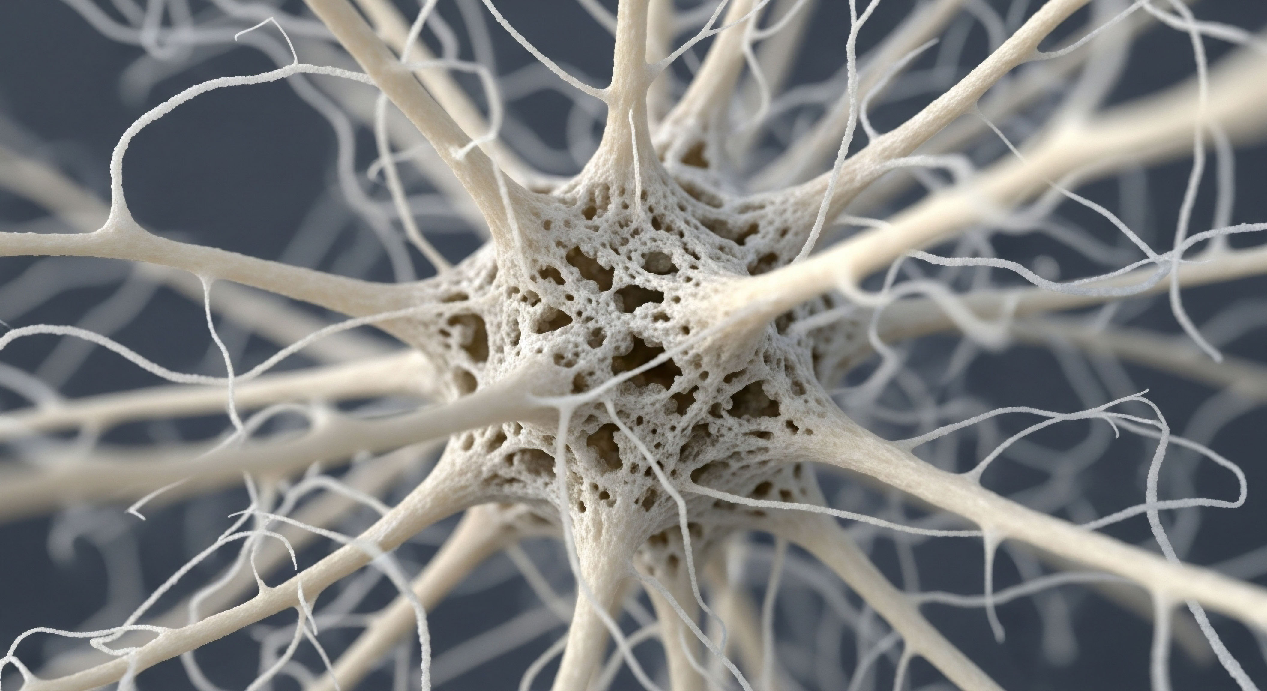
The feeling of strength, of being solid and capable in your own body, is deeply connected to the silent, tireless work happening within your bones. Your skeletal framework is a living, dynamic system, a biological marvel that is constantly renewing itself. This process, known as bone remodeling, is a delicate balance between two types of cells.
Osteoclasts work to break down and remove old bone tissue, while osteoblasts are responsible for building new bone in its place. For most of your early adult life, this system operates in a state of equilibrium, ensuring your skeleton remains strong and resilient.
The primary conductors of this intricate orchestra are your hormones, specifically estrogen and testosterone. These powerful chemical messengers are fundamental to maintaining skeletal integrity. They act as a governing influence, primarily by moderating the activity of osteoclasts. By keeping bone resorption in check, they ensure that the rate of new bone formation is sufficient to maintain or even increase your bone mass. This hormonal protection is a cornerstone of your structural health.
Your body’s hormonal state directly governs the continuous process of bone renewal.
As you move through different life stages, particularly during perimenopause, menopause, or andropause, the natural decline in these key hormones disrupts this carefully maintained balance. With lower levels of estrogen and testosterone, the restraining signal on osteoclasts weakens. This allows bone resorption to outpace bone formation, leading to a net loss of bone tissue over time.
The experience of this internal shift can manifest as a subtle feeling of increased fragility or a documented change in bone density scans. Understanding this biological mechanism is the first step toward addressing the root cause of age-related bone loss and exploring therapeutic pathways to restore skeletal resilience.

The Foundation of Hormonal Support
Traditional hormone replacement therapy directly addresses this underlying hormonal deficit. The goal of these protocols is to reintroduce the specific hormones that have declined, thereby restoring the body’s natural ability to regulate bone remodeling. For women, this typically involves estrogen, which is the most critical hormone for protecting against bone loss.
For men, testosterone serves a similar protective function. By replenishing these hormones, biochemical recalibration aims to re-establish the systemic environment that supports skeletal health, slowing the rate of bone breakdown and preserving the structural integrity you already possess.


Intermediate
Moving beyond the foundational understanding of hormonal influence, we can examine the specific mechanisms through which different therapeutic interventions support bone health. Both traditional hormone replacement and peptide therapies aim to improve skeletal integrity, yet they achieve this through distinct biological pathways. One works by restoring a systemic regulatory function, while the other provides a targeted stimulatory signal.

Hormone Replacement the Direct Approach to Preservation
Hormonal optimization protocols function by replenishing the body’s diminished supply of key hormones like estrogen and testosterone. This approach is a form of systemic support. By restoring these hormones to more youthful levels, the therapy directly counteracts the primary driver of age-related bone loss which is unchecked osteoclast activity.
Estrogen, for instance, is exceptionally effective at suppressing the signaling molecules that promote the formation and activation of bone-resorbing osteoclasts. Testosterone contributes to bone health through its own direct actions and through its conversion to estrogen within bone tissue.
The clinical application is tailored to the individual’s specific hormonal needs:
- For Women ∞ Protocols often involve bioidentical estrogen, administered via patches, creams, or pellets, to provide a steady state of hormonal support. Progesterone is typically included to protect the uterine lining in women who have not had a hysterectomy. Low-dose testosterone may also be used to support libido, energy, and its own contributions to bone density.
- For Men ∞ Testosterone Replacement Therapy (TRT), usually administered via weekly injections of Testosterone Cypionate, is the standard. This therapy directly addresses low testosterone levels, which are a known contributor to bone loss in men. Ancillary medications like Gonadorelin may be used to maintain testicular function.
Hormone replacement therapy primarily acts as a brake on bone breakdown, preserving existing bone mass.

Peptide Therapy a Targeted Signal for Growth
Peptide therapies represent a different strategy. Peptides are short chains of amino acids that act as precise signaling molecules. Instead of replacing a hormone, certain peptides stimulate the body’s own production of other beneficial compounds. In the context of bone health, the most relevant peptides are growth hormone secretagogues (GHS). This class includes therapies like Sermorelin, Tesamorelin, and the combination of Ipamorelin and CJC-1295.
These peptides work by signaling the pituitary gland to release more Growth Hormone (GH). The increased GH levels then travel to the liver and other tissues, stimulating the production of Insulin-like Growth Factor 1 (IGF-1). IGF-1 is a potent anabolic factor, meaning it promotes growth and building activities throughout the body. Within the skeletal system, IGF-1 directly stimulates osteoblasts, the cells responsible for forming new bone tissue. This pathway effectively encourages the ‘building’ side of the bone remodeling equation.
Another peptide, BPC-157, is recognized for its profound healing properties. While not a primary bone density agent, it has demonstrated a significant ability to accelerate the healing of fractures and soft tissue injuries by promoting blood vessel growth and cellular repair at the site of injury.

Comparing Therapeutic Mechanisms
The following table outlines the distinct approaches of these two therapeutic modalities.
| Feature | Hormone Replacement Therapy (HRT) | Growth Hormone Peptide Therapy |
|---|---|---|
| Therapeutic Agent | Bioidentical Estrogen, Progesterone, Testosterone | Ipamorelin/CJC-1295, Sermorelin, Tesamorelin |
| Primary Mechanism | Directly replaces deficient hormones to restore systemic balance. | Stimulates the pituitary gland to increase natural Growth Hormone and IGF-1 production. |
| Target Cells | Primarily suppresses osteoclast (bone resorption cell) activity. | Primarily stimulates osteoblast (bone formation cell) activity. |
| Primary Effect on Bone | Anti-resorptive (slows bone breakdown). | Anabolic (promotes new bone formation). |
| Administration | Injections, pellets, patches, creams. | Subcutaneous injections. |


Academic
A sophisticated analysis of bone health interventions requires a deep examination of the cellular and molecular signaling pathways that govern skeletal homeostasis. The comparison between traditional endocrine system support and peptide-based strategies reveals two distinct, yet potentially synergistic, approaches to modulating the bone remodeling unit. Their efficacy is rooted in their differential impact on the complex interplay between osteoblasts and osteoclasts.

The Molecular Basis of Hormonal Bone Protection
The protective effect of estrogen on the skeleton is primarily mediated through its influence on the OPG/RANK/RANKL signaling axis. This system is the central regulator of osteoclast differentiation and activation.
- RANKL (Receptor Activator of Nuclear Factor Kappa-B Ligand) is a protein expressed by osteoblasts and other cells. When it binds to its receptor, RANK, on the surface of osteoclast precursor cells, it triggers a signaling cascade that leads to their maturation into active, bone-resorbing osteoclasts.
- OPG (Osteoprotegerin) is also produced by osteoblasts and acts as a soluble decoy receptor. It binds to RANKL, preventing it from interacting with RANK. The ratio of RANKL to OPG is therefore a critical determinant of bone resorption rates.
- Estrogen’s Role ∞ Estrogen signaling within bone cells increases the expression of OPG and decreases the expression of RANKL. This action shifts the RANKL/OPG ratio in favor of OPG, effectively suppressing osteoclastogenesis and reducing bone resorption. The decline of estrogen during menopause removes this crucial brake, leading to a surge in osteoclast activity and accelerated bone loss.

The Anabolic Signaling of the GH/IGF-1 Axis
Growth hormone secretagogue peptides like Ipamorelin/CJC-1295 operate through a different mechanism focused on anabolism. By stimulating pulsatile GH release, they elevate systemic and local levels of IGF-1. IGF-1 is a master regulator of cellular growth and proliferation, with profound effects on osteoblasts.
IGF-1 binds to its receptor (IGF-1R) on osteoblasts, initiating downstream signaling through pathways like the PI3K/Akt cascade. This activation promotes several key osteogenic processes:
- Proliferation ∞ It encourages the division of pre-osteoblast cells, expanding the pool of bone-building cells.
- Differentiation ∞ It guides these precursor cells to mature into fully functional osteoblasts.
- Synthesis ∞ It enhances the production of type 1 collagen and other proteins that form the bone matrix, the very scaffolding upon which minerals are deposited.
Peptide therapies for bone health work by amplifying the body’s innate anabolic signals for tissue generation.
Research in animal models has shown that treatment with GHS agents like ipamorelin can increase bone mineral content. A key finding from these studies is that the increase in bone mass was largely due to an increase in the bone’s cross-sectional area, a process known as periosteal apposition, rather than a significant change in volumetric bone mineral density.
This suggests that GH/IGF-1 signaling promotes the laying down of new bone on the outer surface, leading to larger, geometrically stronger bones, which is a distinct mechanism from the mineralization-preserving effect of estrogen.

Comparative Cellular Signaling Pathways
The following table provides a granular comparison of the signaling cascades initiated by these two classes of therapies.
| Signaling Factor | Estrogen | Insulin-like Growth Factor 1 (IGF-1) |
|---|---|---|
| Primary Source | Ovaries, Adrenals, Adipose Tissue (Replenished by HRT) | Liver and local bone cells (Stimulated by GH peptides) |
| Key Signaling Pathway | Modulation of the OPG/RANK/RANKL axis. | Activation of the IGF-1R and downstream PI3K/Akt/mTOR pathways. |
| Direct Effect on Osteoblasts | Promotes survival and OPG production. | Stimulates proliferation, differentiation, and matrix synthesis. |
| Direct Effect on Osteoclasts | Inhibits differentiation and promotes apoptosis (cell death). | Minimal direct effect; action is primarily through osteoblasts. |
| Resulting Bone Architecture | Preservation of mineral density and microarchitecture by reducing resorption. | Potential increase in bone size (periosteal apposition) and formation. |

References
- Svensson, J. Lall, S. Dickson, S. L. Bengtsson, B. Å. Rømer, J. Ahnfelt-Rønne, I. Ohlsson, C. & Jansson, J. O. (2000). The GH secretagogues ipamorelin and GH-releasing peptide-6 increase bone mineral content in adult female rats. Journal of Endocrinology, 165(3), 569 ∞ 577.
- Riggs, B. L. Khosla, S. & Melton, L. J. 3rd. (2002). Sex steroids and the construction and conservation of the adult skeleton. Endocrine Reviews, 23(3), 279 ∞ 302.
- Welle, S. Thornton, C. Statt, M. & McHenry, B. (1996). Growth hormone and insulin-like growth factor I effects on the synthesis of myofibrillar and sarcoplasmic proteins in healthy men and women. The Journal of Clinical Endocrinology and Metabolism, 81(10), 3523-3527.
- Lindsay, R. Gallagher, J. C. Kleerekoper, M. & Pickar, J. H. (2002). Effect of lower doses of conjugated equine estrogens with and without medroxyprogesterone acetate on bone in early postmenopausal women. JAMA, 287(20), 2668 ∞ 2676.
- Brixen, K. Nielsen, H. K. Mosekilde, L. & Flyvbjerg, A. (1990). A short course of recombinant human growth hormone treatment stimulates osteoblasts and activates bone remodeling in normal human volunteers. The Journal of Bone and Mineral Research, 5(6), 609 ∞ 618.
- Rosen, T. Wilhelmsen, L. Landin-Wilhelmsen, K. Lappas, G. & Bengtsson, B. A. (1997). Increased fracture frequency in adult patients with hypopituitarism and GH deficiency. European Journal of Endocrinology, 137(3), 240-245.
- Appelman-Dijkstra, N. M. Claessen, K. M. Roelfsema, F. Pereira, A. M. & Biermasz, N. R. (2014). Long-term effects of growth hormone (GH) replacement on bone mineral density in patients with adult-onset GH deficiency ∞ a meta-analysis. The Journal of Clinical Endocrinology and Metabolism, 99(11), 4049-4058.
Reflection
The information presented here offers a map of the biological terrain related to your skeletal health. It details the pathways and mechanisms that govern the strength and resilience of your internal framework. This knowledge is a powerful tool, transforming abstract concerns into a clear understanding of the systems at play within your own body.
Your personal health story is unique, written in the language of your symptoms, your lab results, and your goals. Viewing these therapeutic options through the lens of their distinct mechanisms allows for a more informed conversation about your path forward. The ultimate goal is to select the right tools to support your body’s innate capacity for strength and vitality, creating a personalized strategy that aligns with your unique physiology and aspirations for long-term wellness.