
Fundamentals
The question of whether lifestyle changes alone can reverse bone loss from menopause touches upon a deeply personal aspect of health. It speaks to a desire to reclaim control over a body that feels as though it is operating under a new set of rules.
You may feel this shift as a subtle change in your physical resilience or a new awareness of your body’s fragility. This experience is a direct biological reality, rooted in the profound hormonal transition of menopause. Understanding the mechanics of this process is the first step toward informed action.
Your bones are not static structures; they are dynamic, living tissues in a constant state of renewal, a process known as remodeling. This intricate biological dance involves two primary types of cells ∞ osteoclasts, which break down old bone tissue, and osteoblasts, which build new bone tissue. For most of your life, this process is balanced, orchestrated largely by the hormone estrogen.
Estrogen acts as a protective brake on the activity of osteoclasts. It modulates the rate at which bone is resorbed, ensuring that the construction work of the osteoblasts can keep pace. As menopause approaches, the ovaries decrease their production of estrogen. The decline of this essential hormone removes the restraining signal on the osteoclasts.
Consequently, the rate of bone breakdown begins to outpace the rate of bone formation. This systemic change leads to a net loss of bone density and a degradation of bone quality, a condition known as osteopenia, which can progress to osteoporosis. The architecture of your skeleton, once robust and dense, becomes more porous and susceptible to fracture. This process is a silent one, often progressing without any outward signs until a fracture occurs.
The decline in estrogen during menopause disrupts the natural balance of bone renewal, leading to a net loss of bone density.
Lifestyle interventions represent a foundational strategy to counteract this hormonal shift. These are conscious choices that directly influence the biological environment of your skeletal system. Engaging in specific forms of exercise and ensuring proper nutrition provides the necessary signals and resources for your bones to maintain their strength.
These actions are your way of communicating with your cellular machinery, supporting the work of the osteoblasts and providing the raw materials they need to function effectively. While these changes are powerful, their effect is one of slowing the rate of loss and preserving the bone that exists. They are a critical component of a comprehensive strategy for lifelong skeletal health.

What Is the True Nature of Bone?
Your skeletal framework is a metabolically active organ, much like your heart or liver. It possesses a remarkable capacity to adapt to the loads placed upon it. This principle, known as Wolff’s Law, states that bone remodels itself in response to mechanical stress.
When you engage in activities like walking, running, or lifting weights, you are sending a direct message to your bones to become stronger and denser. This mechanical loading stimulates osteoblast activity, encouraging the deposition of new bone tissue. The sensation of physical effort during exercise is the external feeling of this internal biological conversation taking place.
The composition of your bones is also critically dependent on a steady supply of specific nutrients. Calcium is the primary mineral that gives bone its hardness and structural integrity. Vitamin D is essential for your body to absorb that calcium from your diet and deposit it into your skeleton.
Think of calcium as the bricks and vitamin D as the master mason that directs their placement. Without adequate vitamin D, even a calcium-rich diet may fail to support bone health effectively, as the mineral cannot be properly utilized. Your daily dietary choices, therefore, have a direct and cumulative impact on the physical substance of your skeleton.

How Does Menopause Alter This System?
The menopausal transition introduces a systemic challenge to this carefully balanced system. The reduction in circulating estrogen is the primary catalyst for accelerated bone loss. Research indicates that women can lose up to 20% of their bone density in the five to seven years following menopause.
This period represents a window of heightened vulnerability for skeletal health. The loss of estrogen’s protective influence means that the osteoclasts become more numerous and more active, leading to a rapid excavation of bone tissue that the osteoblasts cannot fully replenish.
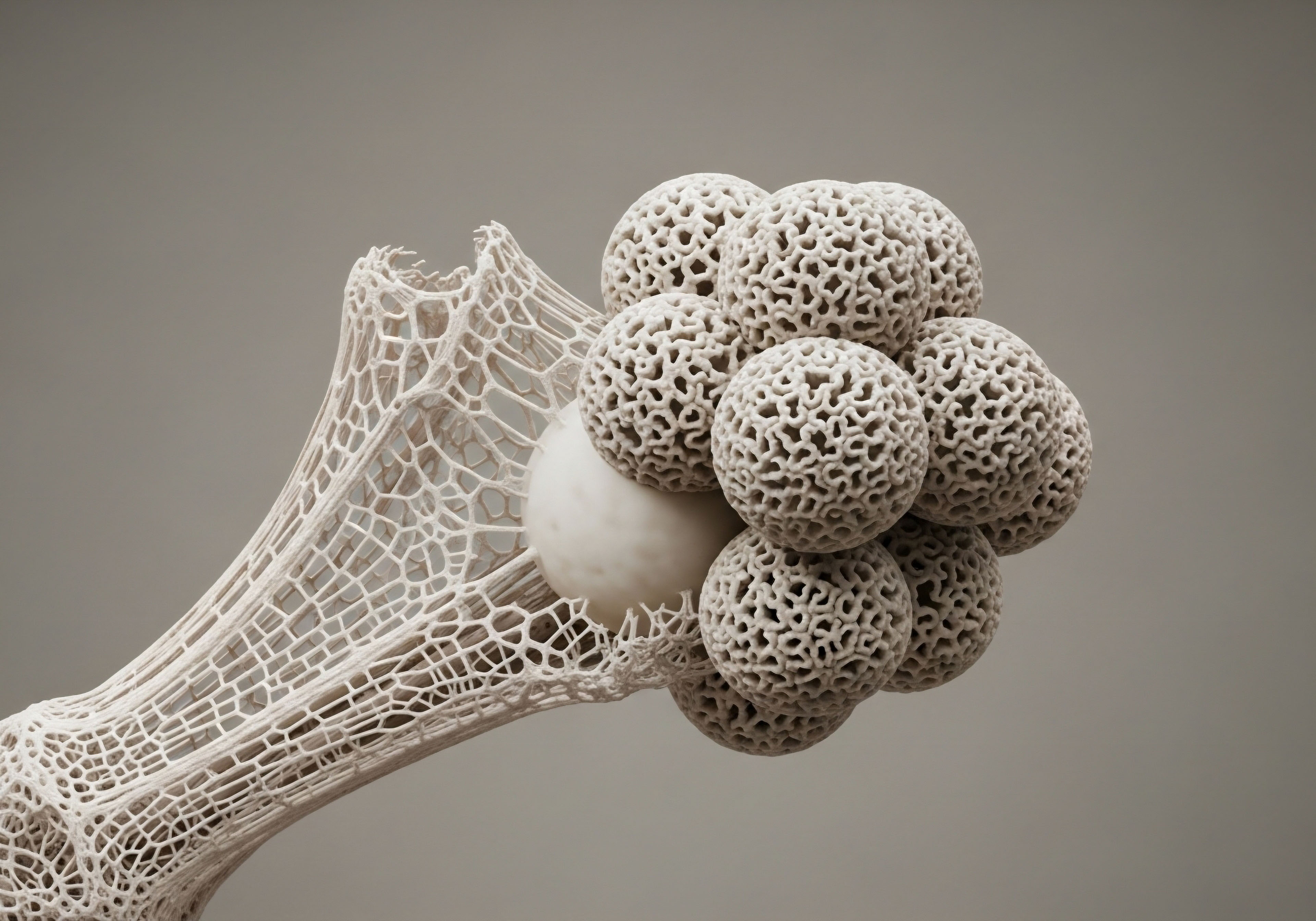
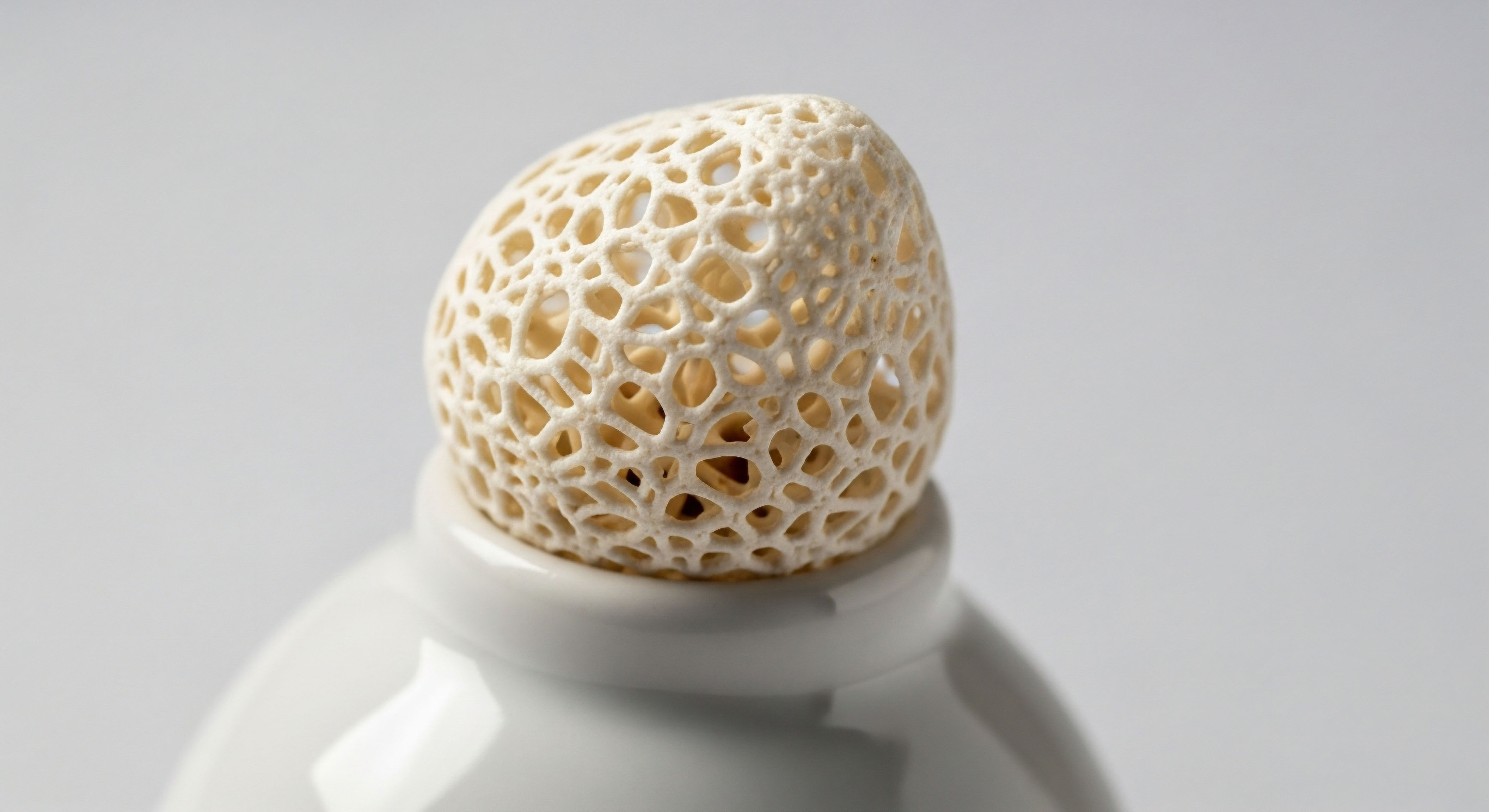
This hormonal change affects the entire skeleton, but certain areas are more vulnerable. The trabecular bone, the spongy, honeycomb-like inner part of bones found in the spine, hip, and wrist, is particularly susceptible to this accelerated resorption. This is why fractures in these areas are so common in postmenopausal women.
The experience of menopause is unique to each individual, yet the underlying biological mechanism of estrogen withdrawal and its impact on bone remodeling is a shared reality. Addressing bone health during this time requires a proactive and informed approach that acknowledges the profound nature of this hormonal shift.


Intermediate
Moving beyond the fundamentals, a more detailed examination of lifestyle interventions reveals their specific mechanisms and their inherent limitations. While these strategies are indispensable for mitigating bone loss, the question of reversal requires a nuanced understanding of clinical realities. Lifestyle modifications alone cannot fully restore bone density that has already been lost due to the profound hormonal shifts of menopause.
Their primary role is to slow the rate of decline, preserve existing bone architecture, and reduce the risk of fracture. Achieving a more significant improvement in bone mineral density often necessitates a combination of these foundational habits with targeted clinical protocols designed to recalibrate the body’s endocrine system.
The efficacy of lifestyle changes is best understood by viewing them as powerful inputs into a complex biological system. Exercise provides the mechanical stimulus for bone formation, while nutrition supplies the essential building blocks. However, these inputs are being introduced into a hormonal environment that has fundamentally changed.
The absence of sufficient estrogen means the baseline rate of bone resorption remains elevated. Therefore, even the most dedicated lifestyle program is working against a strong biological current. This is where a clinical perspective becomes essential, offering tools that can address the underlying hormonal imbalance directly, creating a more favorable environment for bone health to be maintained and potentially improved.

A Closer Look at Exercise Protocols
To effectively stimulate bone, exercise must be specific and progressive. Different types of exercise confer distinct benefits to the skeletal system. A well-rounded program incorporates elements of mechanical loading, muscular strengthening, and balance training to create a comprehensive defense against bone fragility. The goal is to send clear, powerful signals to the osteoblasts to increase their activity.
Weight-bearing exercises are those in which your bones and muscles work against gravity to keep you upright. High-impact versions of these exercises, such as jumping, have shown potential for stimulating bone growth, particularly in premenopausal women. For postmenopausal women, the results can be more varied, but activities like brisk walking, jogging, and dancing remain beneficial.
Resistance training, which involves moving your body against an opposing force, such as weights or resistance bands, directly strengthens the muscles that pull on the bones, which in turn stimulates bone growth at the site of muscle attachment. Balance exercises, such as tai chi or yoga, are also important for preventing falls, which are the primary cause of fractures in individuals with osteoporosis.
| Exercise Type | Primary Mechanism | Examples | Key Benefit |
|---|---|---|---|
| Weight-Bearing (High-Impact) | Direct mechanical loading from ground reaction forces. | Jumping, running, high-impact aerobics. | Stimulates osteoblast activity throughout the skeleton. |
| Weight-Bearing (Low-Impact) | Sustained force of gravity on the skeleton. | Walking, elliptical training, stair climbing. | Maintains bone density with lower stress on joints. |
| Resistance Training | Muscles pulling on bone insertion points. | Lifting weights, using resistance bands, bodyweight exercises. | Targets specific bone sites for increased density. |
| Balance & Flexibility | Improves proprioception and stability. | Yoga, Tai Chi, standing on one leg. | Reduces the risk of falls and subsequent fractures. |

Nutritional Architecture for Skeletal Support
A diet optimized for bone health provides the essential minerals and vitamins required for the continuous process of bone remodeling. While calcium and vitamin D are the most recognized nutrients, a broader spectrum of minerals and vitamins contributes to the structural integrity of the skeleton. The following table outlines the key nutritional components for maintaining bone health after menopause.
A targeted exercise regimen and a nutrient-dense diet form the cornerstones of managing postmenopausal bone health.
| Nutrient | Recommended Daily Intake (Women 50+) | Primary Function | Dietary Sources |
|---|---|---|---|
| Calcium | 1,200 mg | Provides the primary mineral component of bone structure. | Dairy products, fortified plant milks, leafy greens, tofu. |
| Vitamin D | 800-1,000 IU | Facilitates the absorption of calcium from the intestine. | Fatty fish, egg yolks, fortified foods, sun exposure. |
| Magnesium | 320 mg | Contributes to the bone crystal lattice and influences osteoblasts. | Nuts, seeds, whole grains, dark chocolate. |
| Vitamin K | 90 mcg | Activates proteins involved in bone mineralization. | Leafy green vegetables, broccoli, Brussels sprouts. |
| Protein | ~1.0-1.2 g/kg body weight | Forms the collagen matrix that provides bone with flexibility. | Lean meats, poultry, fish, legumes, dairy. |

The Limitations of Lifestyle and the Role of Clinical Support
It is important to approach the potential of lifestyle changes with a clear and realistic perspective. For a woman who has already experienced significant bone loss, diet and exercise alone are unlikely to restore her bone mineral density to premenopausal levels.
These interventions are exceptionally effective at slowing further loss and are a non-negotiable part of any treatment plan. However, the biological reality of an estrogen-deficient state means that the process of bone resorption remains fundamentally upregulated. This is where modern clinical protocols can offer a significant advantage.
Therapies such as hormonal optimization can address the root cause of the accelerated bone loss by restoring the protective effects of key hormones. For instance, estrogen therapy can directly reduce bone resorption by inhibiting osteoclast activity, effectively re-establishing the balance that was lost during menopause. This approach, when combined with a robust lifestyle program, creates a synergistic effect, providing both the foundational support and the systemic recalibration needed for optimal skeletal health.


Academic
An academic exploration of menopausal bone loss requires a shift in perspective from individual interventions to the underlying systemic dysregulation. The process is initiated by the functional senescence of the ovaries and the subsequent decline in the production of 17β-estradiol, the most potent human estrogen.
This event triggers a cascade of downstream effects that extend far beyond the reproductive system, fundamentally altering the homeostatic mechanisms that govern skeletal integrity. The core of the issue lies within the disruption of the delicate equilibrium between bone resorption by osteoclasts and bone formation by osteoblasts.
Estrogen exerts a powerful regulatory influence on this process, primarily by promoting osteoclast apoptosis (programmed cell death) and inhibiting their differentiation from hematopoietic precursors. The withdrawal of this hormonal signal leads to an increase in the lifespan and activity of osteoclasts, resulting in a state of high-turnover bone loss.
This process is mediated by a complex interplay of cytokines and signaling pathways. Estrogen deficiency leads to an upregulation of several pro-resorptive cytokines, including Receptor Activator of Nuclear Factor kappa-B Ligand (RANKL), Interleukin-1 (IL-1), Interleukin-6 (IL-6), and Tumor Necrosis Factor-alpha (TNF-α). RANKL, in particular, is a critical mediator of osteoclastogenesis.
By binding to its receptor, RANK, on the surface of osteoclast precursors, it drives their differentiation and activation. Estrogen normally suppresses the production of RANKL by osteoblasts and other stromal cells. Its absence, therefore, allows for an unchecked increase in RANKL expression, tipping the balance decisively in favor of bone resorption. This molecular-level understanding clarifies why lifestyle changes, while beneficial, may be insufficient to fully counteract the powerful biological drive toward bone degradation in a hypoestrogenic state.

Can High Impact Exercise Truly Recapitulate Bone Strength?
The concept of using mechanical loading to offset hormonal bone loss is grounded in the principles of mechanotransduction, the process by which cells convert physical forces into biochemical signals. High-impact exercises, such as jumping, generate ground reaction forces that are transmitted through the skeleton, stimulating osteocytes embedded within the bone matrix.
These osteocytes act as the primary mechanosensors of bone, and in response to mechanical strain, they orchestrate the adaptive response of osteoblasts and osteoclasts. However, the efficacy of this stimulus is significantly modulated by the prevailing hormonal environment.
Research into the effects of high-impact exercise in postmenopausal women has yielded mixed results. Some studies demonstrate modest gains or maintenance of bone mineral density (BMD), while others show no significant effect compared to control groups. This variability can be attributed to several factors.
The hypoestrogenic state itself may blunt the anabolic response to mechanical loading, a phenomenon referred to as skeletal resistance. Furthermore, the baseline bone density and lifestyle factors of the participants can influence the outcomes.
A randomized controlled study investigating a jumping program in postmenopausal women noted that factors such as postmenopausal hypoestrogenism and lifestyle variations during the study period may have prevented substantial gains in bone strength. This suggests that while mechanical loading is a valid and necessary input, its ability to drive significant bone formation is compromised in the absence of the permissive signaling provided by estrogen.
The hypoestrogenic environment of menopause may create a form of skeletal resistance, blunting the bone-building response to even well-designed exercise programs.

The Systemic View Endocrine Interconnectivity
A comprehensive academic view considers bone health within the broader context of the integrated endocrine system. Hormones do not operate in isolation. The menopausal transition affects the Hypothalamic-Pituitary-Adrenal (HPA) axis and influences the secretion of other hormones relevant to bone metabolism, including androgens and growth hormone.
While estrogen is the dominant regulator of bone health in women, testosterone also plays an anabolic role in the skeleton. Adrenal and ovarian production of androgens declines with age, contributing further to the net catabolic state of the postmenopausal skeleton.
Moreover, the Growth Hormone/Insulin-like Growth Factor-1 (GH/IGF-1) axis, a critical regulator of somatic growth and tissue maintenance, also declines with age in a process known as somatopause. GH stimulates the liver to produce IGF-1, which has direct anabolic effects on bone by promoting osteoblast proliferation and collagen synthesis.
The age-related decline in this axis exacerbates the bone loss initiated by estrogen deficiency. This systems-biology perspective reveals that postmenopausal osteoporosis is a multifactorial process. It underscores the rationale for therapeutic approaches that look beyond single-pathway interventions.
For instance, hormonal optimization protocols that address deficiencies in both estrogen and testosterone, or the use of growth hormone secretagogues like Sermorelin or Ipamorelin, aim to restore a more favorable systemic milieu for bone health. These advanced clinical strategies, grounded in a deep understanding of endocrine interconnectivity, represent the next frontier in managing and potentially improving skeletal integrity throughout the aging process.
Ultimately, the evidence indicates that lifestyle interventions are a foundational and essential component of preserving bone health during and after menopause. They provide the necessary mechanical and nutritional inputs for skeletal maintenance. However, due to the powerful systemic effects of estrogen withdrawal on cytokine signaling and osteoclast activity, these changes alone are generally insufficient to reverse significant bone loss.
A truly effective strategy often requires the integration of these lifestyle measures with clinical therapies that address the underlying hormonal dysregulation, thereby creating a synergistic effect that offers the most robust protection against fracture and the preservation of long-term skeletal function.

What Are the Clinical Endpoints for Success?
In a clinical research context, the success of an intervention for osteoporosis is measured by specific, quantifiable endpoints. The primary endpoint is typically the incidence of fragility fractures, as this is the ultimate clinical consequence of the disease.
Secondary endpoints often include changes in bone mineral density (BMD), as measured by dual-energy X-ray absorptiometry (DXA), and changes in bone turnover markers (BTMs) in the blood or urine. BTMs provide a dynamic picture of bone remodeling by measuring products of osteoclast (e.g. CTX) and osteoblast (e.g. P1NP) activity.
When evaluating lifestyle interventions alone, studies often show a stabilization or a modest increase in BMD, typically in the range of 1-2% over one to two years. They may also show a favorable shift in BTMs, with a slight decrease in resorption markers and an increase in formation markers.
In contrast, potent pharmacological interventions, such as bisphosphonates or hormonal therapies, can produce more substantial increases in BMD and more dramatic reductions in fracture risk, often in the range of 40-70% for vertebral fractures. This quantitative comparison highlights the distinct roles of these different approaches. Lifestyle changes are a low-risk, foundational strategy for risk reduction and health maintenance, while clinical therapies are potent tools for the treatment of established osteoporosis and the significant reversal of fracture risk.
- Bone Mineral Density (BMD) ∞ A static measurement of the mineral content per unit area of bone, providing an assessment of fracture risk.
- Bone Turnover Markers (BTMs) ∞ Dynamic indicators of the rate of bone resorption and formation, offering insight into the current state of bone metabolism.
- Fracture Incidence ∞ The ultimate clinical outcome, representing the true measure of a therapy’s efficacy in preventing the primary complication of osteoporosis.

References
- Endocrine Society. “Menopause and Bone Loss.” 24 January 2022. endocrine.org.
- OB-GYN Associates of Marietta. “7 Tips to Combat Bone Loss After Menopause.” Accessed July 2024. obgynmarietta.com.
- Sri Ramakrishna Hospital. “Can Lifestyle Changes Help Prevent Osteoporosis In Women?” 9 July 2025. sriramakrishnahospital.com.
- Al-Daghri, Nasser M. et al. “Menopause Osteoporosis and Bone Intervention Using Lifestyle Exercise ∞ A Randomized Controlled Study.” Applied Sciences, vol. 12, no. 19, 2022, p. 9581.
- National Health Service. “Food for healthy bones.” Accessed July 2024. nhs.uk.
- Bassey, E.J. et al. “The effect of exercise on the bones of postmenopausal women.” Journal of Bone and Mineral Research, vol. 7, no. 10, 1992, pp. 1117-23.

Reflection

Charting Your Personal Path Forward
You have now explored the intricate biological landscape of menopausal bone loss, from its hormonal origins to the specific actions you can take to protect your skeletal health. This knowledge is more than just information; it is the raw material for constructing your own personalized wellness protocol.
The journey through menopause and beyond is a highly individual one. The way your body responds to lifestyle changes, the specific rate of your bone loss, and your personal risk profile are all unique to you. The data and mechanisms discussed here provide a map, yet you are the one who must navigate the territory of your own physiology.
Consider the information presented not as a set of rigid rules, but as a framework for a more informed conversation with your body and with your healthcare provider. What sensations do you notice during and after different types of exercise? How does your body feel when you prioritize nutrient-dense foods?
This internal feedback is a valuable source of data. This understanding empowers you to ask more precise questions, to seek out solutions that align with your biology, and to become an active co-creator in your long-term health. The path forward is one of proactive engagement, where each choice is a deliberate step toward building a more resilient and vital future.


