

Fundamentals
You may feel a persistent sense of dissonance in your body. You follow wellness advice, you try to manage stress, and yet you experience symptoms like irregular cycles, challenges with conception, or a general feeling that your internal systems are not synchronized.
Your lab results might even come back within the “normal” range, leaving you with more questions than answers. This experience is valid, and the explanation may reside at a level deeper than standard blood panels can reveal.
The conversation about reproductive health often begins with the ovaries and uterus, yet the master regulator of the entire operation, the conductor of your body’s metabolic orchestra, is the thyroid gland. Its influence extends into every cell, including those responsible for creating and sustaining life.
Understanding your own biology is the first step toward reclaiming vitality. The journey into hormonal health starts with appreciating the profound connection between your thyroid and your reproductive system. This connection is not merely incidental; it is a foundational element of your physiological design.
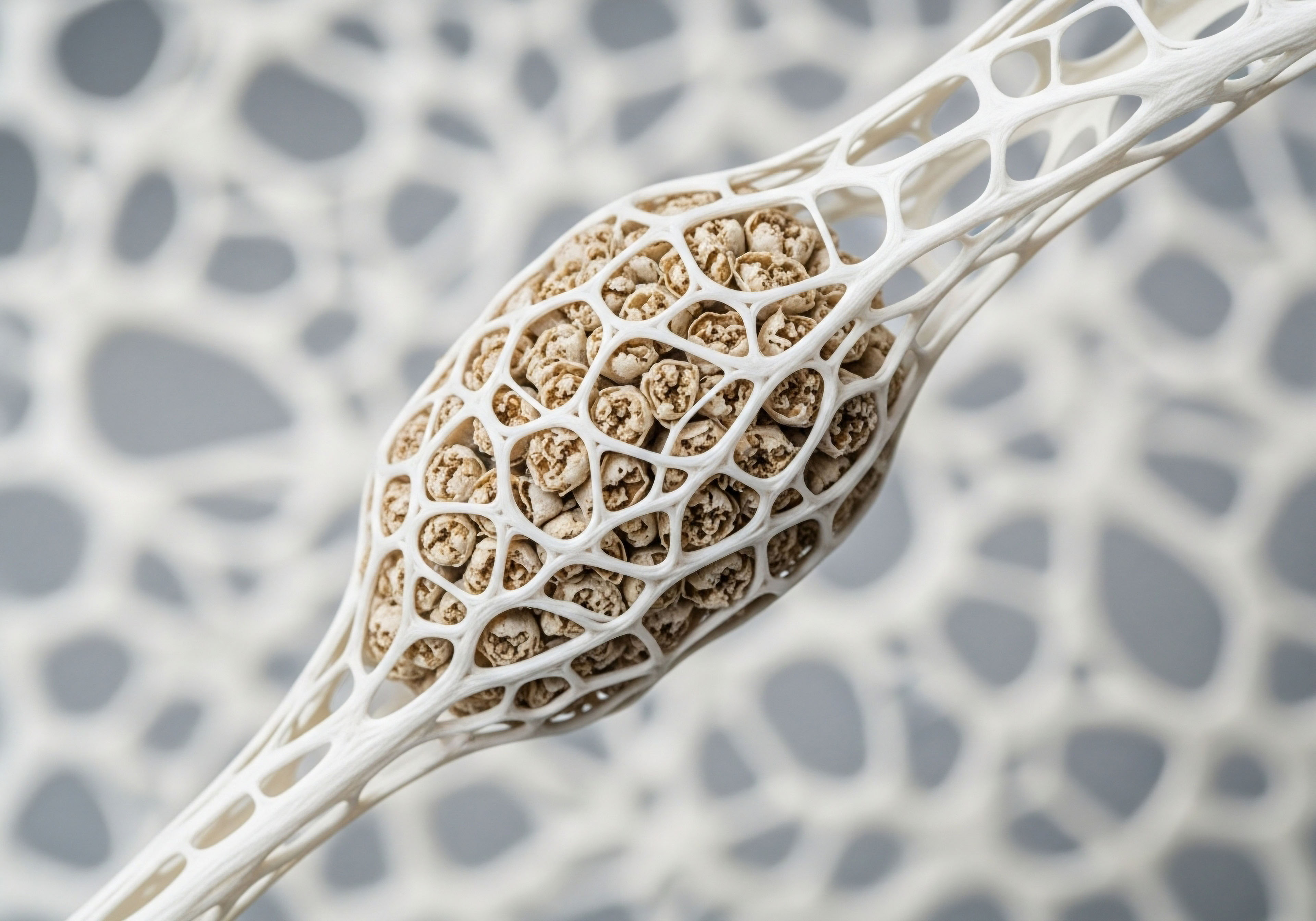
The thyroid gland produces hormones that set the metabolic pace for every single cell in your body. Think of it as the power utility for a city. If the power supply is inconsistent or weak, every home, office, and streetlamp will function poorly. In the same way, if thyroid hormone signaling is impaired, the energy-intensive processes of ovulation, fertilization, and implantation can be compromised.

The Body’s Internal Messaging Service
Your endocrine system operates as a sophisticated communication network, using hormones as chemical messengers. At the top of the command chain for thyroid function is the hypothalamus in your brain, which sends a signal called Thyrotropin-Releasing Hormone (TRH) to the pituitary gland. The pituitary, in turn, releases Thyroid-Stimulating Hormone (TSH).
This is the signal that tells your thyroid gland, located in your neck, to produce its hormones. This entire feedback loop is known as the Hypothalamic-Pituitary-Thyroid (HPT) axis.
The primary hormone produced by the thyroid gland is Thyroxine, or T4. You can think of T4 as a stable, reserve form of the hormone. It travels through the bloodstream to every tissue in your body. T4 itself is largely inactive. Its power must be unlocked.
For a cell to use the energy signal, it must first convert T4 into the much more potent, active form ∞ Triiodothyronine, or T3. This conversion is the most important event in thyroid physiology, because it is T3 that overwhelmingly carries out the work of the thyroid hormone at the cellular level.
The true measure of thyroid health is how effectively your cells can access and use the active T3 hormone.
This conversion from T4 to T3 does not happen in the thyroid gland itself but inside the cells of target tissues throughout the body, including the tissues of the female reproductive system. The ovaries, the uterus, and even the placenta have their own machinery to perform this critical activation step.
This localized process ensures that a tissue can fine-tune its own metabolic rate according to its specific needs. It is this cellular-level activity that holds a key to understanding reproductive wellness.

The Lock and Key a Cellular Dialogue
Once T3 is activated within a cell, it must deliver its message. It does this by binding to specialized proteins called thyroid hormone receptors (TRs). These receptors are located in the nucleus of the cell, the very command center that houses its genetic blueprint.
The binding of T3 to its receptor is like a key fitting into a lock. This action turns on specific genes, instructing the cell to perform its designated functions more efficiently. In the context of fertility, this means telling an ovarian follicle to mature properly, signaling the uterine lining to become receptive to an embryo, or supporting the healthy development of the placenta.
The presence of these receptors in reproductive tissues is a profound statement of biological intent. The body has explicitly designed the reproductive system to listen and respond to thyroid hormone. When this signaling is robust, the system functions with precision.
When the signal is weak or distorted, either because of poor T4-to-T3 conversion or because the receptors are not functioning correctly, the reproductive process can falter. This can happen even when the amount of T4 and TSH in the blood appears normal. The problem lies within the cell, at the site of activation and reception. This is the essence of impaired cellular thyroid signaling, and it is a pivotal piece of the fertility puzzle.


Intermediate
The distinction between systemic hormone levels and cellular action is where a more sophisticated understanding of fertility begins. Many individuals feel the frustration of being told their thyroid is “fine” based on a TSH test alone, yet they continue to struggle with reproductive challenges.
The reality is that the journey of a thyroid hormone from the gland to its final destination is complex. Its effectiveness is determined by a series of local, tissue-specific events that regulate its power. At the heart of this regulation are a family of enzymes called deiodinases. These enzymes are the gatekeepers of thyroid hormone activation and deactivation, essentially deciding how much active T3 a particular tissue gets to see.
Impaired cellular thyroid signaling is a condition of localized T3 deficiency. The cells of the ovaries or the uterine lining can be starved of active thyroid hormone even when the rest of the body has adequate levels. This cellular hypothyroidism can disrupt the delicate choreography of the menstrual cycle and pregnancy.
Understanding the function of the deiodinase enzymes provides a clear mechanism for how this happens. It moves the conversation from a simple gland-focused model to a more accurate, systems-based view of health.

The Gatekeepers of Cellular Energy
The body uses three main types of deiodinase enzymes (D1, D2, and D3) to manage thyroid hormone activity with remarkable precision. Each has a distinct role and location, working together to control the local concentration of T3.
- Type 1 Deiodinase (D1) is found primarily in the liver and kidneys. It is responsible for producing a significant portion of the circulating T3 in the bloodstream. It also helps clear reverse T3 (rT3), an inactive byproduct, from the system.
- Type 2 Deiodinase (D2) is the primary activating enzyme within specific tissues. It is found in the brain, the pituitary gland, and critically, in reproductive tissues like the ovaries and placenta. D2 performs the crucial conversion of local T4 into T3, essentially turning up the metabolic dial within that specific cell. Its action is what allows the reproductive system to directly manage its own energy supply.
- Type 3 Deiodinase (D3) is the primary deactivating enzyme. It converts T4 into the inactive reverse T3 (rT3) and breaks down active T3 into a weaker form. D3 acts as a protective brake, preventing excessive thyroid stimulation. It is highly active in the placenta to protect the developing fetus from high levels of maternal thyroid hormone.
The balance between D2 and D3 activity within the reproductive tissues is what determines the ultimate strength of the thyroid signal. Optimal fertility depends on robust D2 activity to generate sufficient T3 for processes like follicular growth and a healthy uterine lining, combined with appropriately regulated D3 activity.

How Cellular Thyroid Signaling Becomes Impaired
The delicate activity of these enzymes can be disrupted by a number of physiological stressors. This disruption is a primary cause of impaired cellular thyroid signaling and can directly influence reproductive health.
Several factors are known to downregulate the activity of the crucial D2 activating enzyme, effectively starving reproductive cells of the T3 they need:
- Chronic Inflammation ∞ Inflammatory messengers called cytokines, often elevated due to stress, infection, or autoimmune conditions, directly suppress D2 function. This is a primary mechanism linking systemic inflammation to reproductive difficulties.
- High Cortisol ∞ Chronic stress leads to elevated levels of the adrenal hormone cortisol. Sustained high cortisol inhibits the conversion of T4 to T3, shifting the conversion pathway towards the inactive rT3.
- Nutrient Deficiencies ∞ The deiodinase enzymes are dependent on specific micronutrients to function correctly. Selenium is the most critical, as it is a core component of the enzymes themselves. Deficiencies in zinc and iron can also impair their activity.
- Toxins ∞ Certain environmental toxins, including heavy metals and some plastics, have been shown to interfere with deiodinase function and thyroid receptor binding.
When these factors are present, the ratio of T3 to rT3 can shift unfavorably. Even with a normal T4 level, the cell experiences a functional hypothyroidism. This state can manifest as poor egg quality, a thin uterine lining, luteal phase defects, or failed implantation, all of which are common features of unexplained infertility.
A healthy reproductive system requires not just the presence of thyroid hormone, but the carefully orchestrated local conversion of T4 to T3 within the reproductive tissues themselves.

Consequences for the Reproductive Axis
Impaired cellular thyroid signaling has direct consequences for the entire Hypothalamic-Pituitary-Gonadal (HPG) axis, the hormonal cascade that governs reproduction. Thyroid hormones are necessary for the normal pulsatile release of Gonadotropin-Releasing Hormone (GnRH) from the hypothalamus. A local deficiency of T3 can disrupt this rhythm, leading to irregular signals to the pituitary.
This, in turn, affects the pituitary’s release of Follicle-Stimulating Hormone (FSH) and Luteinizing Hormone (LH), the two hormones that directly control ovarian function. The result can be anovulation or inconsistent ovulation. Furthermore, T3 acts directly on the granulosa cells that surround the developing egg, supporting their growth and steroid hormone production.
Without adequate T3, follicular development can stall, leading to diminished ovarian reserve or poor oocyte quality. The table below outlines the specific roles of deiodinase enzymes, highlighting their importance in reproductive health.
| Enzyme | Primary Function | Location in Reproductive System | Impact on Fertility |
|---|---|---|---|
| Type 1 Deiodinase (D1) | Contributes to circulating T3; clears rT3 | Primarily liver, kidneys | Indirectly supports overall metabolic health required for reproduction. |
| Type 2 Deiodinase (D2) | Activates T4 to T3 locally | Ovarian granulosa cells, placenta, endometrium | Drives follicular maturation, supports endometrial receptivity and placental function. Its impairment is a direct cause of cellular hypothyroidism in reproductive tissues. |
| Type 3 Deiodinase (D3) | Inactivates T4 and T3 | Placenta, uterus | Protects the fetus from excessive thyroid hormone. Overactivity can lead to localized T3 deficiency and implantation issues. |
Ultimately, a successful pregnancy requires a series of energy-demanding events to unfold in perfect sequence. Impaired cellular thyroid signaling removes the fundamental energetic support for this sequence. Addressing fertility challenges from this perspective requires looking beyond TSH and considering the factors that influence the all-important local conversion of T4 to T3.


Academic
A molecular-level examination of reproductive endocrinology reveals an intricate and bidirectional relationship between thyroid hormones and the female reproductive axis. The influence of thyroid signaling extends far beyond systemic metabolic rate, exerting direct genomic and non-genomic effects within the ovary, uterus, and placenta.
The clinical associations between thyroid dysfunction and infertility are underpinned by these precise cellular mechanisms. Understanding these pathways provides a scientifically robust framework for appreciating how subtle disruptions in local thyroid hormone homeostasis can precipitate significant reproductive pathology, including conditions often categorized as “unexplained infertility.”
The core of this interaction lies in the expression of specific thyroid hormone receptor (TR) isoforms and deiodinase enzymes within reproductive tissues. These components allow the reproductive system to function as a semi-autonomous regulator of its thyroid status, responding to local physiological demands.
The active hormone, 3,5,3′-triiodothyronine (T3), functions as a potent transcription factor, binding to TRs in the cell nucleus to modulate the expression of genes critical for reproductive processes. Pathophysiology arises when this local T3 availability is compromised or when receptor sensitivity is altered.

What Is the Molecular Dialogue between Thyroid Hormones and Ovarian Function?
The ovary is a primary target for thyroid hormone action. Both thyroid hormone receptors (TRα and TRβ) and TSH receptors have been identified on human ovarian surface epithelium and granulosa cells. T3 exerts a direct, pleiotropic influence on folliculogenesis and steroidogenesis.
In vitro studies demonstrate that T3 promotes the FSH-induced growth of preantral follicles, partly through activation of the protein kinase B (Akt) signaling pathway. It acts synergistically with gonadotropins to stimulate the proliferation of granulosa cells, which are essential for nurturing the developing oocyte and producing sex steroids.
Furthermore, thyroid hormones modulate ovarian steroidogenesis. T3 has been shown to increase the expression of key steroidogenic enzymes, including StAR (Steroidogenic Acute Regulatory Protein), which facilitates the transport of cholesterol into the mitochondria, the rate-limiting step in hormone production.
The local conversion of T4 to T3 by Type 2 deiodinase (D2) within granulosa cells ensures a high-T3 microenvironment during follicular development, which is essential for oocyte maturation and competence. Disruption of this local T3 supply, whether through systemic hypothyroidism, nutrient deficiencies impacting D2 function, or genetic polymorphisms in the DIO2 gene, can impair follicle quality and lead to ovulatory dysfunction.

How Does Thyroid Signaling Prepare the Uterus for Implantation?
Successful embryonic implantation requires a receptive endometrium, a time-sensitive state characterized by specific morphological and molecular changes. Thyroid hormones are critical regulators of this process. The expression of both TRs and TSHRs in the human endometrium increases significantly during the mid-secretory phase, coinciding with the “window of implantation.” This suggests a direct role for T3 in preparing the uterine lining for the arriving embryo.
One of the key molecular mediators of endometrial receptivity is Leukemia Inhibitory Factor (LIF), a cytokine essential for blastocyst attachment. Thyroid hormones have been shown to regulate the expression of LIF in endometrial cells. Impaired cellular T3 availability can lead to insufficient LIF production, contributing to implantation failure.
Additionally, T3 influences the expression of other receptivity markers, such as integrins and mucins, and promotes the decidualization process, where endometrial stromal cells transform to support the pregnancy. The intricate crosstalk between thyroid hormones and progesterone is evident here, as progesterone also modulates the expression of deiodinases and TRs in the endometrium, highlighting a coordinated hormonal effort to establish a receptive environment.
| Receptor Isoform | Primary Location in Reproductive System | Known Function in Reproductive Health |
|---|---|---|
| Thyroid Receptor Alpha (TRα) | Ovary (granulosa and theca cells), Uterus (endometrium and myometrium), Placenta | Mediates T3 effects on follicular growth, endometrial proliferation, and placental development. Essential for metabolic regulation within these tissues. |
| Thyroid Receptor Beta (TRβ) | Ovary, Pituitary Gland, Liver | Regulates the HPT axis feedback loop at the pituitary level. In the ovary, it works with TRα to control steroidogenesis and cellular response to gonadotropins. |
| TSH Receptor (TSHR) | Ovarian granulosa cells, Endometrium | While its primary role is in the thyroid gland, its presence in reproductive tissues suggests a potential direct, non-canonical role for TSH in modulating ovarian and endometrial function, independent of T3/T4. |

Subclinical Hypothyroidism and Autoimmunity a Deeper Look
The concept of subclinical hypothyroidism (SCH), defined by an elevated serum TSH with normal free T4 levels, is particularly relevant to reproductive health. SCH is more prevalent in women with infertility, especially of unexplained origin, than in the general population.
From a mechanistic standpoint, an elevated TSH represents the pituitary’s attempt to compensate for perceived low thyroid hormone activity at the tissue level. It is an early indicator that the system is under strain. This state may correlate with reduced D2 activity and compromised local T3 availability in peripheral tissues like the ovary, even before serum T4 levels decline.
Studies have associated higher TSH levels, even within the normal range (e.g. >2.5 mIU/L), with poorer outcomes in assisted reproductive technology (ART), including lower fertilization rates and reduced live birth rates.
Autoimmune thyroid disease (AITD), most commonly Hashimoto’s Thyroiditis, adds another layer of complexity. The presence of thyroid peroxidase antibodies (TPOAb) is linked to an increased risk of infertility, miscarriage, and premature ovarian insufficiency, even in euthyroid women (women with normal TSH).
This suggests that the autoimmune process itself, independent of overt thyroid failure, may be detrimental to reproductive function. One hypothesis is that TPOAb positivity is a marker of a generalized autoimmune imbalance that could impair implantation. Another is that the inflammatory environment associated with AITD directly disrupts ovarian and endometrial function, possibly by inhibiting deiodinase activity and interfering with receptor signaling.
This highlights that a comprehensive assessment of thyroid health in the context of fertility must include not just TSH and T4, but also an evaluation of thyroid antibodies to understand the full picture of cellular and immune system function.

References
- Aghajanova, L. et al. “Paracrine interactions of thyroid hormones and thyroid stimulation hormone in the female reproductive tract have an impact on female fertility.” Frontiers in Endocrinology, vol. 3, 2012, p. 33.
- Bianco, A. C. et al. “Deiodinases ∞ implications of the local control of thyroid hormone action.” Journal of Clinical Investigation, vol. 116, no. 10, 2006, pp. 2571-2579.
- Dittrich, R. et al. “Thyroid hormone receptors and their role in female reproduction.” Journal of Reproductive Immunology, vol. 90, no. 1, 2011, pp. 58-66.
- Poppe, K. et al. “The role of thyroid dysfunction in female infertility and in assisted reproduction.” Seminars in Reproductive Medicine, vol. 26, no. 4, 2008, pp. 313-321.
- Unuane, D. and K. Poppe. “Why and when to screen for thyroid disorders in infertile women.” Journal of Endocrinological Investigation, vol. 45, no. 10, 2022, pp. 1837-1846.
- Krassas, G. E. et al. “Thyroid function and human reproductive health.” Endocrine Reviews, vol. 31, no. 5, 2010, pp. 702-755.
- Maraka, S. et al. “Subclinical Hypothyroidism in Women Planning Conception and During Pregnancy ∞ A Guideline.” The Journal of Clinical Endocrinology & Metabolism, vol. 102, no. 3, 2017, pp. 782-797.
- Zhang, J. and L. A. Allison. “Nuclear Import and Export of the Thyroid Hormone Receptor.” Vitamins and Hormones, vol. 106, 2018, pp. 45-66.
- Quintino-Moro, A. et al. “Distribution of thyroid hormone and thyrotropin receptors in reproductive tissues of adult female rabbits.” Systems Biology in Reproductive Medicine, vol. 62, no. 5, 2016, pp. 337-345.
- Cavaliere, H. et al. “The thyroid in reproductive medicine.” Journal of Endocrinological Investigation, vol. 40, no. 9, 2017, pp. 917-924.

Reflection
Your body is a cohesive whole, a network of systems in constant communication. The information presented here offers a new map to understand one of its most fundamental dialogues ∞ the one between your metabolic rhythm and your reproductive potential. This knowledge is not meant to provide a simple answer, but to illuminate a new set of questions.
It shifts the focus from a place of confusion to one of informed curiosity. What is the energetic status of my cells? Are there underlying stressors, inflammatory signals, or nutritional gaps that might be disrupting this delicate cellular conversation? The path forward is one of partnership with your own physiology.
This understanding is the first, most powerful step in a personalized health journey, empowering you to ask deeper questions and seek solutions that honor the intricate design of your own biological systems.