

Fundamentals
The moments and months following a heart attack, a myocardial infarction, represent a profound biological crossroads. The body, in its innate wisdom, rushes to mend the damaged tissue. This healing process, while essential for survival, can create a new set of challenges.
The very structure of the heart muscle begins to change in a process called cardiac remodeling. You may feel this as a shift in your physical capacity, a new awareness of your heartbeat, or a sense of fatigue that settles deep in your bones.
These experiences are the outward expression of a complex cellular and structural transformation occurring within your chest. Understanding this process is the first step toward actively participating in your own recovery and reclaiming a sense of vitality.
Following an infarction, the heart muscle that was deprived of oxygen is replaced by scar tissue. This scar is strong, yet it is functionally different from the contractile muscle it replaces. The remaining healthy heart muscle must work harder to compensate, leading to a gradual enlargement and change in the shape of the heart’s chambers, particularly the left ventricle.
This is the essence of post-infarction remodeling. It is a compensatory mechanism that, over time, can become maladaptive, contributing to the progression of heart failure. The central question for both individuals and clinicians becomes how to guide this healing process toward a more functional and stable outcome. How can we support the heart’s own attempts at repair while mitigating the long-term consequences of structural change?

The Body’s System for Growth and Repair
Our bodies possess an intricate internal communication network dedicated to growth, maintenance, and repair, orchestrated largely by the endocrine system. At the center of this network is the growth hormone (GH) axis. From a young age, this system drives our development, and in adulthood, it shifts to a primary role of metabolic regulation and tissue regeneration.
The pituitary gland, a small structure at thebase of the brain, releases growth hormone in pulses. This GH then travels through the bloodstream, signaling to other organs, most notably the liver, to produce insulin-like growth factor 1 (IGF-1). Together, GH and IGF-1 influence cellular activity throughout the body, promoting cell growth, reproduction, and regeneration. It is this fundamental biological system, designed for repair, that has become a focus of intense scientific inquiry in the context of cardiac health.
The heart’s structural changes after a myocardial infarction are a natural healing response that can lead to long-term functional challenges.
The signals that control this powerful axis originate one level higher, in the hypothalamus. The hypothalamus releases growth hormone-releasing hormone (GHRH), the primary signal that tells the pituitary to produce and release GH. This entire system operates on a sophisticated feedback loop, ensuring that levels of these powerful molecules are kept in careful balance.
When researchers began to consider how to support the heart’s recovery, they looked to this existing, powerful system for clues. The initial thought was logical ∞ if the heart is damaged, perhaps augmenting the body’s primary repair system could help. This line of thinking opened the door to exploring how manipulating the GH axis might influence the trajectory of cardiac remodeling after an injury.

Introducing Growth Hormone Releasing Peptides
Directly administering growth hormone can be a blunt instrument, with potential side effects and complications. This led scientists to explore a more refined approach ∞ using smaller molecules called peptides to influence the body’s own production of GH. Growth hormone-releasing peptides (GHRPs) and GHRH analogues are synthetic molecules designed to interact with this system in a targeted way.
Some, like Sermorelin, are analogues of the body’s natural GHRH. Others, like Ipamorelin and Hexarelin, work on a parallel receptor system, the ghrelin receptor, to stimulate GH release. These peptides offer a way to gently and rhythmically prompt the pituitary gland, mimicking the body’s natural pulsatile release of growth hormone.
The investigation into these molecules has revealed a story far more intricate and targeted than first imagined, pointing toward mechanisms that work directly on the heart tissue itself. This discovery has shifted the conversation from general systemic support to specific, localized cardiac repair.


Intermediate
The exploration of growth hormone-related therapies for cardiac repair has evolved significantly. Initial studies using direct administration of recombinant growth hormone (GH) yielded mixed results. While some animal studies showed benefits like improved cardiac performance and reduced dilation of the left ventricle, others revealed potential adverse effects or a lack of efficacy, particularly with large infarctions.
This inconsistency pointed to a complex reality ∞ flooding the system with high levels of GH might not be the optimal strategy. The body’s hormonal systems are built on pulsatility and precise feedback, and a continuous high-level signal can sometimes lead to receptor downregulation or unintended consequences, including negative impacts on the remodeling process itself. This clinical uncertainty prompted a deeper investigation into the underlying mechanisms and a search for a more sophisticated therapeutic tool.
The turning point came with the study of GHRH agonists, peptides that stimulate the body’s own GHRH receptors. Researchers hypothesized that these molecules could provide a more nuanced signal. A pivotal study involving a GHRH agonist named JI-38 in a rat model of myocardial infarction produced a remarkable finding.
The animals treated with the GHRH agonist showed a marked attenuation of cardiac functional decline and remodeling. Crucially, these profound benefits occurred without any significant elevation in the circulating blood levels of either growth hormone or IGF-1. This was a watershed moment. The positive effects on the heart were not being driven by the systemic GH axis. The benefit was originating from a different, more direct source.

What Is the Direct Cardiac GHRH Receptor Pathway?
The unexpected results from the JI-38 study led to a groundbreaking discovery ∞ the heart has its own GHRH receptors. For decades, the GHRH receptor was thought to exist primarily in the pituitary gland. The revelation that cardiac myocytes, the muscle cells of the heart, also express these receptors changed the entire paradigm.
It meant that GHRH and its agonists could communicate directly with the heart tissue, bypassing the pituitary-liver axis altogether. This local signaling pathway represents a private communication channel, allowing for targeted action right at the site of injury. Activation of these cardiac GHRH receptors initiates a cascade of intracellular signals specifically geared toward cell survival, repair, and regeneration. It is a system of localized control, offering a way to support the heart from within.
The discovery of GHRH receptors on heart cells revealed a direct pathway for therapeutic intervention, independent of the systemic growth hormone axis.
This direct pathway explains why GHRH agonists can be effective where high-dose GH was not. Instead of a systemic flood, the agonist acts like a key in a specific lock on the heart cells themselves. This targeted activation triggers several beneficial processes simultaneously.
Studies have shown that activating these receptors helps protect existing cardiomyocytes from apoptosis, the programmed cell death that occurs in the oxygen-starved region surrounding the infarct. Furthermore, this signaling appears to stimulate the proliferation of the heart’s own pool of cardiac precursor cells, providing the raw materials for repair and regeneration. This mechanism represents a far more elegant biological solution, leveraging the heart’s intrinsic capacity for healing.

Comparing Systemic GH and Local GHRH Agonist Effects
The distinction between systemic GH therapy and targeted GHRH agonist therapy is fundamental to understanding their potential roles in post-infarction care. The table below outlines the key differences in their mechanisms and observed effects.
| Feature | Systemic Growth Hormone (GH) Therapy | Targeted GHRH Agonist Therapy |
|---|---|---|
| Primary Mechanism |
Increases circulating levels of GH and IGF-1 system-wide, affecting all tissues with corresponding receptors. |
Directly activates GHRH receptors present on cardiac myocytes and progenitor cells, initiating local signaling pathways. |
| Effect on GH/IGF-1 Levels |
Markedly elevates systemic GH and IGF-1 levels, which can be measured in blood tests. |
Produces therapeutic effects with little to no increase in systemic GH or IGF-1 levels. |
| Observed Cardiac Benefits |
Variable and sometimes conflicting results in studies; potential for hypertrophy and improved function but also risk of adverse remodeling. |
Consistently shown to attenuate ventricular remodeling, reduce infarct size, and improve cardiac function in animal models. |
| Cellular Action |
General anabolic and growth-promoting effects on a wide range of cells. |
Promotes cardiomyocyte survival (anti-apoptotic), reduces local inflammation, and stimulates proliferation of cardiac precursor cells. |
| Potential Side Effects |
Associated with risks of systemic GH excess, such as insulin resistance, fluid retention, and joint pain. |
The localized action suggests a more favorable safety profile with fewer systemic side effects. |

What Are the Key Peptides in This Class?
While the research has focused on experimental GHRH agonists like JI-38 and MR-409, the findings have broader implications for other peptides that interact with the growth hormone secretagogue system. This includes peptides that, while developed for other purposes, may share some of these cardioprotective mechanisms.
- Sermorelin ∞ A synthetic analogue of the first 29 amino acids of human GHRH. It is used clinically to stimulate the pituitary’s own GH production and may also interact with cardiac GHRH receptors.
- Tesamorelin ∞ A stabilized GHRH analogue with a longer half-life. Its primary indication is for lipodystrophy, but its action as a potent GHRH agonist makes its potential cardiac effects an area of interest.
- Hexarelin ∞ A synthetic growth hormone secretagogue that acts on the ghrelin receptor (GHSR). Interestingly, these receptors are also found in the heart, and studies have shown Hexarelin to exert direct cardioprotective effects, suggesting another local signaling pathway for cardiac repair.
- Ipamorelin / CJC-1295 ∞ This combination involves a GHRP (Ipamorelin) and a GHRH analogue (CJC-1295). The GHRH component works on the GHRH receptor, while Ipamorelin acts on the ghrelin receptor, creating a synergistic pulse of GH release from the pituitary. The GHRH component could also theoretically engage the local cardiac receptors.
The understanding of these peptides is moving from a purely pituitary-focused model to a dual-action model ∞ one that acknowledges both the systemic effects via the pituitary and the potentially more significant direct, local effects on the heart tissue itself. This dual capability is what makes them such a compelling subject of study for cardiac recovery.


Academic
The therapeutic potential of growth hormone-releasing hormone agonists (GHRH-A) in post-myocardial infarction (MI) cardiac remodeling is predicated on the activation of a local, GHRH-receptor (GHRH-R) mediated signaling axis within the myocardium. This paradigm is a significant departure from earlier strategies focused on the systemic elevation of GH/IGF-1.
The evidence demonstrates that GHRH-A can reverse adverse remodeling and improve cardiac function in chronic MI settings, an effect that is nullified by the co-administration of a GHRH-R antagonist. This confirms that the therapeutic action is receptor-dependent and largely independent of the canonical hypothalamic-pituitary-somatotropic axis. The academic exploration of this phenomenon delves into the specific molecular cascades that are initiated by this receptor activation within the cardiac microenvironment.

Molecular Mechanisms of GHRH-A Cardioprotection
Upon binding of a GHRH agonist to its G-protein coupled receptor on the cardiomyocyte cell membrane, a series of downstream signaling events are triggered. These pathways collectively counter the pathological processes that define adverse cardiac remodeling ∞ apoptosis, fibrosis, and inflammation.

Inhibition of Apoptotic Pathways
In the ischemic penumbra surrounding an infarct, cardiomyocytes undergo significant stress, leading to programmed cell death or apoptosis. GHRH-A treatment has been shown to directly counter this. Gene expression studies reveal that GHRH-A administration leads to the downregulation of pro-apoptotic molecules like Bax and a concurrent upregulation of anti-apoptotic proteins such as Bcl-2.
This shift in the Bax/Bcl-2 ratio is a critical determinant of cell fate. The signaling likely proceeds through activation of protein kinase A (PKA) and subsequent phosphorylation of downstream effectors that stabilize the mitochondrial membrane and inhibit the release of cytochrome c, a key step in the intrinsic apoptotic cascade. By preserving viable myocytes in the border zone, GHRH agonists help maintain the structural and functional integrity of the ventricle.

Modulation of the Inflammatory Response
The acute inflammatory response post-MI is necessary for clearing necrotic debris, but a prolonged or excessive inflammatory state contributes to further tissue damage and adverse remodeling. GHRH agonists appear to be potent modulators of this response.
Studies using the agonist MR-409 demonstrated a significant reduction in the plasma levels of key pro-inflammatory cytokines, including Interleukin-2 (IL-2), Interleukin-6 (IL-6), and Tumor Necrosis Factor-alpha (TNF-α), just one week post-MI. This suggests an early and powerful anti-inflammatory effect.
The mechanism may involve the inhibition of the NF-κB (nuclear factor kappa-light-chain-enhancer of activated B cells) signaling pathway, a master regulator of inflammatory gene expression. By tempering the inflammatory milieu, GHRH agonists create a more permissive environment for effective healing and repair.
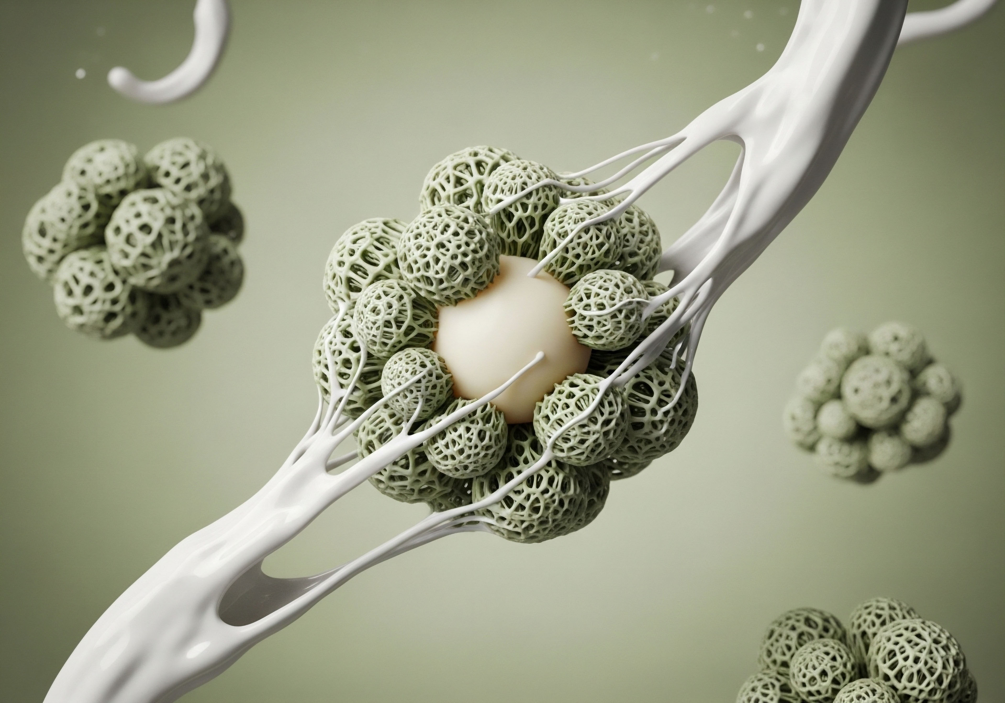
GHRH agonists orchestrate a multi-faceted cellular response, simultaneously limiting cell death, inflammation, and fibrosis while promoting regeneration.
Attenuation of Cardiac Fibrosis
Fibrosis, the excessive deposition of extracellular matrix proteins, leads to stiffening of the ventricle and diastolic dysfunction. The transforming growth factor-beta (TGF-β) pathway is a primary driver of cardiac fibrosis. Research on the broader GH axis has shown that it can inhibit TGF-β signaling.
GHRH agonists likely exert a similar, if not more direct, anti-fibrotic effect. By activating local receptors, they may interfere with the TGF-β signaling cascade, potentially by inhibiting the phosphorylation of downstream mediators like Smad proteins.
Gene expression analyses in GHRH-A treated hearts show a downregulation of pro-fibrotic systems, contributing to a reduction in collagen deposition and preservation of a more compliant myocardial texture. This action is critical for preventing the long-term stiffening that characterizes heart failure with preserved ejection fraction.

How Does GHRH Agonist Therapy Stimulate Cardiac Regeneration?
Perhaps the most profound finding is the ability of GHRH agonists to stimulate regenerative processes within the heart. The adult heart has a very limited, but definite, capacity for regeneration, driven by a resident population of cardiac stem or progenitor cells (CSCs). GHRH-R is expressed on these CSCs.
In vivo studies show that treatment with a GHRH-A leads to a marked increase in myocyte and non-myocyte mitosis and a significant increase in the number of c-kit positive cells, a marker for a population of cardiac progenitor cells.
Ex vivo experiments confirm that GHRH agonists directly stimulate CSC proliferation, an effect that is blocked by a GHRH antagonist. This indicates that GHRH-A therapy effectively “wakes up” the heart’s own dormant repair machinery.
The activated CSCs can then differentiate into new cardiomyocytes, endothelial cells, and smooth muscle cells, contributing to both the replacement of lost muscle and the formation of new blood vessels (neovascularization) to improve perfusion to the damaged area. This dual effect of myogenesis and angiogenesis results in a measurable reduction in the overall infarct size, even when treatment is initiated in a chronic setting, long after the initial injury.
The table below summarizes the key molecular and cellular targets of GHRH agonists in the post-MI heart.
| Cellular Process | Molecular Target/Pathway | Therapeutic Outcome |
|---|---|---|
| Cardiomyocyte Survival |
Upregulation of Bcl-2; Downregulation of Bax; PKA pathway activation. |
Reduced apoptosis in the infarct border zone; Preservation of contractile tissue. |
| Inflammation |
Decreased expression of TNF-α, IL-2, IL-6; Probable inhibition of NF-κB signaling. |
Modulation of the post-MI inflammatory response; Reduced secondary tissue damage. |
| Fibrosis |
Inhibition of pro-fibrotic systems; Potential interference with TGF-β/Smad signaling. |
Decreased collagen deposition; Attenuation of ventricular stiffening. |
| Regeneration |
Activation of GHRH-R on c-kit+ cardiac stem cells; Upregulation of Bone Morphogenetic Proteins (BMPs). |
Increased proliferation and differentiation of CSCs; Neovascularization and myogenesis; Reduction in infarct size. |

References
- Bagnato, G. et al. “Cardiac and peripheral actions of growth hormone and its releasing peptides ∞ Relevance for the treatment of cardiomyopathies.” Cardiovascular Research, vol. 56, no. 3, 2002, pp. 342-355.
- Bagno, A. et al. “Cardioprotective effects of growth hormone-releasing hormone agonist after myocardial infarction.” Proceedings of the National Academy of Sciences, vol. 106, no. 7, 2009, pp. 2359-2364.
- Kanashiro-Takeuchi, R. M. et al. “Activation of growth hormone releasing hormone (GHRH) receptor stimulates cardiac reverse remodeling after myocardial infarction (MI).” Proceedings of the National Academy of Sciences, vol. 109, no. 2, 2012, pp. 559-563.
- Kanashiro-Takeuchi, R. M. et al. “New therapeutic approach to heart failure due to myocardial infarction based on targeting growth hormone-releasing hormone receptor.” Oncotarget, vol. 6, no. 11, 2015, pp. 8386-8399.
- Volpe, C. et al. “Electrophysiologic Effects of Growth Hormone Post-Myocardial Infarction.” Journal of Clinical Medicine, vol. 8, no. 10, 2019, p. 1599.

Reflection
The journey from understanding a symptom to deciphering a cellular mechanism is a powerful one. The knowledge that the heart contains its own specific receptors for repair signals, and that these can be selectively activated, reframes the narrative of recovery. It shifts the perspective from one of passive healing to one of active, targeted biological support.
The science discussed here opens a door to a future where medicine may work more intimately with the body’s own regenerative design. As you consider your own health, contemplate the intricate systems operating constantly within you. The capacity for healing is woven into our very biology. Understanding these pathways is the first step, but the true journey lies in applying that knowledge to a personalized path toward wellness, guided by a deep respect for the body’s innate intelligence and potential.

Glossary

myocardial infarction

cardiac remodeling

growth hormone

growth hormone-releasing hormone

growth hormone-releasing

ghrh receptors

ghrh agonists

ghrh agonist

ghrh receptor

apoptosis
targeted ghrh agonist therapy
fibrosis

anti-inflammatory
