


Fundamentals
You may feel it as a persistent fatigue that sleep does not seem to touch, a mental fog that clouds your thinking, or a frustrating battle with your body’s metabolism that feels disconnected from your efforts with diet and exercise. These experiences are common, and they often point toward a communication breakdown deep within your body.
Your biology is a system of immense complexity, built upon trillions of individual cells, each one needing to send and receive signals with perfect clarity. When this signaling becomes muffled or distorted, the vibrant health you seek remains just out of reach. The journey to understanding this begins at the most foundational level of your physiology ∞ the cell membrane.
Every single cell in your body is encased in this delicate, intelligent barrier. It is a dynamic, fluid structure known as the phospholipid bilayer. Picture a vast, flexible mosaic made of countless individual molecules called phospholipids. Each of these molecules has a head that is attracted to water and two tails that are repelled by it.
They arrange themselves into two layers, with the water-loving heads facing outward toward the watery environment of the body and inward toward the cell’s watery interior, while the water-repelling tails face each other, creating a fatty, protective core. This structure is what gives the membrane its integrity, acting as a sophisticated gatekeeper that controls everything that enters and leaves the cell.
The quality of this membrane is directly constructed from the dietary fats you consume. The fats in your food are broken down and repurposed to become the very building blocks of these cellular barriers. This is where the concept of fat quality becomes profoundly important. There are different families of fats, each with a unique molecular structure that imparts specific physical properties to the cell membranes they help create.
- Saturated Fats ∞ These fats, found in animal products and some tropical oils, have straight, rigid structures. When they are incorporated into the cell membrane, they pack together tightly, making the membrane more stiff and less fluid.
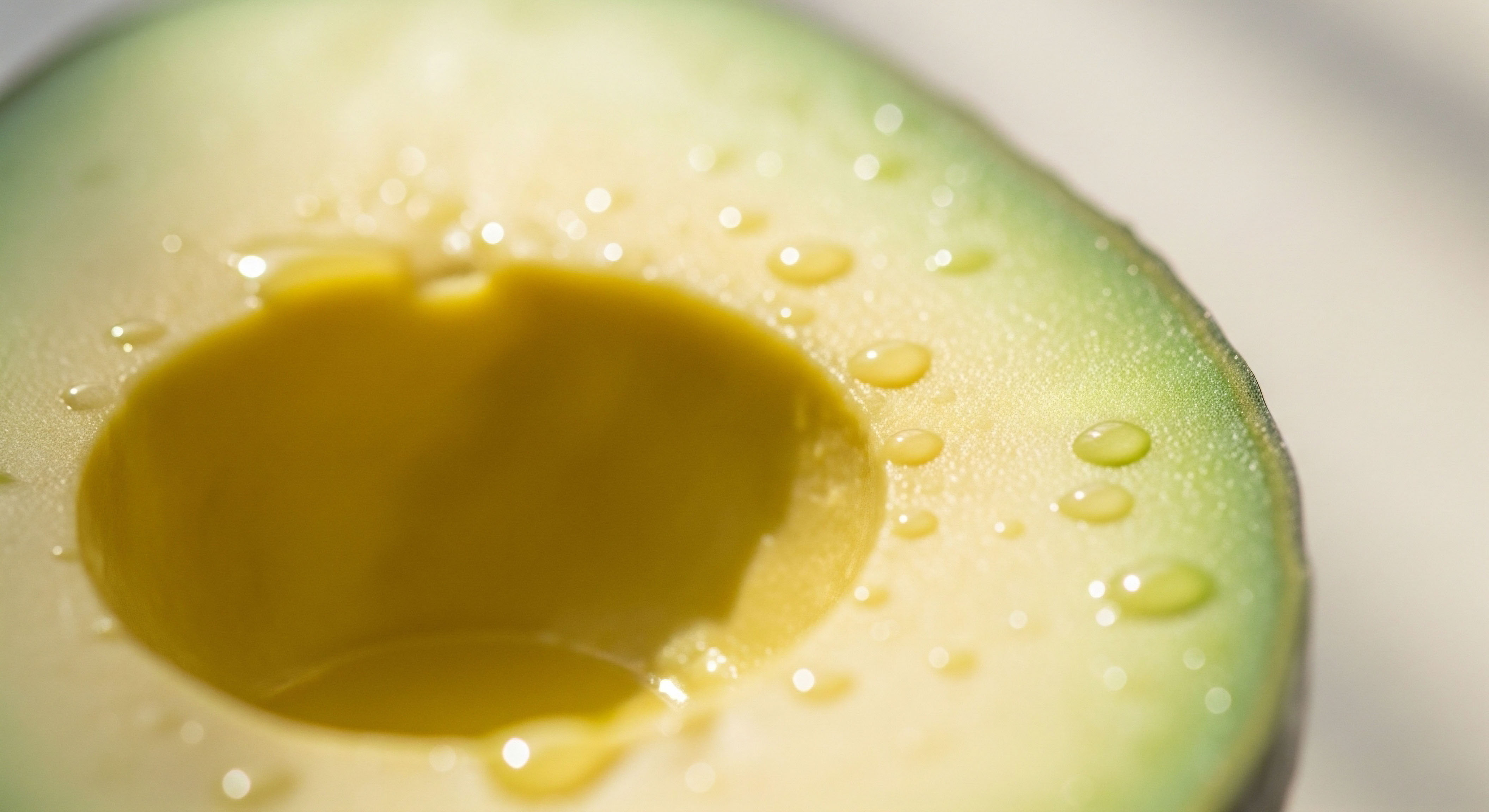
- Monounsaturated Fats (MUFAs) ∞ Found in foods like olive oil and avocados, these fats have a single bend in their structure. This kink prevents them from packing as tightly as saturated fats, which contributes to a more fluid and flexible membrane.
- Polyunsaturated Fats (PUFAs) ∞ This family, which includes omega-3s (from fatty fish, flaxseeds) and omega-6s (from many vegetable oils), has multiple bends. Their presence in the cell membrane dramatically increases its fluidity, making it more pliable and responsive.

The Receptors Embedded Within
Embedded within this fluid sea of the cell membrane are your hormone receptors. These are highly specialized proteins that act as docking stations or antennas, each designed to receive a specific hormonal signal. Think of a hormone like testosterone or insulin as a key, and its receptor as the corresponding lock.
When the hormone (the key) binds to its receptor (the lock), it triggers a cascade of events inside the cell, instructing it on what to do. This is the fundamental mechanism of hormonal action. The cell might be instructed to burn sugar for energy, build new muscle tissue, or regulate mood. The efficiency of this entire process hinges on the concept of receptor sensitivity.
The physical structure of your cell membranes, built from the fats you eat, dictates how effectively your hormones can communicate their vital messages.
Receptor sensitivity describes how well a receptor can “hear” its hormonal signal. High sensitivity means the cellular machinery is responsive; a small amount of hormone produces a robust and appropriate effect. Low sensitivity, often called resistance, means the receptor is deafened to the signal.
The body must then produce more and more of the hormone to get the same job done, leading to hormonal imbalances and a host of metabolic problems. This is precisely where dietary fat quality exerts its powerful influence. A cell membrane constructed from an abundance of saturated and processed trans fats becomes stiff and viscous.
In this rigid environment, receptor proteins can become stuck or unable to change their shape properly. Their ability to move, bind with their hormone, and transmit a clear signal is physically impaired. Conversely, a membrane rich in unsaturated fats, particularly omega-3s, is fluid and flexible.
This suppleness allows receptors to move freely, adopt the optimal shape for binding, and communicate efficiently with the interior of the cell. This physical reality at the cellular level connects the food on your plate directly to the hormonal symptoms you may be experiencing. Your dietary choices are a constant act of architectural renovation for your entire body.


Intermediate
Understanding that dietary fats build your cell membranes is the first step. The next layer of comprehension involves the sophisticated structures within the membrane that orchestrate hormonal signaling. The cell membrane is not a uniform sea of lipids. It contains highly organized microdomains known as lipid rafts.
These are small, dynamic platforms enriched in specific lipids like cholesterol and sphingolipids, along with certain types of saturated fats. These rafts float within the more fluid membrane like a log floating in a lake. Their purpose is to act as powerful organizing centers, gathering specific receptors and signaling proteins together to make cellular communication more efficient and rapid.
Many critical hormone receptors, including certain types of estrogen receptors and a vast family of proteins called G-Protein Coupled Receptors (GPCRs), are preferentially located within these lipid rafts. By concentrating the necessary components in one place, the cell ensures that when a hormone binds to its receptor, the subsequent steps in the signaling cascade can occur almost instantaneously.
The integrity and composition of these lipid rafts are exquisitely sensitive to the balance of fats in your diet. A diet that provides the right building blocks, including sufficient cholesterol and a healthy ratio of fatty acids, supports the formation of stable, functional rafts. An imbalanced intake, particularly one high in inflammatory omega-6 fats or deficient in omega-3s, can disrupt the structure of these rafts, causing receptors to drift away from their necessary co-factors and silencing their signals.

G-Protein Coupled Receptors the Master Switches
GPCRs represent one of the largest and most important families of receptors in the human body. They are the targets for a vast number of hormones and neurotransmitters, including adrenaline, dopamine, serotonin, and the hormones that trigger the release of testosterone and estrogen, like Luteinizing Hormone (LH).
The function of a GPCR is a physical process. Upon binding its hormone, the receptor must physically change its three-dimensional shape and then move laterally through the fluid membrane to connect with its partner, a G-protein. This interaction activates the G-protein, which then initiates a downstream signaling cascade inside the cell.
This entire process is critically dependent on membrane fluidity. Imagine trying to move and turn around in a room packed with solid furniture versus a room with plenty of open space. A rigid, viscous cell membrane, resulting from a diet high in saturated or trans fats, physically restricts the movement and conformational changes of GPCRs.
This slows down signaling and reduces receptor sensitivity. Conversely, a fluid membrane rich in PUFAs allows these receptors to move and interact freely, ensuring a swift and efficient response to hormonal cues. This has direct implications for everything from your stress response to the regulation of your reproductive hormones.

A Clinical Case Study Insulin Resistance
The development of insulin resistance provides a clear and powerful example of how dietary fat quality directly impacts hormonal health. The insulin receptor’s job is to signal the cell to take up glucose from the bloodstream. In a state of insulin resistance, the cells become deaf to insulin’s message, leading to high blood sugar and a cascade of metabolic issues. The quality of dietary fat is a primary driver of this dysfunction.
A diet high in saturated fats leads to their incorporation into cell membranes, particularly in muscle and fat cells. This increases membrane rigidity, which directly impairs the function of the insulin receptor. The receptor’s ability to activate its internal machinery, a process called autophosphorylation, is diminished.
Furthermore, the translocation of glucose transporters (like GLUT4) to the cell surface, which is the final step in getting sugar into the cell, is also hindered. In contrast, omega-3 fatty acids, like EPA and DHA from fish oil, have been shown to improve insulin sensitivity through several mechanisms. They increase membrane fluidity, which enhances the insulin receptor’s function. They also exert potent anti-inflammatory effects, reducing the cellular stress that contributes to insulin resistance.
| Fatty Acid Type | Primary Dietary Sources | Effect on Membrane Fluidity | Impact on Receptor Sensitivity |
|---|---|---|---|
| Saturated Fatty Acids (SFA) | Red meat, butter, coconut oil | Decreases fluidity (more rigid) | Tends to decrease sensitivity, especially for insulin receptors. Can impair receptor movement. |
| Monounsaturated Fatty Acids (MUFA) | Olive oil, avocados, nuts | Increases fluidity | Generally supportive of healthy receptor function and sensitivity. |
| Omega-6 PUFA | Many vegetable oils (soy, corn, sunflower) | Increases fluidity | Can be pro-inflammatory in excess, which may indirectly decrease receptor sensitivity. |
| Omega-3 PUFA (EPA/DHA) | Fatty fish, fish oil, algae | Significantly increases fluidity | Improves sensitivity for many receptors, including insulin. Reduces inflammation. |
| Trans Fats | Processed foods, hydrogenated oils | Drastically decreases fluidity (very rigid) | Severely impairs receptor function and is strongly linked to insulin resistance and systemic inflammation. |

How Does This Relate to Hormone Optimization Protocols?
For individuals undergoing hormonal optimization, such as Testosterone Replacement Therapy (TRT) for men or women, understanding these principles is of great importance. The effectiveness of these protocols relies on the body’s ability to respond to the administered hormones. If a patient’s cellular environment is compromised by poor membrane health and widespread receptor desensitization, the clinical outcomes of therapy may be suboptimal.
Optimizing dietary fat quality is a foundational strategy that works synergistically with hormonal therapies. By creating fluid, responsive cell membranes, you ensure that the hormonal signals being introduced are received clearly and efficiently, maximizing the benefits of the protocol while potentially minimizing the required dosage. It is a way of preparing the soil before planting the seed.


Academic
A granular examination of the relationship between dietary lipids and hormone receptor function requires a focus on the biophysical and biochemical sequelae of fatty acid incorporation into the phospholipid bilayer. The functional state of a hormone receptor is not merely a product of its amino acid sequence but is profoundly modulated by the allosteric influence of its surrounding lipid environment.
The cell membrane is a complex solvent, and its composition directly dictates the conformational ensemble of embedded integral membrane proteins, including receptors.
Polyunsaturated fatty acids (PUFAs), particularly the omega-3 species eicosapentaenoic acid (EPA) and docosahexaenoic acid (DHA), are of particular interest. Their defining structural feature is the presence of multiple double bonds, which introduce kinks into the acyl chains. When esterified into membrane phospholipids, these kinks create steric hindrance, preventing the tight packing of adjacent lipid molecules.
This increases the free volume within the hydrophobic core of the membrane, resulting in a quantifiable increase in membrane fluidity. This can be measured using techniques like fluorescence polarization anisotropy with probes such as diphenylhexatriene (DPH). Studies have demonstrated a direct correlation between the incorporation of PUFAs into erythrocyte membranes and increased membrane fluidity, which in turn correlates with enhanced insulin binding.

Modulation of Lipid Raft-Mediated Signaling
The lipid raft hypothesis provides a critical framework for understanding the spatial organization of signal transduction. These domains, enriched in cholesterol and sphingomyelin, function as platforms for the assembly of signaling complexes. The localization of a receptor within a lipid raft can dramatically alter its functional state.
For instance, certain estrogen receptor (ER) populations, specifically membrane-associated ERα and ERβ, require localization to lipid rafts to initiate rapid, non-genomic signaling cascades through tyrosine kinases like Src. The integrity of these rafts is essential for this process.
Dietary fatty acids modulate raft stability and composition. While saturated fatty acids and cholesterol are primary components that promote raft formation, the surrounding phospholipid sea, rich in PUFAs, creates the necessary phase separation. An excessive intake of certain fatty acids or an imbalanced omega-6 to omega-3 ratio can alter raft integrity, causing dissociation of receptor-effector complexes.
This disrupts the stoichiometry of signaling and desensitizes the cell to hormonal stimuli. For example, cholesterol depletion from rafts has been shown to completely abolish 17β-estradiol-induced signaling in human platelets, demonstrating the physical necessity of the raft structure for hormonal action.
The specific fatty acid composition of membrane phospholipids acts as a direct allosteric modulator, influencing the conformational equilibrium of G-protein coupled receptors.

What Is the Biophysical Impact on G-Protein Coupled Receptor Activation?
The activation of a G-protein coupled receptor (GPCR) is a dynamic process involving a transition between inactive and active conformational states. This transition is governed by a complex energy landscape that is influenced by the surrounding lipid bilayer. The membrane does not simply act as a passive scaffold; it exerts lateral pressure on the transmembrane helices of the receptor, stabilizing certain conformations over others.
Molecular dynamics simulations and biophysical studies using reconstituted nanodiscs have shown that increased membrane fluidity, induced by phospholipids with unsaturated acyl chains, can shift the conformational equilibrium of a GPCR toward its active state. This “pre-activates” the receptor to a certain degree, lowering the energy barrier for full activation upon ligand binding.
The fluid environment allows for the subtle yet critical outward tilting of transmembrane helices (like TM6 in the β2-adrenergic receptor) that is required to open the intracellular cavity for G-protein docking. In a rigid membrane, this movement is energetically penalized, leading to a blunted signaling response. Therefore, dietary fat quality directly translates into the modulation of the thermodynamic landscape of receptor activation.
| Fatty Acid/Lipid | Molecular Structure | Effect on Membrane Biophysics | Documented Impact on Receptor Signaling |
|---|---|---|---|
| Palmitic Acid (C16:0) | Saturated, 16 carbons | Increases acyl chain order; promotes membrane rigidity and lipid raft formation. | Associated with impaired insulin receptor kinase activity and induction of endoplasmic reticulum stress. |
| Oleic Acid (C18:1 n-9) | Monounsaturated, 18 carbons | Introduces a single kink; increases membrane fluidity compared to SFA. | Generally neutral to beneficial; can improve insulin sensitivity by displacing SFAs from membranes. |
| Linoleic Acid (C18:2 n-6) | Polyunsaturated, 18 carbons | Increases membrane fluidity; precursor to arachidonic acid (AA). | Can be pro-inflammatory when AA is converted to prostaglandins/leukotrienes, indirectly causing receptor desensitization. |
| Docosahexaenoic Acid (DHA; C22:6 n-3) | Polyunsaturated, 22 carbons | Highly flexible; dramatically increases membrane fluidity and disrupts ordered raft domains. | Enhances insulin receptor signaling; promotes anti-inflammatory resolvins; essential for retinal rhodopsin (a GPCR) function. |
| Cholesterol | Sterol ring structure | Acts as a fluidity buffer; essential for the formation and stability of lipid rafts. | Critical for the function of many raft-dependent receptors, including some GPCRs and membrane estrogen receptors. |

How Does Systemic Inflammation Connect Diet and Receptor Health?
The biochemical link between dietary fats and systemic inflammation provides another layer of mechanism. Diets with a high ratio of omega-6 to omega-3 PUFAs promote a pro-inflammatory state. The omega-6 PUFA arachidonic acid (AA) is the substrate for enzymes that produce inflammatory eicosanoids like prostaglandin E2 and leukotriene B4. In contrast, the omega-3 PUFAs EPA and DHA are precursors to anti-inflammatory and pro-resolving mediators like resolvins and protectins.
Chronic low-grade inflammation, driven by this dietary imbalance, directly impairs hormone receptor function through several pathways. Inflammatory cytokines, such as TNF-α and IL-6, can activate intracellular kinases (e.g. JNK, IKK) that phosphorylate serine/threonine residues on insulin receptor substrate proteins (IRS-1).
This serine phosphorylation inhibits the normal tyrosine phosphorylation required for insulin signal propagation, effectively creating a state of post-receptor insulin resistance. This demonstrates that the influence of dietary fat extends beyond the biophysical properties of the membrane and engages with the complex inflammatory signaling networks that regulate metabolic homeostasis.

References
- Abbott, S. K. Else, P. L. Atkins, T. A. & Hulbert, A. J. (2012). Fatty acid composition of membrane bilayers ∞ importance of diet and tissue type. Lipids, 47(5), 431 ∞ 442.
- Angelina, M. Siracusa, J. & Gurevich, V. V. (2022). The structural basis of G protein-coupled receptor signaling. Journal of Biological Chemistry, 298(3), 101629.
- Corsetto, P. A. Montorfano, G. Klersy, C. Massimino, L. D’Addario, S. Berra, B. & Rizzo, A. M. (2017). Fatty acid profile of the erythrocyte membrane as a biomarker of cardiovascular risk and mortality in a cohort of patients with peripheral arterial disease. Journal of Human Nutrition and Dietetics, 30(5), 626 ∞ 633.
- Dawaliby, R. Trubbia, C. Gervasi, N. & Prezeau, L. (2016). Phospholipids ∞ Key players in G protein-coupled receptor signaling. World Journal of Biological Chemistry, 7(1), 1 ∞ 12.
- Hulbert, A. J. & Else, P. L. (2004). Membranes as metabolic pacemakers. Journal of Experimental Biology, 207(Pt 14), 2271 ∞ 2281.
- Mollinedo, F. & Gajate, C. (2020). Lipid rafts as major platforms for signaling regulation in cancer. Journal of Lipid Research, 61(5), 603 ∞ 621.
- O’Connell, M. J. (2013). The G-protein-coupled receptor GPR120 ∞ a new therapeutic target for diabetes? Drug Discovery Today, 18(1-2), 11 ∞ 16.
- Stillwell, W. & Wassall, S. R. (2003). Docosahexaenoic acid ∞ membrane properties of a unique fatty acid. Chemistry and Physics of Lipids, 126(1), 1 ∞ 27.
- Torres-Díaz, L. et al. (2020). The role of lipid rafts in the signaling of dopamine receptors. International Journal of Molecular Sciences, 21(23), 8968.
- Yeagle, P. L. (1989). Lipid regulation of cell membrane structure and function. The FASEB Journal, 3(7), 1833 ∞ 1842.
Reflection
The information presented here provides a biological basis for experiences that can often feel abstract and disconnected. The architecture of your health is being built and rebuilt with every meal. The quality of the fats you choose to consume is not a passive act but an active instruction to your body, a direct form of biological communication that tells your cells how to function.
This knowledge shifts the perspective from one of restriction to one of construction. You are the architect of your own physiology.
Consider the communication network within you. Are your cellular antennas receiving their signals with clarity, or is there static on the line? The path toward hormonal balance and metabolic efficiency is paved with the building blocks you provide. This understanding is the starting point. Applying it to your unique biology, your personal health history, and your future goals is the next phase of the journey, a collaborative process of recalibrating your system from the cell membrane up.