

Fundamentals
You have arrived at a pivotal point in your personal health narrative. The decision to explore growth hormone peptide therapy comes from a desire to reclaim a state of vitality, to feel your body functioning with the efficiency and resilience you know is possible.
You may be experiencing a subtle slowing down, a change in body composition that diet and exercise alone cannot seem to correct, or a decline in recovery and sleep quality. These are not isolated symptoms; they are signals from a complex, interconnected biological system. Your body is communicating, and understanding its language is the first step toward true optimization.
The journey into hormonal optimization protocols begins with a foundational concept ∞ your body is a dynamic network of information. Hormones are the primary messengers in this network, carrying instructions from one group of cells to another, orchestrating everything from your energy levels to your mood and metabolic rate.
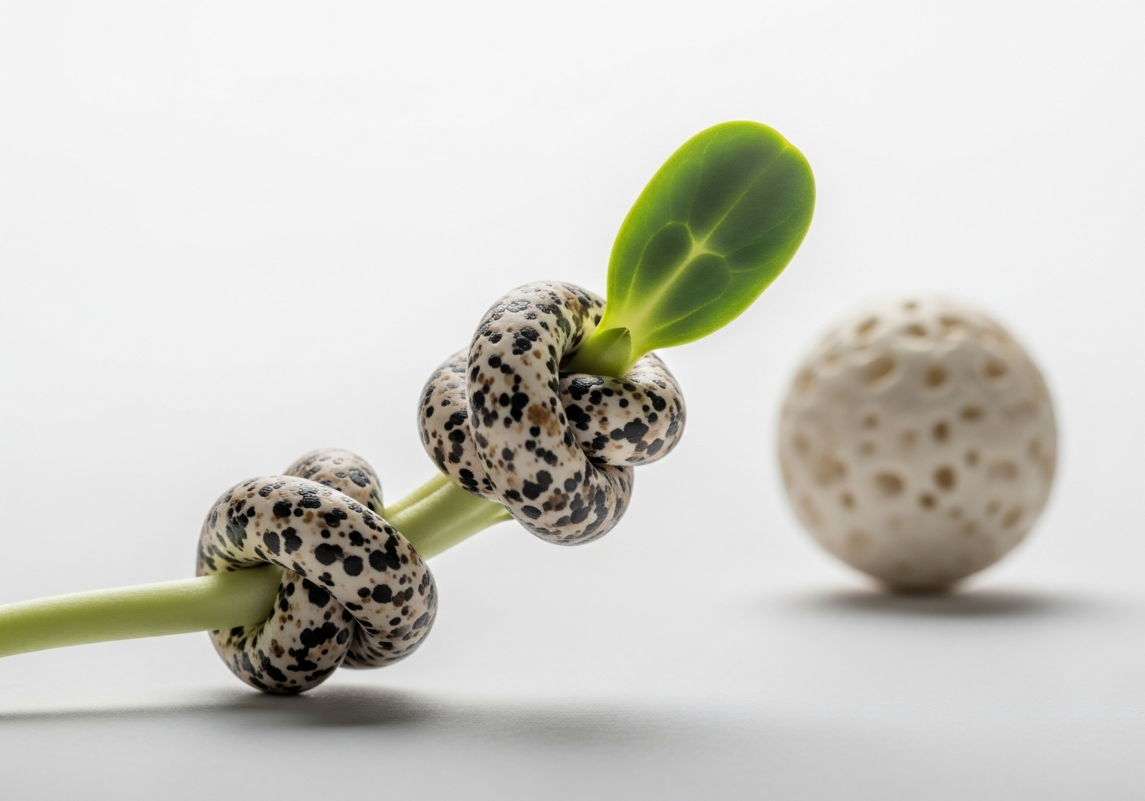
Growth hormone (GH) is a master conductor in this orchestra, directing processes of repair, regeneration, and growth. Growth hormone peptides, such as Sermorelin or Ipamorelin, are sophisticated tools designed to work with your body’s natural rhythms. They act as precise signals, prompting your pituitary gland to release its own endogenous growth hormone. This approach honors the body’s innate intelligence, aiming to restore a youthful pattern of hormonal communication.

The Cellular Architecture of Wellness
To understand how to support this process, we must look at the very structure of our cells. Every cell in your body is encased in a membrane, a dynamic barrier composed primarily of lipids, or fats. This membrane is the gatekeeper of cellular communication.
It contains the receptors that receive hormonal messages, including the signals from growth hormone. The composition of this membrane, which is directly influenced by the dietary fats you consume, determines its fluidity and the efficiency of its receptors. A rigid, poorly constructed cell membrane can muffle hormonal signals, much like a faulty antenna struggling to pick up a clear radio broadcast.
A fluid, well-formed membrane allows for crisp, clear communication, ensuring that the messages sent by your peptide protocol are received loud and clear.
Your dietary fat intake directly builds the cellular infrastructure that determines how well your body listens and responds to hormonal signals.
Dietary fats are more than just a source of calories; they are critical structural and signaling molecules. Moving beyond outdated classifications of “good” and “bad” fats allows us to appreciate their distinct biological roles. The fats you eat are incorporated into your cell membranes, they become the precursors for inflammatory or anti-inflammatory molecules, and they directly influence the hormonal environment of your bloodstream.

A Primer on Dietary Fat Families
Understanding the different families of fats is essential for anyone undertaking a sophisticated wellness protocol. Each type has a unique chemical structure and a corresponding unique function within your physiology.
- Saturated Fatty Acids (SFAs) Found in animal fats, coconut oil, and palm oil, SFAs are a stable source of energy and play a role in the synthesis of key hormones, including testosterone. They contribute to the structural integrity of cell membranes.
- Monounsaturated Fatty Acids (MUFAs) Abundant in olive oil, avocados, and nuts, MUFAs are known for their role in supporting cardiovascular health and improving insulin sensitivity. Their presence in cell membranes enhances fluidity and signaling.
- Polyunsaturated Fatty Acids (PUFAs) This family includes two critical sub-groups, omega-6 and omega-3, whose balance is paramount for metabolic health.
- Omega-6 PUFAs Found in many vegetable oils (like soybean, corn, and sunflower oil) and processed foods, they are precursors to signaling molecules that can promote inflammation when consumed in excess relative to omega-3s.
- Omega-3 PUFAs Found in fatty fish (salmon, mackerel, sardines), flaxseeds, and walnuts, these fats are precursors to powerful anti-inflammatory molecules. They significantly enhance cell membrane fluidity and are critical for optimal receptor function.
The question of how your diet affects your peptide therapy is therefore a question of cellular logistics. Are you providing your body with the raw materials it needs to build a responsive, efficient communication network? The choices you make at every meal contribute to the biological environment in which your therapy operates, directly influencing its potential for success.


Intermediate
Advancing our understanding requires a closer look at the precise biochemical conversations occurring within your body. The efficacy of growth hormone peptide therapy, which uses agents like CJC-1295 and Ipamorelin to stimulate natural GH pulses, is profoundly influenced by the metabolic state at the moment of administration.
One of the most powerful modulators of this state is the level of circulating free fatty acids (FFAs) in your bloodstream. These molecules, liberated from stored body fat or absorbed from recent meals, can act as a potent brake on growth hormone secretion.
This biological reality presents a critical strategic consideration for any individual on a peptide protocol. A meal high in dietary fat, consumed too close to your scheduled injection, will lead to a transient spike in plasma FFAs. These elevated FFAs send a direct inhibitory signal to the pituitary gland, the very organ your peptide is designed to stimulate.
The result is a blunted response. The carefully timed signal from the peptide is effectively dampened, and the potential for a robust, therapeutic pulse of growth hormone is diminished. This interaction underscores the necessity of aligning your dietary patterns with your therapeutic schedule to create a permissive metabolic environment.

The Critical Interplay of Insulin and Free Fatty Acids
The relationship between dietary choices and GH release is governed by a delicate interplay between several key hormones and metabolites. Insulin, the hormone responsible for nutrient storage, shares an inverse relationship with growth hormone. A meal rich in both refined carbohydrates and fats triggers a significant release of insulin.
This insulin surge works to clear glucose and fats from the blood, but it also independently suppresses GH secretion. When high insulin is combined with high FFAs, the inhibitory effect on the pituitary is compounded.
The timing and composition of your meals create a specific metabolic window that can either amplify or mute the signal from your peptide therapy.
Therefore, optimizing your protocol involves managing both the type of fat you consume and the timing of your meals. The goal is to ensure that at the moment of your peptide injection, both insulin and FFA levels are relatively low.
This is why many protocols advise administration on an empty stomach, such as first thing in the morning or at least two to three hours after your last meal. Taking a dose before fasted cardiovascular exercise can be particularly effective, as this is a time when insulin is low and the body is primed to utilize FFAs for energy, further clearing them from the bloodstream and opening the window for a powerful GH pulse.

How Do Different Fat Profiles Influence the Hormonal Environment?
The type of fat you consume regularly has a cumulative effect on your baseline metabolic health, which in turn affects the efficacy of your peptide therapy. A diet’s overall fatty acid profile shapes insulin sensitivity, systemic inflammation, and cell membrane composition over time.
A diet chronically high in processed omega-6 fatty acids and certain saturated fats can contribute to a state of low-grade systemic inflammation and insulin resistance. This creates a challenging metabolic backdrop for peptide therapy, as the body’s signaling pathways are already under strain.
Conversely, a diet rich in omega-3 PUFAs and MUFAs helps to quell inflammation, improve insulin sensitivity, and build highly fluid and responsive cell membranes. This creates a biological environment that is far more receptive to the subtle prompts of GH secretagogues.
| Dietary Fat Profile | Primary Sources | Impact on Free Fatty Acids (FFAs) | Effect on Insulin Sensitivity | Influence on Systemic Inflammation | Overall Impact on Peptide Efficacy |
|---|---|---|---|---|---|
| High Omega-3 PUFA | Fatty fish, flaxseed, walnuts, chia seeds |
Can lower circulating triglycerides; promotes efficient FFA utilization. |
Generally improves insulin sensitivity over time. |
Strongly anti-inflammatory, reducing metabolic noise. |
Highly Favorable. Creates a receptive, low-inflammation environment and enhances cell receptor function. |
| High Monounsaturated (MUFA) | Olive oil, avocados, almonds, macadamia nuts |
Neutral to favorable impact on FFA levels. |
Significantly improves insulin sensitivity. |
Anti-inflammatory, though less potent than omega-3s. |
Favorable. Supports the metabolic conditions needed for a robust GH pulse. |
| High Saturated (SFA) | Red meat, butter, coconut oil, full-fat dairy |
Can acutely raise FFA levels after meals. |
Can reduce insulin sensitivity when consumed in excess, particularly with high sugar intake. |
Can be pro-inflammatory depending on the specific acid and overall dietary context. |
Context-Dependent. Requires careful timing and moderation to avoid blunting GH pulses. |
| High Omega-6 PUFA | Soybean oil, corn oil, safflower oil, processed foods |
No direct negative impact on acute FFA levels. |
Can worsen insulin resistance over time if imbalanced with omega-3s. |
Strongly pro-inflammatory when the omega-6 to omega-3 ratio is high. |
Unfavorable. Contributes to a chronic state of inflammation that interferes with optimal hormonal signaling. |

Practical Dietary Strategies for Protocol Optimization
Translating this science into actionable steps is straightforward. The following strategies can help ensure your dietary choices support, rather than hinder, your therapeutic goals.
- Time Your Injections Strategically Administer your peptide dose in a fasted state. The most common and effective times are upon waking (at least 30-60 minutes before your first meal) or immediately before bed (at least 2-3 hours after your last meal).
- Avoid High-Fat Meals Pre-Injection Do not consume a meal, particularly one high in fat, within the 2-3 hour window before your injection. This will prevent the acute spike in FFAs that blunts the pituitary’s response.
- Prioritize Omega-3 and Monounsaturated Fats Structure your overall diet around sources of MUFAs and omega-3s. Use olive oil as your primary cooking fat, incorporate avocados and nuts into your meals, and consume fatty fish several times per week.
- Manage Your Omega-6 to Omega-3 Ratio Actively reduce your intake of processed vegetable oils and packaged foods. This simple step can dramatically lower systemic inflammation and improve your body’s overall hormonal signaling capacity.
- Consider Post-Workout Dosing for Anabolism For individuals focused on muscle accrual, administering a peptide dose immediately following a resistance training workout can be effective. During this window, muscle cells are highly insulin-sensitive, and the body is primed to use nutrients for repair, creating a powerful anabolic environment when combined with a GH pulse.


Academic
A granular analysis of the interaction between lipid metabolism and somatotropic axis function reveals a sophisticated, multi-layered regulatory system. The inhibitory influence of free fatty acids on growth hormone secretion is not a simple, monolithic process.
It is a finely tuned feedback mechanism executed through at least two distinct and synergistic pathways ∞ direct modulation of pituitary somatotrophs and indirect control via hypothalamic neuropeptides. Understanding these pathways is critical for appreciating the profound impact of dietary composition on the pharmacodynamics of GH secretagogues.
The primary regulator of the somatotropic axis is the dynamic balance between hypothalamic Growth Hormone-Releasing Hormone (GHRH), which is stimulatory, and Somatostatin (SST), which is inhibitory. Peptides like Sermorelin and Tesamorelin are GHRH analogs; they function by activating the GHRH receptor on pituitary somatotrophs to stimulate GH synthesis and release.
Other peptides, like Ipamorelin, and oral secretagogues, like MK-677, are ghrelin mimetics, activating the GHSR receptor, which also stimulates GH release while simultaneously suppressing somatostatin’s influence. The success of these therapies hinges on their ability to tip this balance in favor of GHRH and away from somatostatin.

The Hypothalamic Pathway the Somatostatin Clamp
Elevated plasma FFAs exert a powerful influence at the hypothalamic level by increasing somatostatin tone. Research has demonstrated that lipid infusions in human subjects augment the inhibitory signal of SST, effectively creating a “somatostatin clamp” that suppresses GH pulsatility. FFAs appear to directly stimulate the SST-secreting neurons in the periventricular nucleus of the hypothalamus.
This increased SST release into the hypophyseal portal system then acts on the pituitary, making the somatotrophs less responsive to the stimulatory signal of GHRH. Consequently, even in the presence of a therapeutic dose of a GHRH-analog peptide, the heightened inhibitory environment prevents a robust secretory response.
This mechanism is a classic negative feedback loop ∞ high circulating energy substrates (FFAs) signal to the brain that the body is in a fed state, thus downregulating the pro-growth, mobilizing signals of GH.

What Is the Direct Impact on the Pituitary?
Beyond the hypothalamic effect, compelling in-vitro evidence from studies using cultured rat anterior pituitary cells shows that FFAs can directly inhibit GH secretion at the pituitary level. In these experiments, the introduction of fatty acids like oleic and caprylic acid to the cell culture medium suppressed both basal and GHRH-stimulated GH release.
This suggests that FFAs can interfere with the intracellular signaling cascade that follows GHRH receptor activation. The precise molecular mechanism may involve alterations in second messenger systems like cyclic AMP (cAMP) or modulation of ion channel activity, effectively uncoupling the receptor’s activation from the final process of GH vesicle exocytosis. This direct pituitary action means that even if the hypothalamic somatostatin signal were bypassed, high FFA levels could still independently impair the efficacy of a GHRH-based peptide therapy.
The dual inhibitory action of free fatty acids, acting at both the hypothalamus and the pituitary, creates a formidable biological barrier to peptide-stimulated growth hormone release.
The clinical significance of this dual blockade was starkly illustrated in studies where lipid-heparin infusions were used to artificially raise FFA levels in healthy subjects. In one such study, the GHRH-induced GH secretory peak was almost completely abolished in the presence of high FFAs. This demonstrates the potent and clinically relevant nature of this inhibitory feedback.
| Study Focus | Methodology | Key Finding | Implication for Peptide Therapy |
|---|---|---|---|
| GHRH Response Under Lipid Infusion |
Intravenous infusion of a lipid-heparin solution in healthy male subjects, followed by a bolus of GHRH. |
The peak GH response to GHRH was reduced by approximately 71% (from 28.9 ng/mL to 8.4 ng/mL) during lipid infusion compared to saline control. |
Demonstrates that acute elevations in FFAs, similar to those after a high-fat meal, can severely blunt the effect of GHRH-analog peptides. |
| Direct Pituitary Action In Vitro |
Monolayer cultures of rat anterior pituitary cells exposed to FFAs (oleic and caprylic acid) and then stimulated with GHRH. |
FFAs inhibited both basal and GHRH-stimulated GH release directly at the cellular level, without affecting prolactin release, indicating a specific effect. |
The inhibitory mechanism is not solely dependent on the brain; it also occurs at the target tissue of the peptide, the pituitary gland itself. |
| Somatostatin Tone Investigation |
Comparing GH response to GHRH alone vs. GHRH plus Arginine (an SST suppressor) in subjects with high FFAs. |
High FFA levels were associated with increased somatostatin tone, which was a primary contributor to reduced GH secretion. |
Highlights the importance of managing factors that increase somatostatin, with high FFAs being a primary, diet-related example. |
| Ghrelin and FFA Interaction |
Investigation of ghrelin levels and GH dynamics in patient populations with metabolic dysregulation and high FFAs. |
Reduced ghrelin levels and impaired GH response were linked to excess FFAs, suggesting FFAs can disrupt multiple stimulatory pathways for GH. |
Ghrelin-mimetic peptides (e.g. Ipamorelin) may also face resistance in an environment of chronically elevated FFAs. |

The Role of Cell Membrane Composition in Receptor Sensitivity
A final layer of complexity involves the chronic impact of dietary fat choices on the biophysical properties of the somatotroph cell membrane. The phospholipid bilayer is not a static structure; its fluidity is determined by the fatty acid tails of its constituent phospholipids.
Diets rich in omega-3 PUFAs lead to their incorporation into these membranes, increasing fluidity. Diets high in saturated fats can lead to more rigid membranes. Membrane fluidity is critical for the proper function of transmembrane proteins, including the GHRH receptor.
A more fluid membrane allows the receptor to undergo the necessary conformational changes upon binding to its ligand, facilitating more efficient signal transduction. Therefore, a long-term dietary strategy emphasizing omega-3 fatty acids may enhance the sensitivity of the pituitary to GHRH-analog peptides, yielding a more robust response over time. This represents a foundational, structural optimization that complements the acute, tactical timing of injections.

References
- Lanzi, Roberto, et al. “Control on Growth Hormone Release by Free Fatty Acids Is Maintained in Acromegaly.” The Journal of Clinical Endocrinology & Metabolism, vol. 84, no. 8, 1999, pp. 2797-802.
- Casanueva, F. F. et al. “Free Fatty Acids Block Growth Hormone (GH) Releasing Hormone-Stimulated GH Secretion in Man Directly at the Pituitary.” The Journal of Clinical Endocrinology & Metabolism, vol. 65, no. 4, 1987, pp. 634-42.
- Lee, E. J. et al. “The Effect of Free Fatty Acids on Growth Hormone (GH)-Releasing Hormone-Mediated GH Secretion in Man.” The Journal of Clinical Endocrinology & Metabolism, vol. 66, no. 1, 1988, pp. 203-6.
- Grinspoon, S. et al. “Metabolic Regulation of Growth Hormone by Free Fatty Acids, Somatostatin, and Ghrelin in HIV-Lipodystrophy.” American Journal of Physiology-Endocrinology and Metabolism, vol. 286, no. 3, 2004, pp. E425-33.
- Veldhuis, Johannes D. and Cyril Y. Bowers. “Integrating GHS-R/Ghrelin and GHRH/GHRH-R Signaling Pathways ∞ A Model of How Two Hormones May Subserve One Function.” Reviews in Endocrine and Metabolic Disorders, vol. 11, no. 3, 2010, pp. 157-66.
- Skeik, N. and A. A. Shubrook. “Effects of Omega 3 Fatty Acids and Vitamin E on Hormones Involved in Carbohydrate and Lipid Metabolism in Men.” The American Journal of the Medical Sciences, vol. 305, no. 6, 1993, pp. 361-6.
- Goodman, H. Maurice. “The Role of Growth Hormone in Fat Mobilization.” Designing Foods ∞ Animal Product Options in the Marketplace, National Academies Press (US), 1988.
- J. A. G. M. van der Lely, et al. “The Somatotropic Axis in Obesity.” The Journal of Clinical Endocrinology & Metabolism, vol. 82, no. 1, 1997, pp. 31-35.

Reflection
The information presented here provides a map of the intricate biological landscape you are navigating. It details the molecular conversations, the feedback loops, and the profound connections between what you consume and how your body responds. This knowledge is a powerful tool, shifting your perspective from that of a passive recipient of therapy to an active, informed participant in your own physiological recalibration.
Consider the architecture of your daily life. How do your nutritional patterns align with the rhythm of your protocol? Where are the opportunities to create a more receptive environment for these signals of repair and regeneration? This journey is one of deep listening ∞ paying attention to the subtle feedback from your own body as you apply these principles.
The ultimate goal is to move beyond a rigid set of rules and cultivate an intuitive understanding of your unique system, fostering a collaborative partnership with your own biology to unlock your full potential for vitality and function.